Loading video…


 14 Concepts
 25.7K views
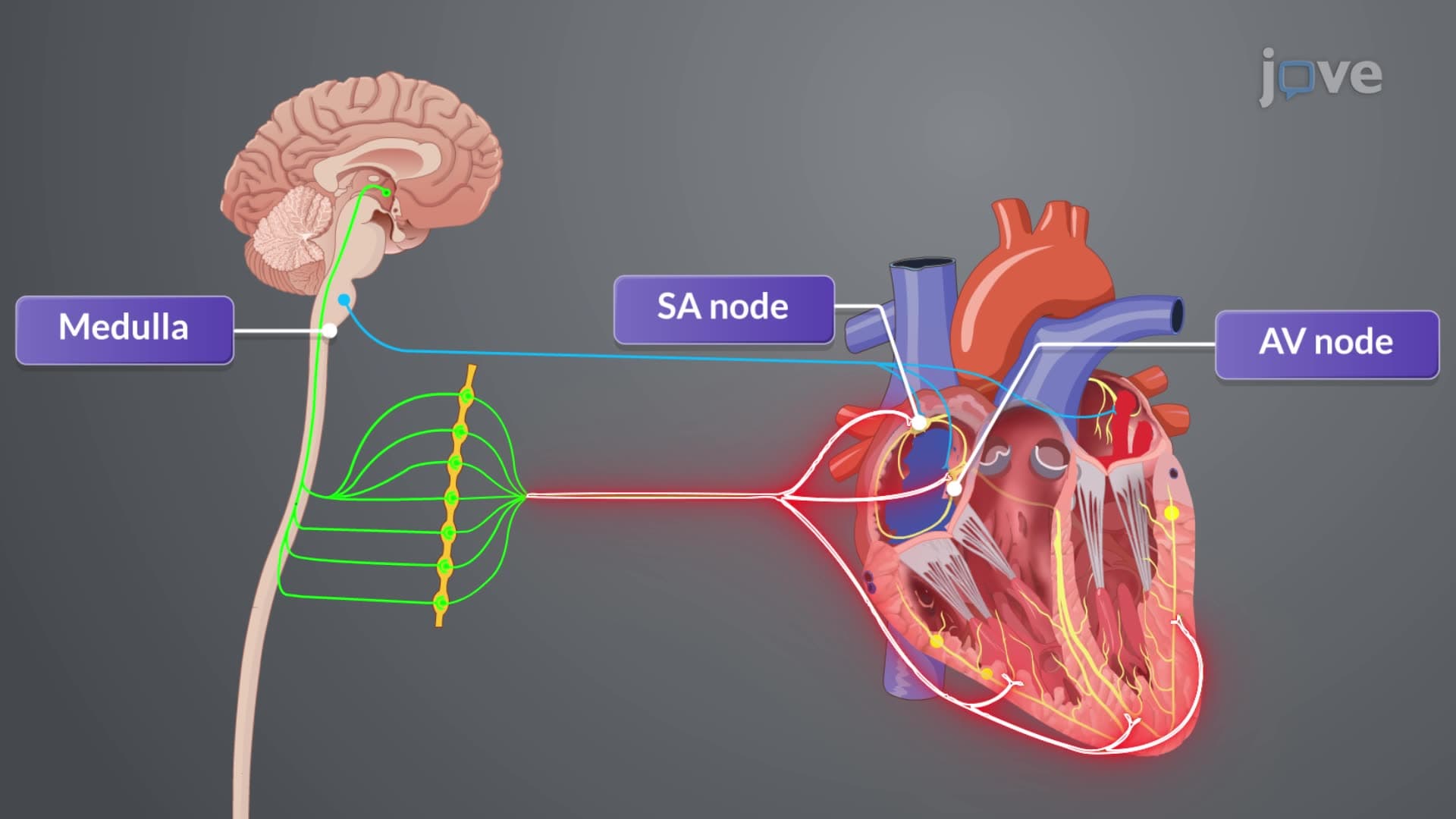

Video Summary: Cardiac Output I Effect of Explained
Ever wonder why your heart pounds during a marathon but slows during meditation? The cardiac output i effect of demonstrates how your cardiovascular system dynamically adjusts blood flow-from 5 liters per minute at rest to over 25 liters during intense exercise. Consider how emergency room physicians at Johns Hopkins use cardiac output measurements to assess critical patients' heart function and guide life-saving interventions. This fundamental concept explains the intricate balance between heart rate and stroke volume that keeps every cell in your body properly oxygenated. Watch the full video on JoVE Coach to master this concept with expert-led visuals and step-by-step explanations.
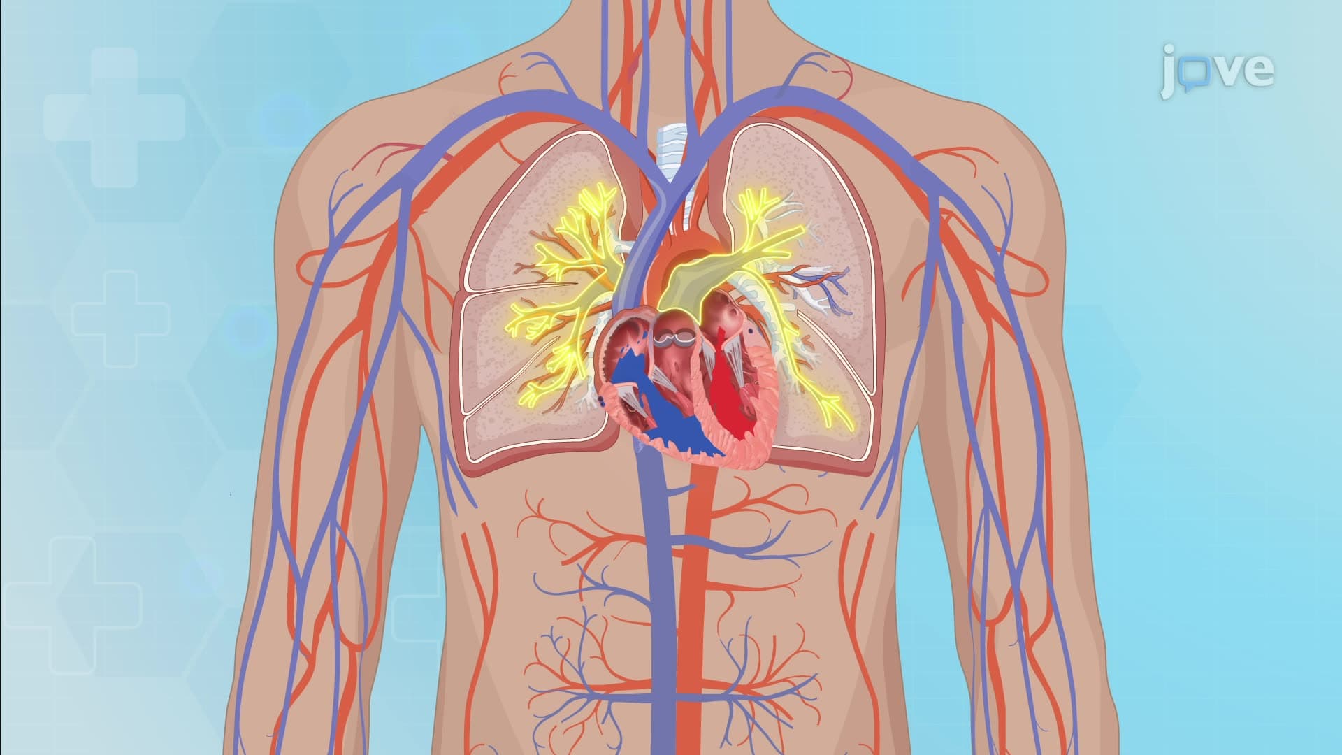
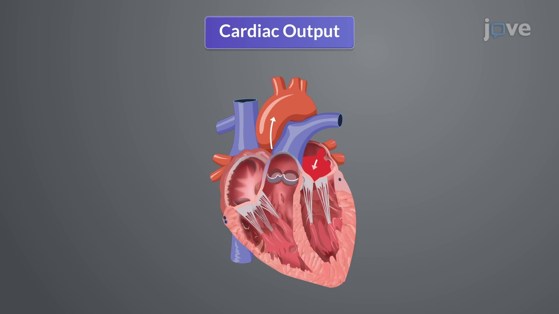
The cardiac output i effect of represents one of physiology's most elegant regulatory mechanisms, ensuring your cardiovascular system meets tissue demands whether you're sleeping, studying, or sprinting. Cardiac output, measured in liters per minute, equals heart rate multiplied by stroke volume-the amount of blood ejected with each heartbeat. A healthy adult's resting cardiac output averages 5 liters per minute, but this can triple during intense physical activity.
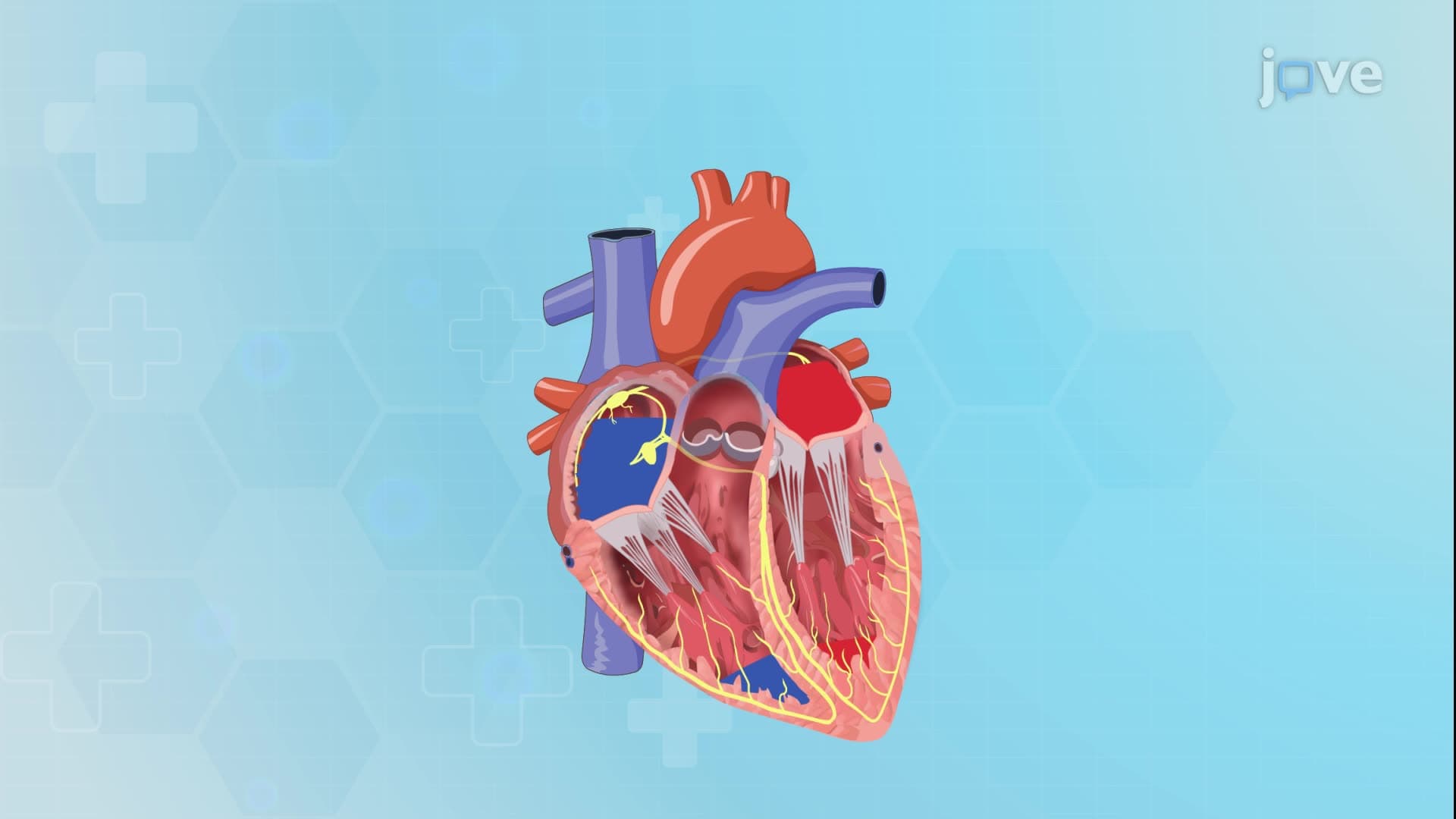
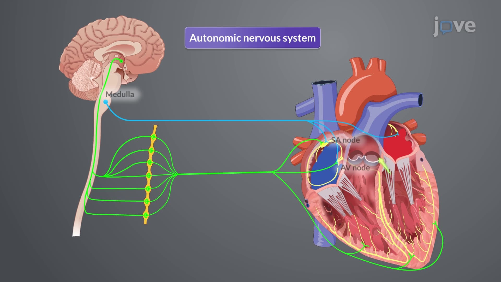
The sinoatrial (SA) node, your heart's natural pacemaker, receives constant input from both parasympathetic and sympathetic divisions. The parasympathetic system, operating through the vagus nerve, releases acetylcholine to slow heart rate-explaining why relaxation techniques lower your pulse. Conversely, sympathetic activation releases norepinephrine and epinephrine, binding to beta-1 receptors in cardiac muscle to increase both heart rate and contractility. This system becomes crucial during emergencies; paramedics at Houston Methodist Hospital rely on understanding these pathways when administering medications like atropine or epinephrine.
Specialized pressure sensors called baroreceptors, located in the aortic arch and carotid sinuses, continuously monitor blood pressure changes. When blood pressure rises (hypertension), these receptors signal the medulla oblongata to increase parasympathetic activity, reducing heart rate and cardiac output. During hypotension, decreased baroreceptor firing triggers sympathetic responses, elevating heart rate and vasoconstriction to restore adequate perfusion pressure.
For MCAT preparation, focus on how hormones like thyroid hormone and cortisol affect cardiac output long-term, while catecholamines provide immediate responses. AP Biology students should understand how cardiac output regulation exemplifies negative feedback mechanisms. In college physiology courses, you'll encounter advanced concepts like the Frank-Starling mechanism, which adjusts stroke volume based on venous return. Nursing students preparing for NCLEX exams must recognize abnormal cardiac output patterns in conditions like heart failure or shock, where compensatory mechanisms may become overwhelmed.
Related Micro-courses