Loading subject page…


25,669 views

Video Summary: Cardiovascular System Abnormal Findings I Explained
Did you know that a simple inspection of a patient's fingernails can reveal life-threatening heart conditions? Cardiovascular system abnormal findings I encompasses critical physical examination techniques that help identify heart disease, from detecting cyanosis in nail beds to recognizing jugular vein distension. At Johns Hopkins Hospital, emergency physicians routinely use these assessment skills to quickly identify patients experiencing heart failure or endocarditis. Watch the full video on JoVE Coach to master this concept with expert-led visuals and step-by-step explanations.
The systematic identification of cardiovascular abnormalities through physical examination represents a cornerstone skill in clinical medicine. These findings provide immediate insights into cardiac function, circulation status, and potential underlying pathology without requiring expensive diagnostic equipment.
The initial visual assessment reveals numerous cardiovascular clues. Xanthelasma, those characteristic yellowish deposits around the eyelids, signals hyperlipidemia-a major risk factor for coronary artery disease affecting over 38% of American adults. Cyanosis, particularly central cyanosis observed in the lips and tongue, indicates inadequate oxygenation often stemming from congenital heart defects or severe heart failure.
Nail findings provide particularly valuable information. Clubbing, where fingertips become enlarged and nail beds curve, develops in chronic hypoxic conditions like cyanotic congenital heart disease. Splinter hemorrhages-thin, dark lines under the nails-suggest infective endocarditis, a serious infection of heart valves that affects approximately 15,000 Americans annually.
Jugular venous distention (JVD) assessment in the Semi-Fowler's position (45-degree elevation) provides direct insight into right heart pressures. When visible above the sternal angle in this position, JVD indicates elevated right atrial pressure, commonly seen in heart failure, which affects over 6 million Americans.
Lower extremity examination reveals circulation adequacy through multiple indicators. Pitting edema, where finger pressure leaves temporary indentations, suggests fluid retention from heart failure. Arterial insufficiency manifests as leg ulcers, particularly around the ankles, and rubor-the dependent redness that occurs when poorly perfused legs are lowered.
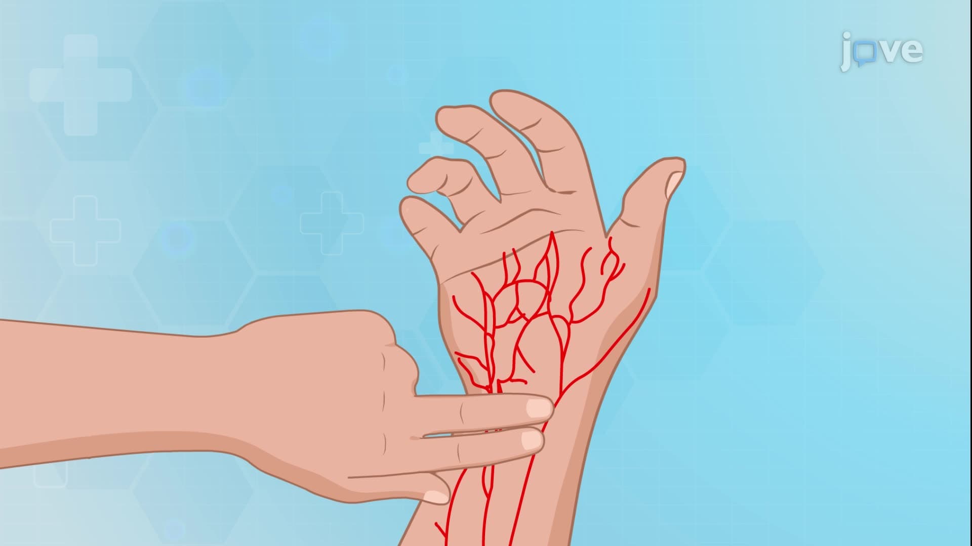
Pulse assessment extends beyond simple rate counting. Thready pulses indicate decreased stroke volume, while pulsus alternans-alternating strong and weak beats-suggests severe left ventricular dysfunction. Pulse deficits, where the apical heart rate exceeds the peripheral pulse rate, indicate atrial fibrillation or other arrhythmias.
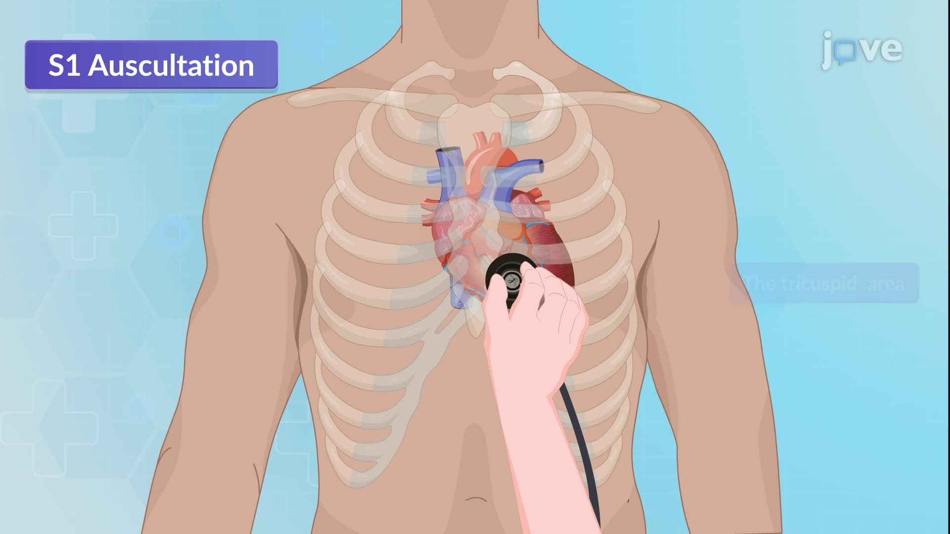
Precordial palpation identifies structural abnormalities. The point of maximal impulse (PMI) normally occurs at the fifth intercostal space in the midclavicular line. Lateral displacement suggests left ventricular enlargement, while heaves and thrills indicate turbulent blood flow from valvular disease or septal defects.
These examination skills prove essential for MCAT preparation, AP Biology coursework, and nursing school clinical rotations, providing foundational knowledge for healthcare career paths.
Related Micro-courses