Loading subject page…

2,643 views

Video Summary: Anatomy Genitourinary System I Kidneys Explained
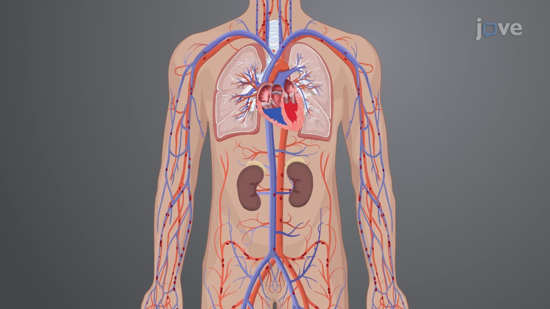
Every minute, your kidneys filter about 1.2 liters of blood-that's roughly 1,700 liters daily, equivalent to filtering your entire blood volume 400 times! The anatomy genitourinary system I kidneys reveals how these bean-shaped organs in your retroperitoneal space accomplish this remarkable feat through specialized structures like nephrons and renal pyramids. Consider how kidney disease affects over 37 million Americans, making understanding renal anatomy crucial for healthcare professionals. Watch the full video on JoVE Coach to master this concept with expert-led visuals and step-by-step explanations.
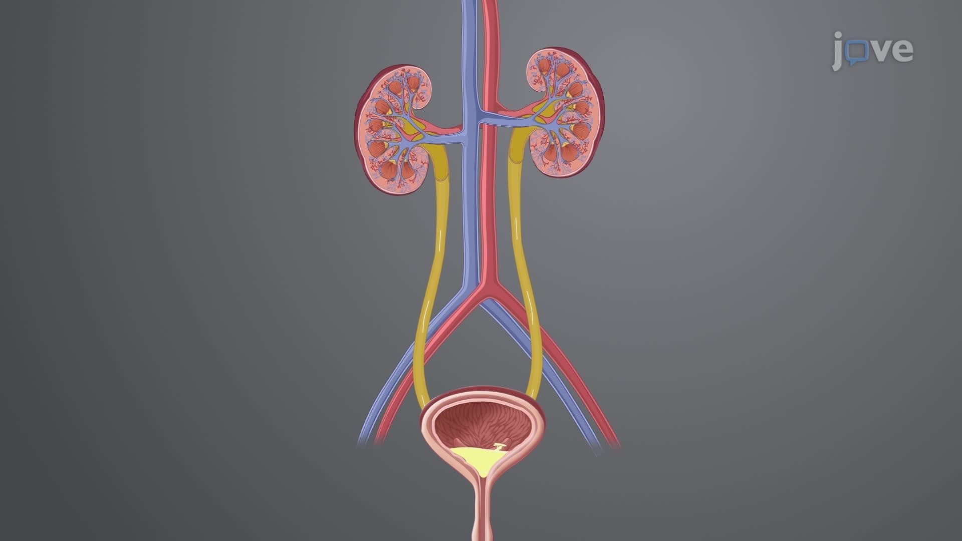
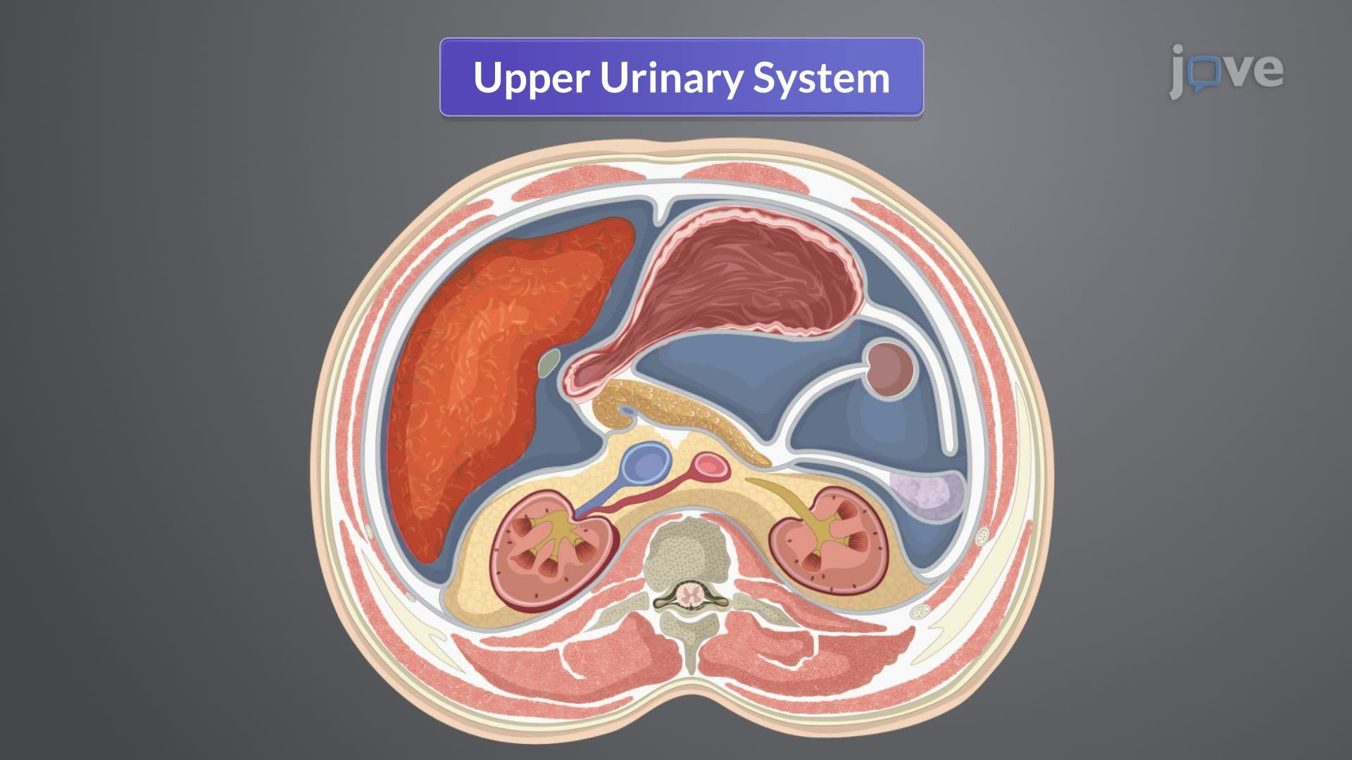
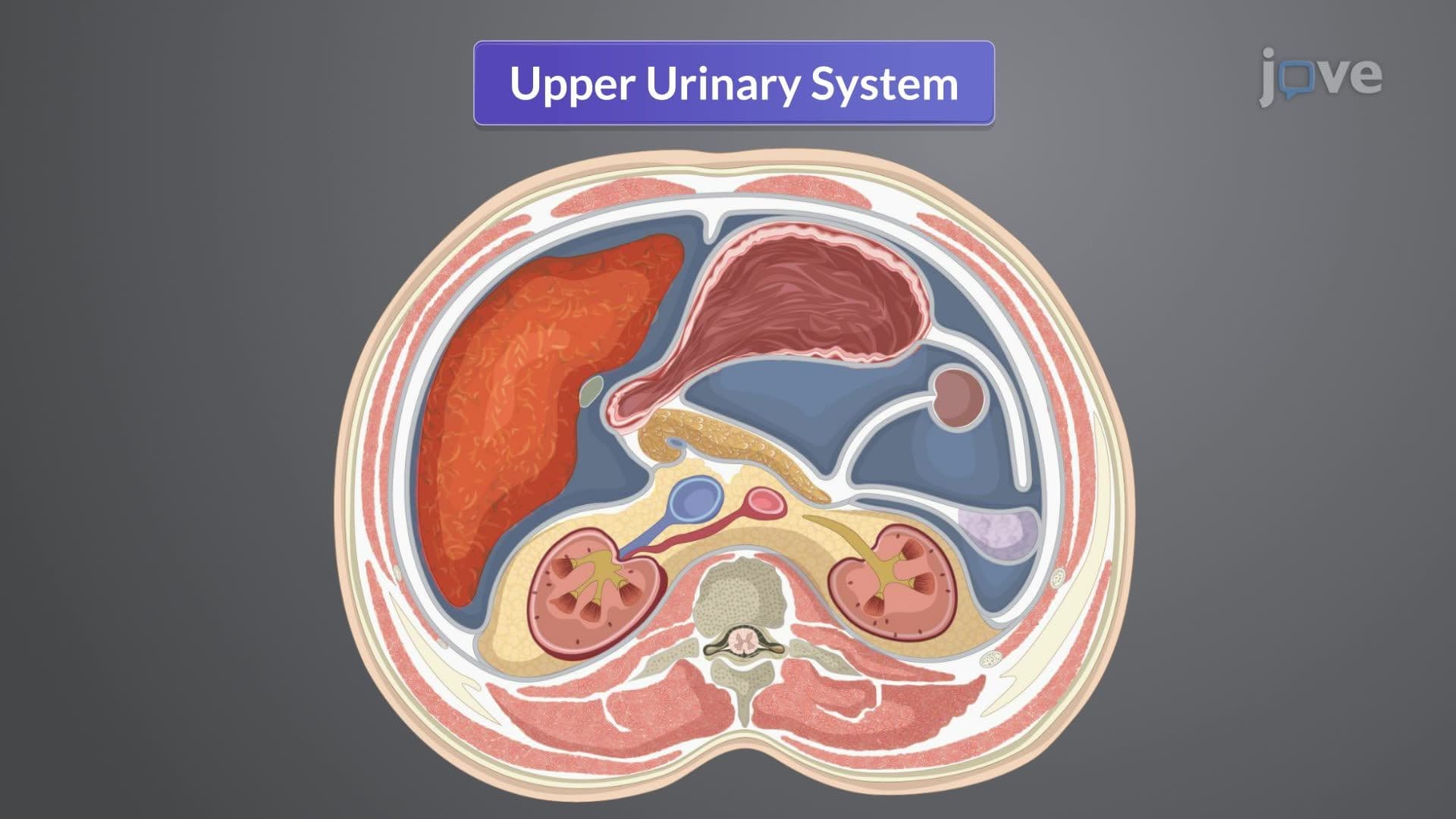
The human kidney represents one of anatomy's most sophisticated filtration systems, strategically positioned in the retroperitoneal space behind the peritoneum. This location provides stability while allowing the kidneys to move slightly with breathing-a design feature that prevents tearing during normal body movements.
Each kidney sits encased in a tough renal capsule, surrounded by protective fat called perinephric fat. This cushioning system proves critical during contact sports injuries-a common concern for high school athletes. The retroperitoneal location means kidneys are vulnerable to blunt force trauma from car accidents, making understanding their anatomy essential for emergency medicine professionals taking the MCAT or USMLE exams.
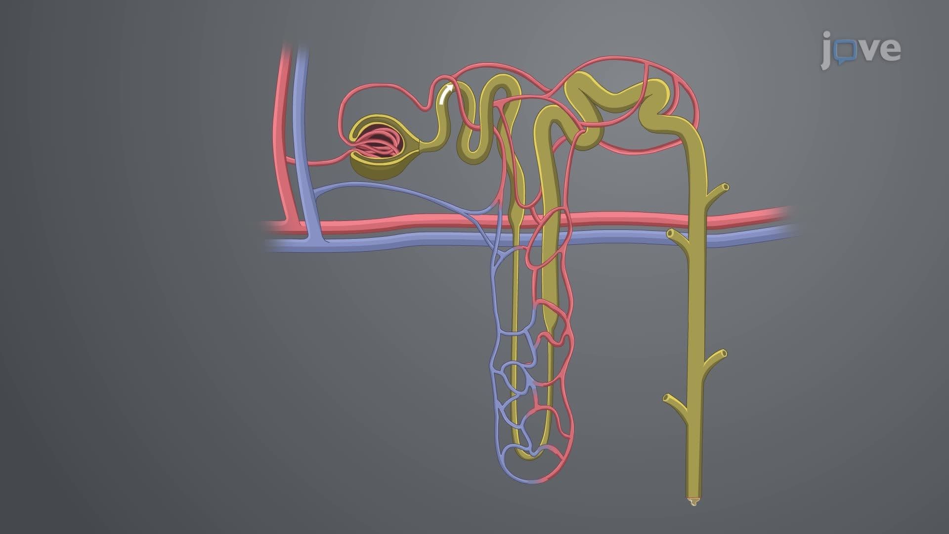
The kidney's architecture divides into two distinct regions with specialized functions. The outer cortex houses approximately one million nephrons per kidney-microscopic filtering units that perform the actual work of blood purification. Students preparing for AP Biology often struggle with nephron components, but understanding their spatial arrangement within the cortex helps clarify their coordinated function.
The inner medulla contains 8-12 renal pyramids, cone-shaped structures that channel filtered substances toward collection points. These pyramids create the kidney's characteristic striated appearance on cross-section, a detail frequently tested on college anatomy midterms and nursing entrance exams like the HESI A2.
The ureters demonstrate elegant anatomical engineering-25-30 centimeter tubes with three distinct layers enabling efficient urine transport. The innermost mucosa provides a smooth surface for fluid flow, while the middle muscular layer creates peristaltic waves pushing urine toward the bladder. The outer adventitia anchors ureters to surrounding tissues, preventing displacement during movement. This three-layer structure appears regularly on NCLEX questions, as nurses must understand normal anatomy to recognize pathological changes like kidney stones or urinary tract infections affecting millions of Americans annually.
Related Micro-courses