Loading subject page…

2,643 views

Video Summary: Anatomy Genitourinary System Ii Bladder Guide
Did you know your bladder can stretch to hold nearly a full water bottle's worth of urine? The anatomy genitourinary system ii bladder reveals how this remarkable muscular organ, positioned behind your pubic bone, manages urine storage through four specialized layers and works with the urethra to control elimination. Medical students at Johns Hopkins University study these structures extensively when learning urological procedures. Watch the full video on JoVE Coach to master this concept with expert-led visuals and step-by-step explanations.
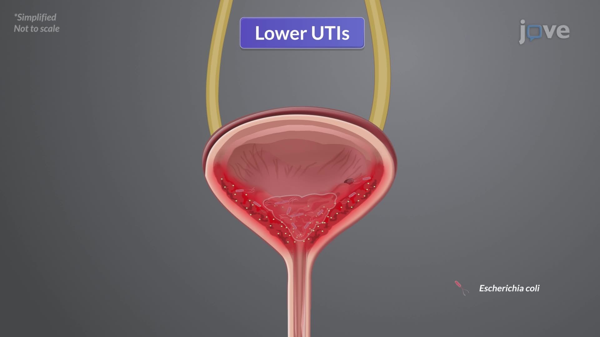
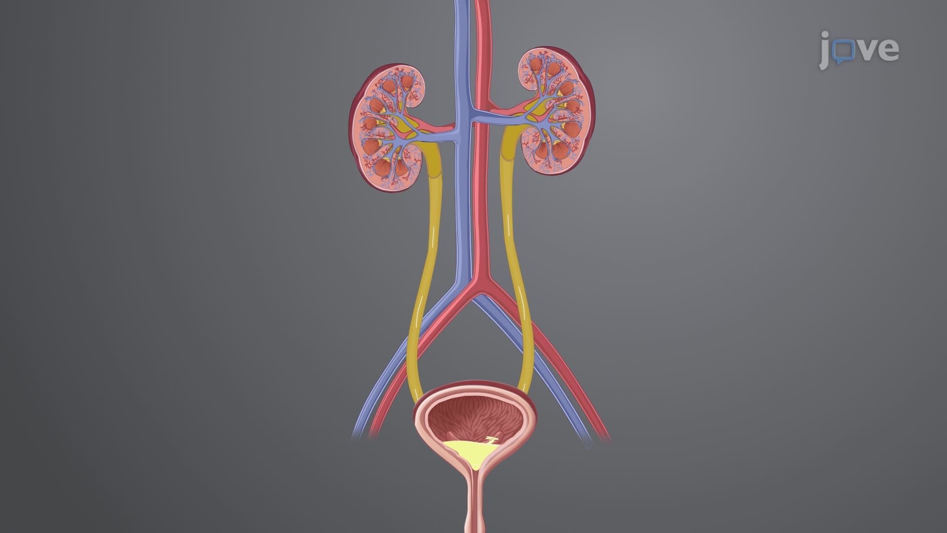
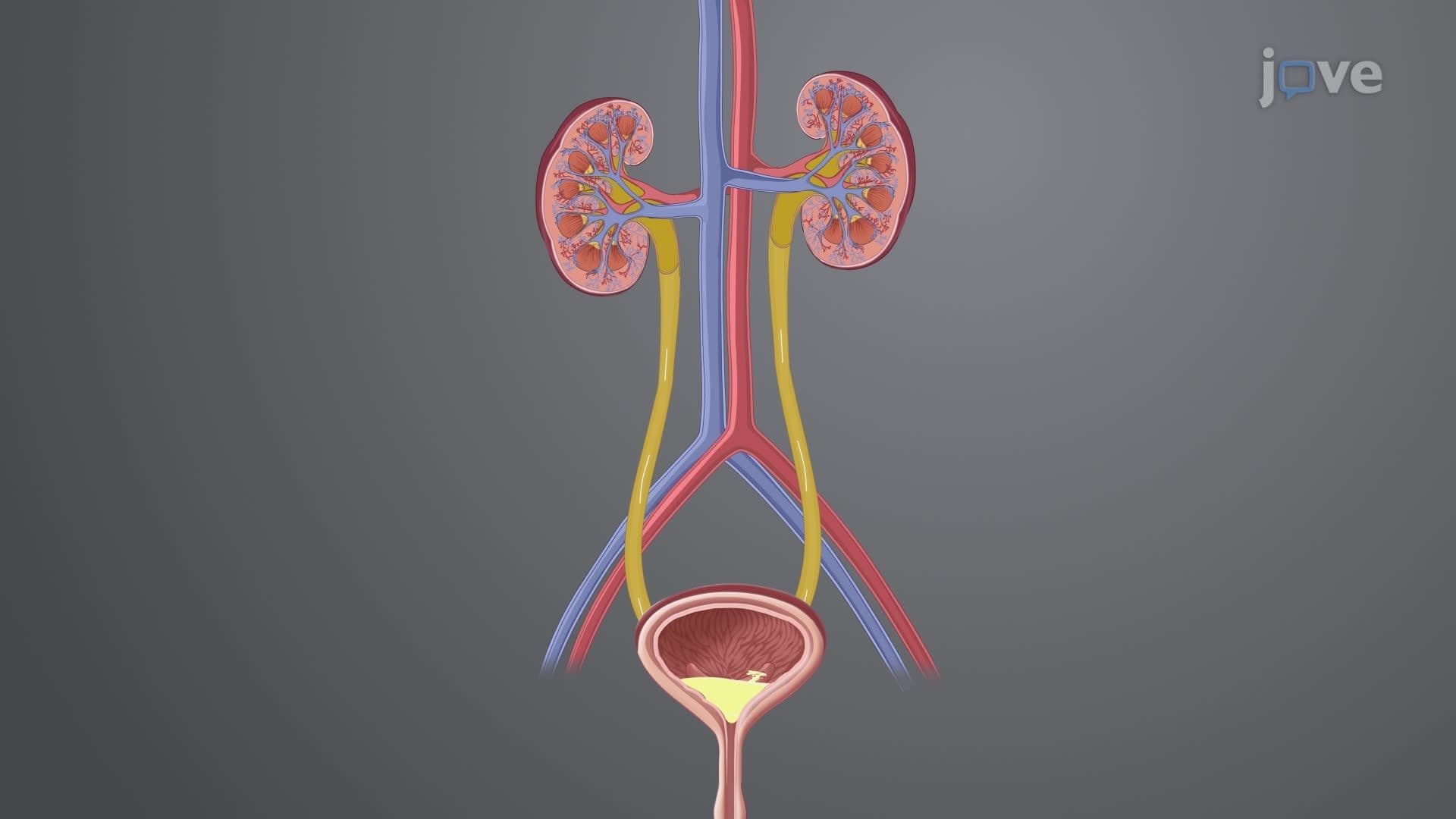
The lower urinary system represents a masterpiece of biological engineering, where form perfectly follows function. The bladder serves as the body's primary urine reservoir, strategically positioned in the pelvis behind the protective pubic symphysis. This location shields the organ from external trauma while maintaining optimal anatomical relationships with surrounding structures.
The bladder's four-layered wall structure demonstrates remarkable specialization. The outermost adventitia provides structural support and houses major blood vessels, while the powerful detrusor muscle-composed of smooth muscle fibers arranged in multiple directions-generates the contractile force necessary for complete bladder emptying. The submucosa layer contains the intricate network of blood vessels and nerve fibers that coordinate bladder function, and the innermost urothelium creates an impermeable barrier that prevents urine components from being reabsorbed into the bloodstream.
Students preparing for the MCAT or AP Biology exams should note how the urothelium's transitional epithelium allows for significant stretching without losing barrier function-a concept frequently tested in cellular biology sections.
Male and female lower urinary systems exhibit distinct anatomical differences with important clinical implications. In females, the short 2.5-5 centimeter urethra increases susceptibility to urinary tract infections, a concept relevant for nursing students taking the NCLEX exam. The female bladder's position anterior to the vagina and uterus creates unique considerations during pregnancy and childbirth.
Male anatomy presents greater complexity, with the 20-25 centimeter urethra passing through the prostate gland-a relationship crucial for understanding benign prostatic hyperplasia, commonly studied in college anatomy and physiology courses. The trigone region, visible in both sexes, forms a triangular area at the bladder base where the ureters enter and urine exits toward the urethra.
The dual sphincter system exemplifies the body's sophisticated control mechanisms. The involuntary internal urethral sphincter, composed of smooth muscle, maintains continence automatically, while the voluntary external sphincter allows conscious control over urination timing. This coordination between autonomic and somatic nervous systems frequently appears in USMLE Step 1 questions and undergraduate neuroscience coursework.
Related Micro-courses