Loading video…


 19 Concepts
 15.5K views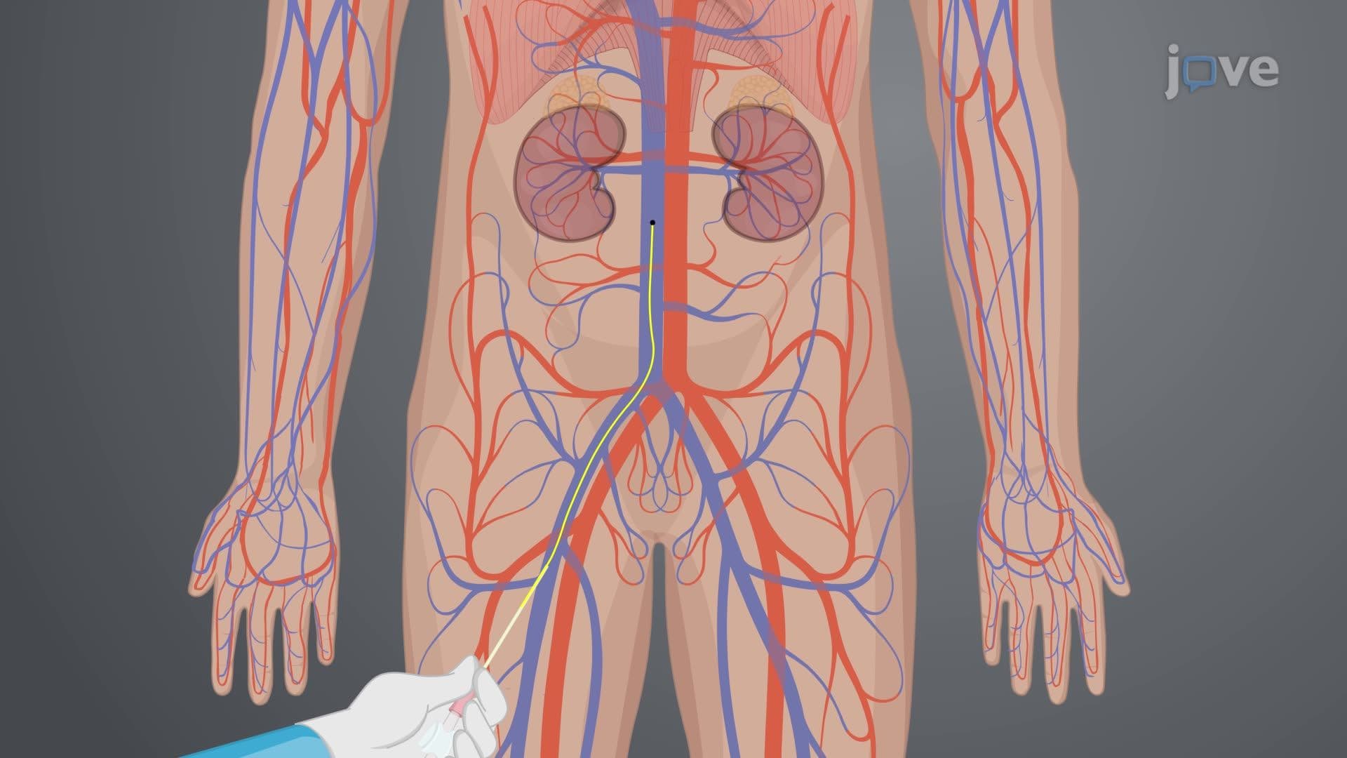

Video Summary: Cardiac Catheterization Ii Right Heart Catheterization Explained
Did you know that right heart catheterization can detect life-threatening pulmonary hypertension that affects over 200,000 Americans annually? Cardiac catheterization ii right heart catheterization is an invasive diagnostic procedure that directly measures pressures in the heart's right chambers and pulmonary arteries. At leading medical centers like Mayo Clinic, cardiologists use this technique to diagnose conditions ranging from heart failure to congenital defects by threading a specialized catheter through veins into the right heart. Watch the full video on JoVE Coach to master this concept with expert-led visuals and step-by-step explanations.
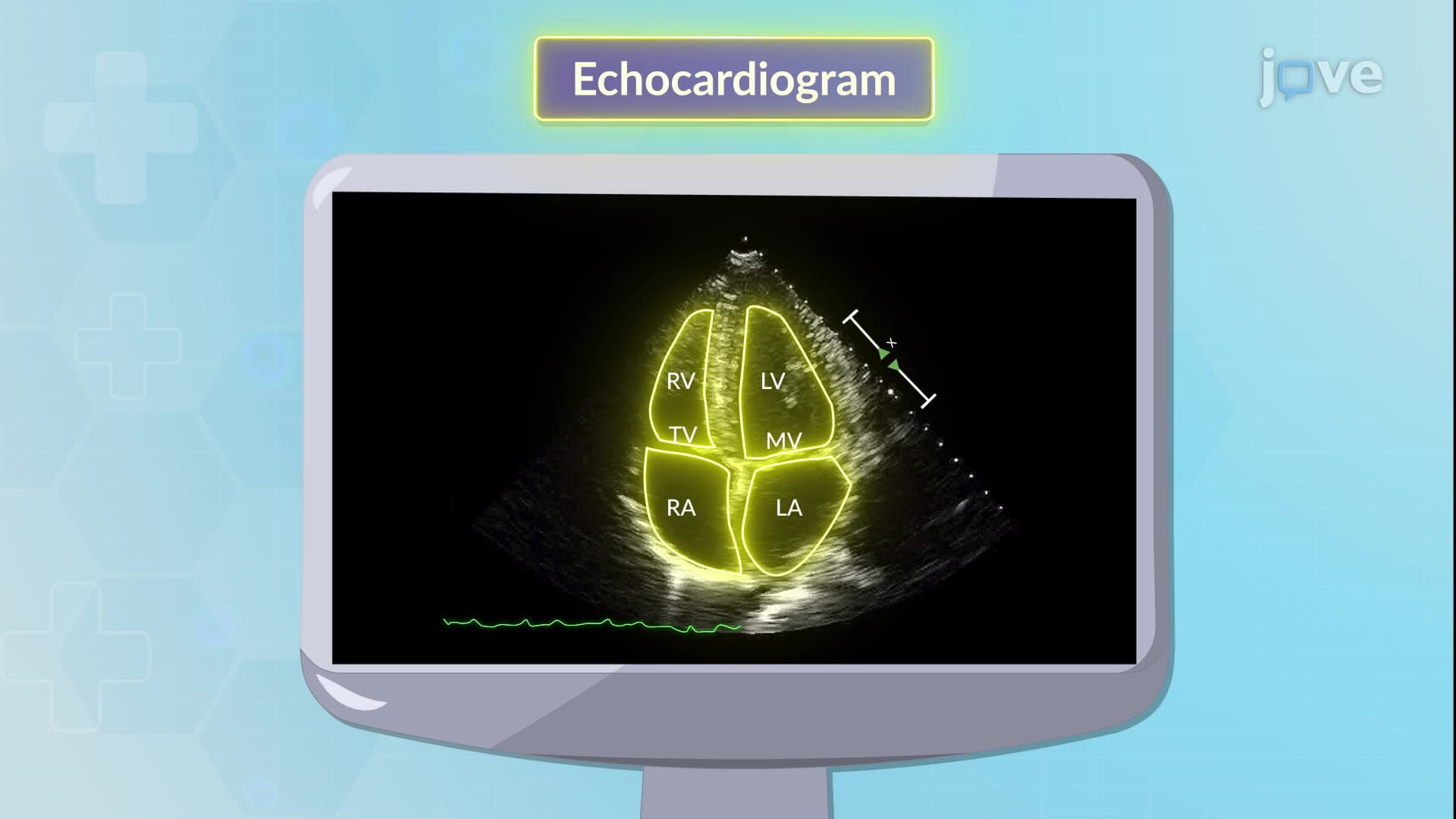
Right heart catheterization represents a cornerstone diagnostic procedure in modern cardiology, providing direct hemodynamic measurements that non-invasive tests cannot achieve. Unlike echocardiography or cardiac MRI, this invasive technique offers real-time pressure data from the heart's right chambers and pulmonary circulation, making it essential for diagnosing conditions like pulmonary arterial hypertension, heart failure, and congenital heart defects.
Healthcare providers at major US medical centers including Cleveland Clinic and Johns Hopkins utilize right heart catheterization when patients present with unexplained dyspnea, suspected pulmonary hypertension, or complex congenital heart disease. The procedure proves particularly valuable in evaluating patients for heart transplantation candidacy, as it provides precise measurements of pulmonary vascular resistance and cardiac output. For students preparing for the MCAT or advanced placement biology exams, understanding these clinical applications demonstrates the practical relevance of cardiovascular physiology concepts.
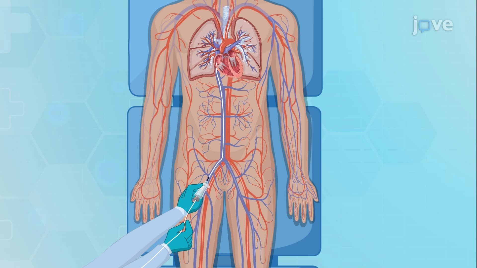
The procedure begins with establishing venous access through carefully selected sites. The femoral vein remains the most common approach due to its large diameter and straight path to the right atrium, while the internal jugular vein offers advantages in patients with peripheral vascular disease. The catheter's journey follows the venous return pathway: from access site through the superior or inferior vena cava, into the right atrium, across the tricuspid valve into the right ventricle, and finally through the pulmonary valve into the pulmonary artery.
Normal right atrial pressure ranges from 2-8 mmHg, while right ventricular systolic pressure typically measures 15-30 mmHg. Elevated readings may indicate conditions such as tricuspid stenosis, pulmonary hypertension, or right heart failure. For college students studying cardiovascular physiology, these pressure relationships illustrate Frank-Starling mechanisms and the impact of afterload on cardiac performance. The procedure also enables calculation of cardiac output using the Fick equation, integrating oxygen consumption data with arteriovenous oxygen differences.
Related Micro-courses