Loading video…


 19 Concepts
 15.5K views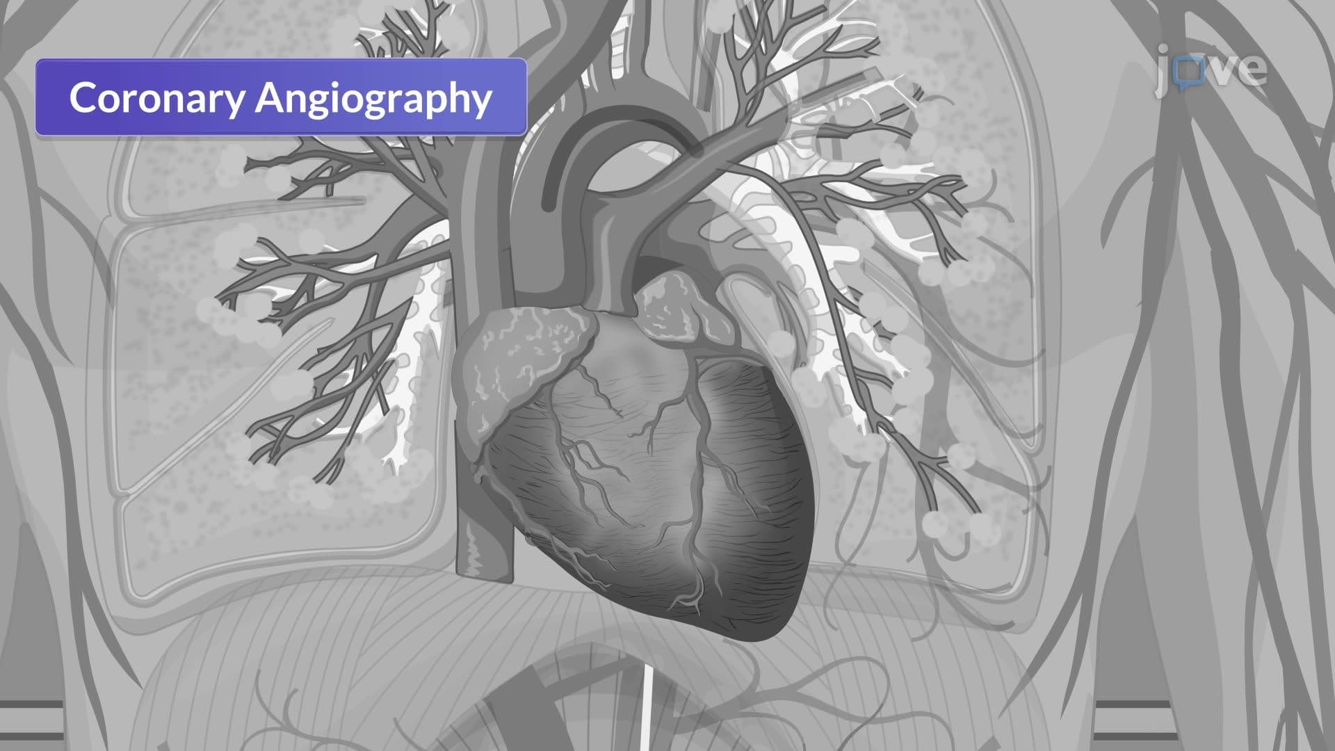

Video Summary: Cardiac Catheterization Iii Left Heart Catheterization Explained
Every year, over 600,000 Americans undergo cardiac catheterization procedures at hospitals like Cleveland Clinic and Mayo Clinic to diagnose life-threatening heart conditions. Cardiac catheterization III left heart catheterization represents a critical diagnostic technique where physicians thread specialized catheters through arteries to evaluate the heart's left ventricle, coronary arteries, and major valves. This procedure combines advanced imaging with real-time assessment to detect blockages, valve dysfunction, and heart muscle problems. Watch the full video on JoVE Coach to master this concept with expert-led visuals and step-by-step explanations.
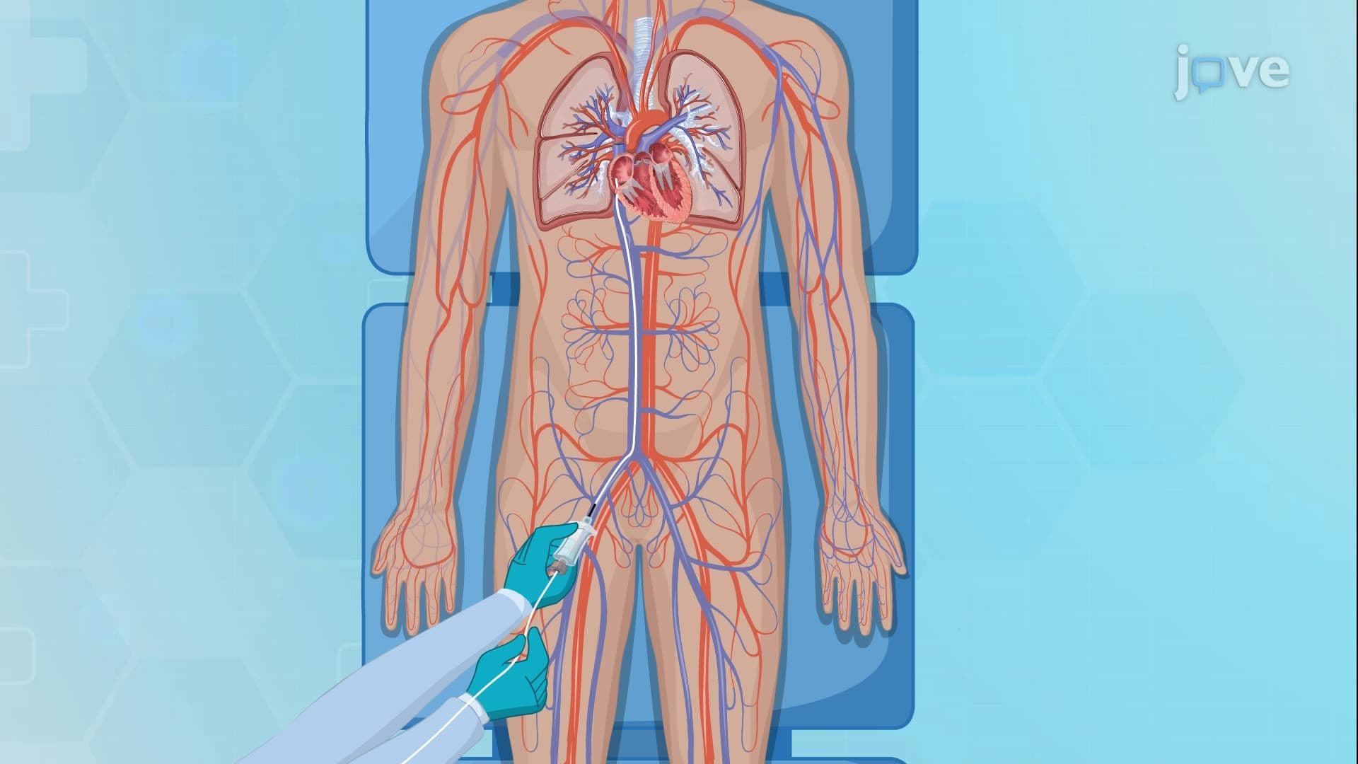
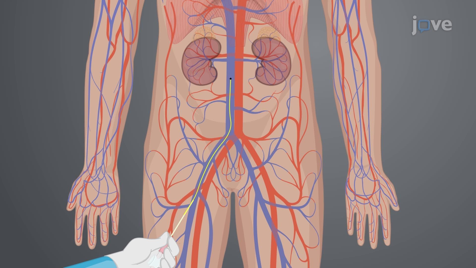
Cardiac catheterization III left heart catheterization stands as one of the most sophisticated diagnostic procedures in modern cardiology, providing cardiologists with unprecedented access to evaluate the heart's left-sided structures. Unlike non-invasive imaging techniques, this procedure offers real-time, high-resolution visualization of cardiac function through direct arterial access. The retrograde approach-meaning the catheter travels backward through the arterial system-allows physicians to reach the left ventricle by threading specialized catheters through peripheral arteries and into the aorta.
The procedure typically utilizes three primary arterial access points, each offering distinct advantages. The radial artery approach, increasingly popular at institutions like Johns Hopkins and Massachusetts General Hospital, provides enhanced patient comfort and reduced bleeding complications. The traditional femoral artery access remains the gold standard for complex procedures requiring larger catheter systems, while brachial artery catheterization serves as an alternative when other routes are contraindicated. Students preparing for the MCAT or advanced placement biology exams should understand that catheter selection depends on patient anatomy, procedural complexity, and physician expertise.
The procedure encompasses three primary diagnostic modalities that work synergistically to provide comprehensive cardiac evaluation. Angiography involves injecting radiopaque contrast agents to visualize blood flow patterns through the left heart chambers and aortic arch, revealing structural abnormalities and flow disturbances. Coronary angiography represents the gold standard for detecting coronary artery disease, enabling direct visualization of arterial blockages that could lead to myocardial infarction. Ventriculography assesses left ventricular function by measuring ejection fraction, wall motion abnormalities, and chamber dimensions-critical parameters that determine treatment strategies.
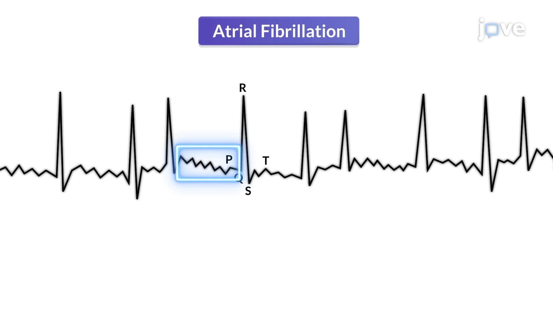
Real-world applications of this procedure extend far beyond diagnostic imaging. At leading cardiac centers like the Cleveland Clinic, left heart catheterization guides treatment decisions for conditions ranging from coronary artery disease to valvular abnormalities. However, the procedure carries inherent risks that require careful monitoring. Catheter manipulation and contrast injection can precipitate myocardial ischemia by temporarily disrupting coronary blood flow, while mechanical irritation may trigger dangerous arrhythmias. Understanding these complications proves essential for students pursuing healthcare careers, as it emphasizes the critical balance between diagnostic benefits and procedural risks in clinical decision-making.
Related Micro-courses