Loading video…


 19 Concepts
 15.5K views

Video Summary: Cardiovascular Imaging Echocardiography Overview
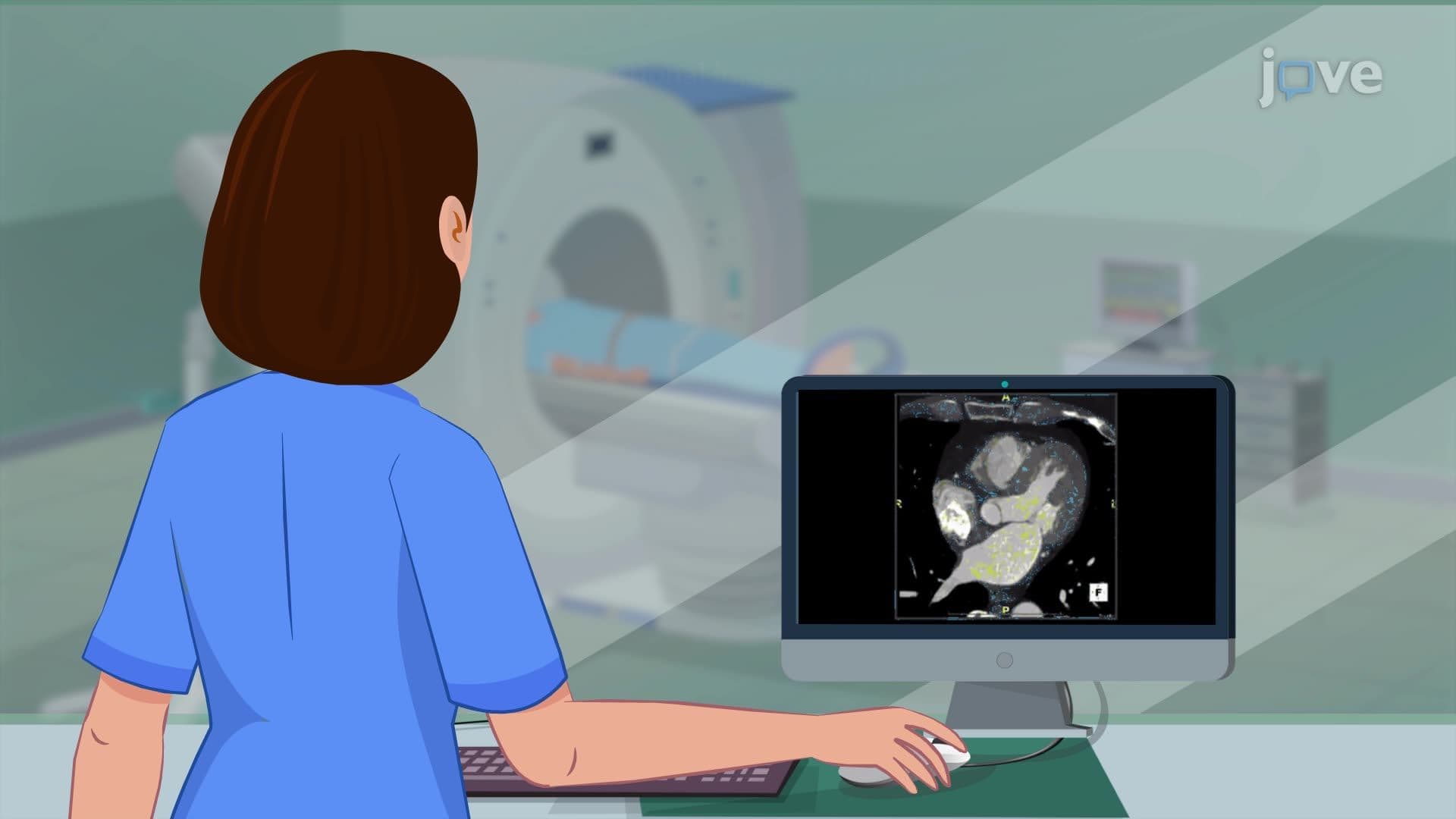
Did you know that over 6 million Americans live with heart failure, yet many cases could be detected early through a simple, painless test? Cardiovascular imaging: echocardiography overview reveals how ultrasound technology creates real-time moving pictures of your beating heart, helping doctors at institutions like Mayo Clinic diagnose conditions from valve problems to heart muscle weakness. This non-invasive procedure uses the same sound wave technology that allows expectant parents to see their unborn babies. Watch the full video on JoVE Coach to master this concept with expert-led visuals and step-by-step explanations.
Echocardiography represents one of the most important non-invasive diagnostic tools in modern cardiology, using ultrasound technology to create detailed, real-time images of cardiac structures. This imaging modality has revolutionized heart disease diagnosis since its introduction in the 1950s, allowing healthcare providers to assess cardiac function without surgical intervention.
The fundamental principle underlying echocardiography involves the transmission of high-frequency sound waves (typically 2-10 MHz) through chest tissues. When these waves encounter different cardiac structures-such as heart valves, chamber walls, and blood-they reflect back at varying intensities and timing. The transducer both emits these sound waves and receives the returning echoes, similar to how bats use echolocation for navigation.
Modern echocardiographic equipment utilizes sophisticated computer algorithms to convert these reflected sound waves into comprehensive visual displays. The Doppler effect, discovered by Austrian physicist Christian Doppler, plays a crucial role in assessing blood flow velocity and direction within cardiac chambers and vessels. Students preparing for the MCAT will recognize this as the same physical principle that causes ambulance sirens to change pitch as they approach and recede.
Echocardiography excels in diagnosing numerous cardiovascular conditions prevalent in the United States. Heart failure, affecting approximately 6.2 million American adults according to the CDC, can be assessed through measurements of ejection fraction-the percentage of blood pumped out of the left ventricle with each heartbeat. Normal ejection fraction ranges from 50-70%, while values below 40% typically indicate systolic heart failure.
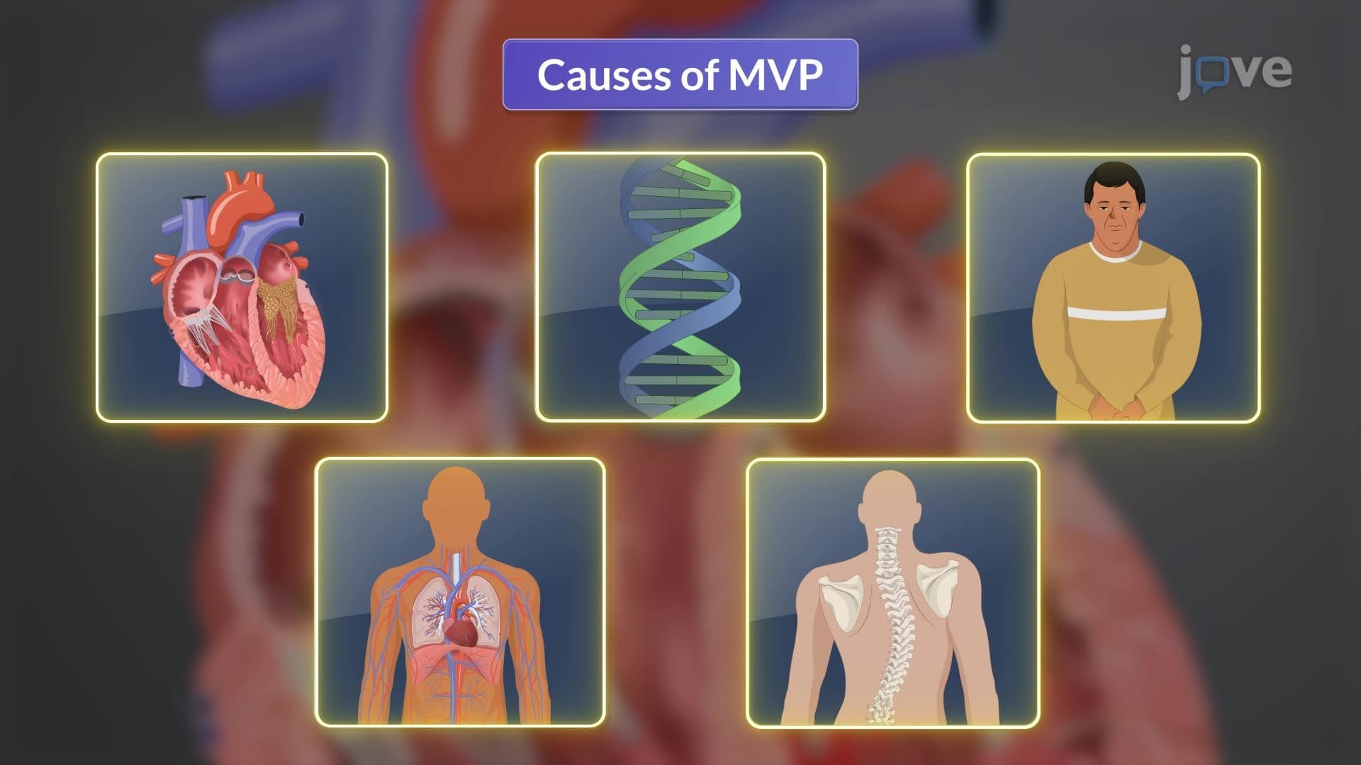
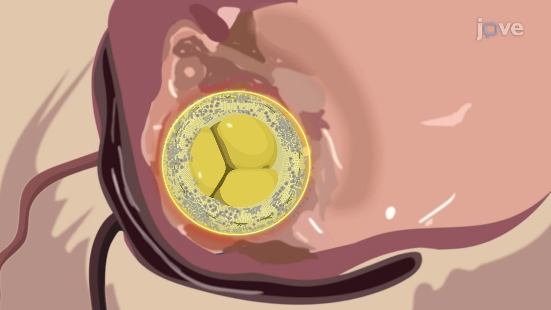
Valve disorders, including mitral valve prolapse (affecting 2-3% of Americans) and aortic stenosis, are readily identified through echocardiographic assessment. The procedure can detect valve regurgitation (backward blood flow), stenosis (narrowing), and structural abnormalities that may require surgical intervention. Major cardiac centers like Cleveland Clinic and Johns Hopkins rely heavily on echocardiographic findings to guide treatment decisions for valve replacement or repair procedures.
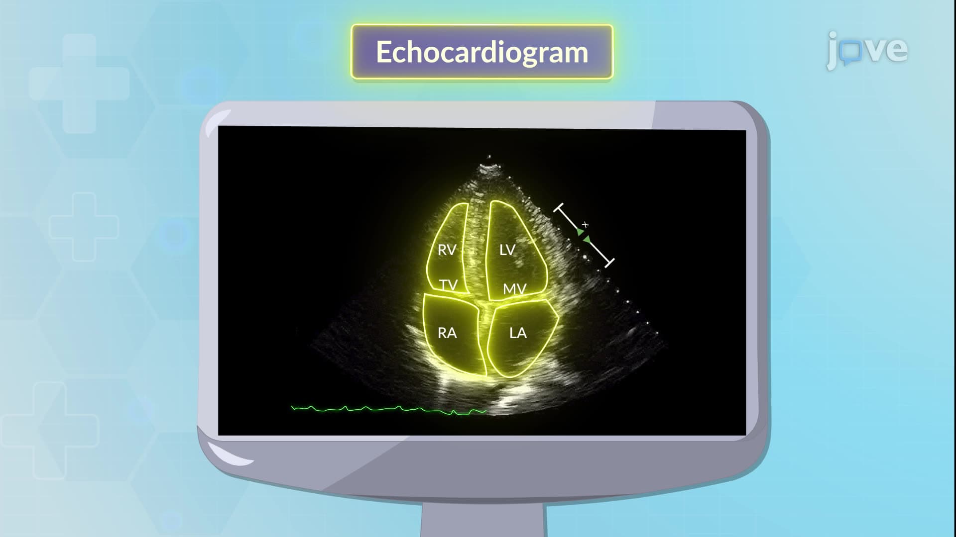
The positioning techniques described in the procedure-including parasternal long-axis, parasternal short-axis, apical four-chamber, and subcostal views-provide comprehensive cardiac assessment from multiple angles. Each view offers unique insights: parasternal views excel for measuring chamber dimensions and wall thickness, while apical views optimize assessment of ventricular function and valve competency.
For students taking AP Biology or preparing for nursing entrance exams like the HESI A2 or TEAS, understanding these anatomical correlations proves essential. The second intercostal space corresponds to the aortic area for auscultation, while the fourth intercostal space aligns with the tricuspid valve region-concepts that appear frequently on the NCLEX-RN examination.
Related Micro-courses