Loading video…


 19 Concepts
 15.5K views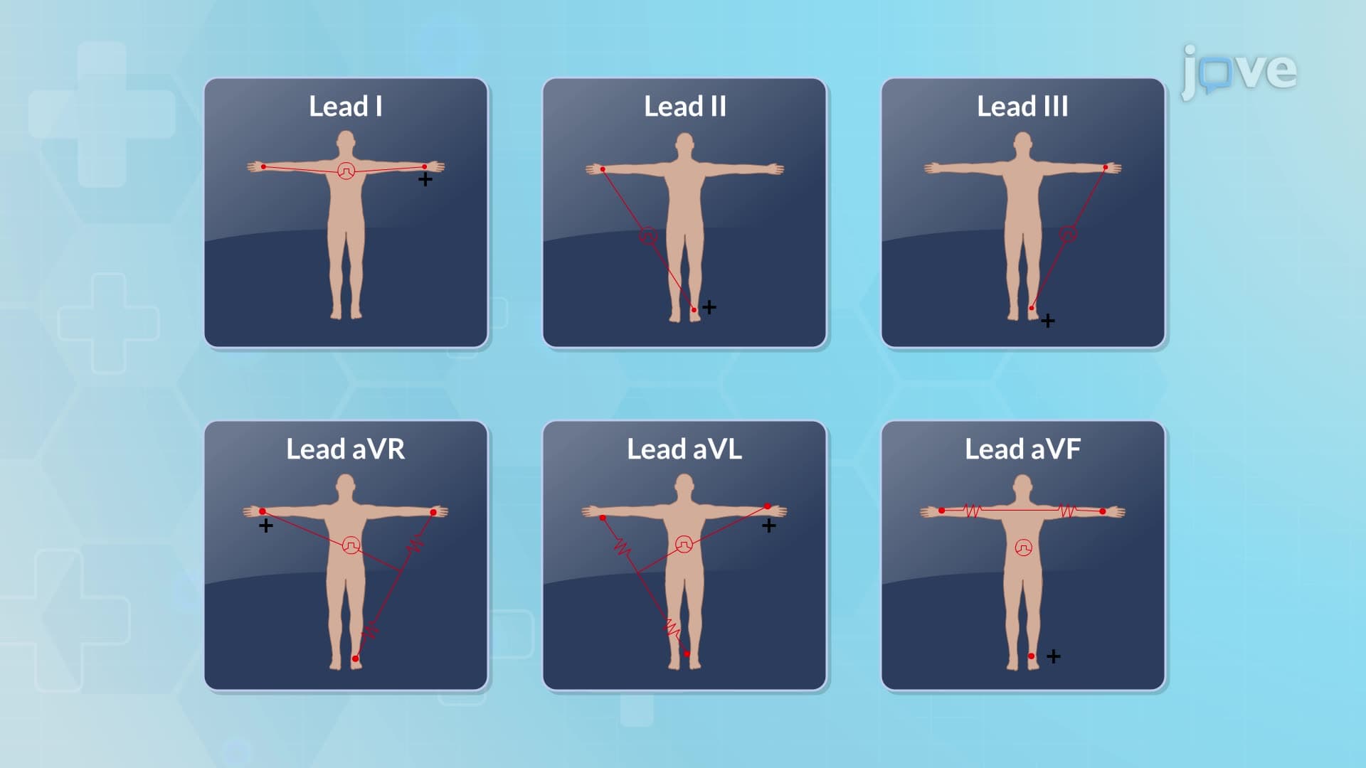

Video Summary: What Is Electrocardiogram Fundamentals
Did you know that every heartbeat generates electrical signals that can be captured and analyzed to diagnose heart conditions? Electrocardiogram fundamentals form the foundation of cardiac diagnostics, involving a standardized 12-lead system that records the heart's electrical activity through strategically placed electrodes. At hospitals like Mayo Clinic, healthcare professionals rely on these precise waveform interpretations daily to assess patient cardiac health. Watch the full video on JoVE Coach to master this concept with expert-led visuals and step-by-step explanations.
Electrocardiogram fundamentals represent the cornerstone of modern cardiac diagnostics, providing healthcare professionals with a non-invasive window into the heart's electrical activity. This sophisticated yet standardized approach has revolutionized cardiovascular medicine since its development, becoming an essential skill for medical professionals across specialties.
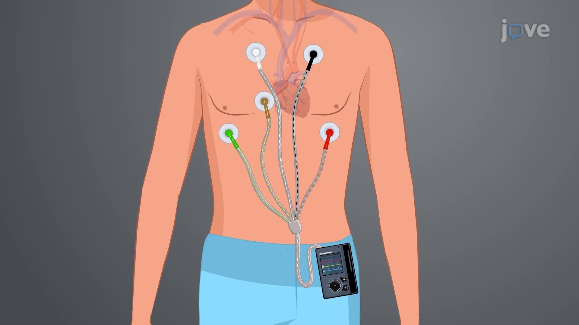
The standard ECG employs a meticulously designed 12-lead configuration that captures electrical activity from multiple cardiac perspectives. The limb electrodes, positioned on the inner wrists and ankles, generate six limb leads that provide frontal plane views of cardiac electrical activity. These include the bipolar limb leads (I, II, III) and the augmented unipolar leads (aVR, aVL, aVF), each offering unique diagnostic insights.
The precordial chest leads (V1-V6) provide horizontal plane perspectives, with each electrode strategically positioned to optimize cardiac electrical signal detection. V1 and V2, positioned at the fourth intercostal spaces, primarily assess right heart and septal activity. V4's placement at the fifth intercostal space on the left midclavicular line targets the cardiac apex, while V5 and V6 evaluate lateral wall activity. This systematic approach ensures comprehensive cardiac assessment, crucial for conditions ranging from myocardial infarctions to arrhythmias commonly studied in AP Biology and college-level anatomy courses.
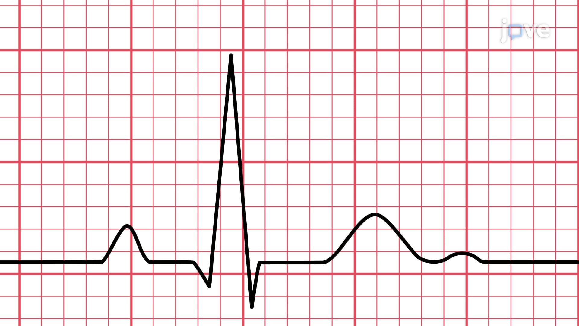
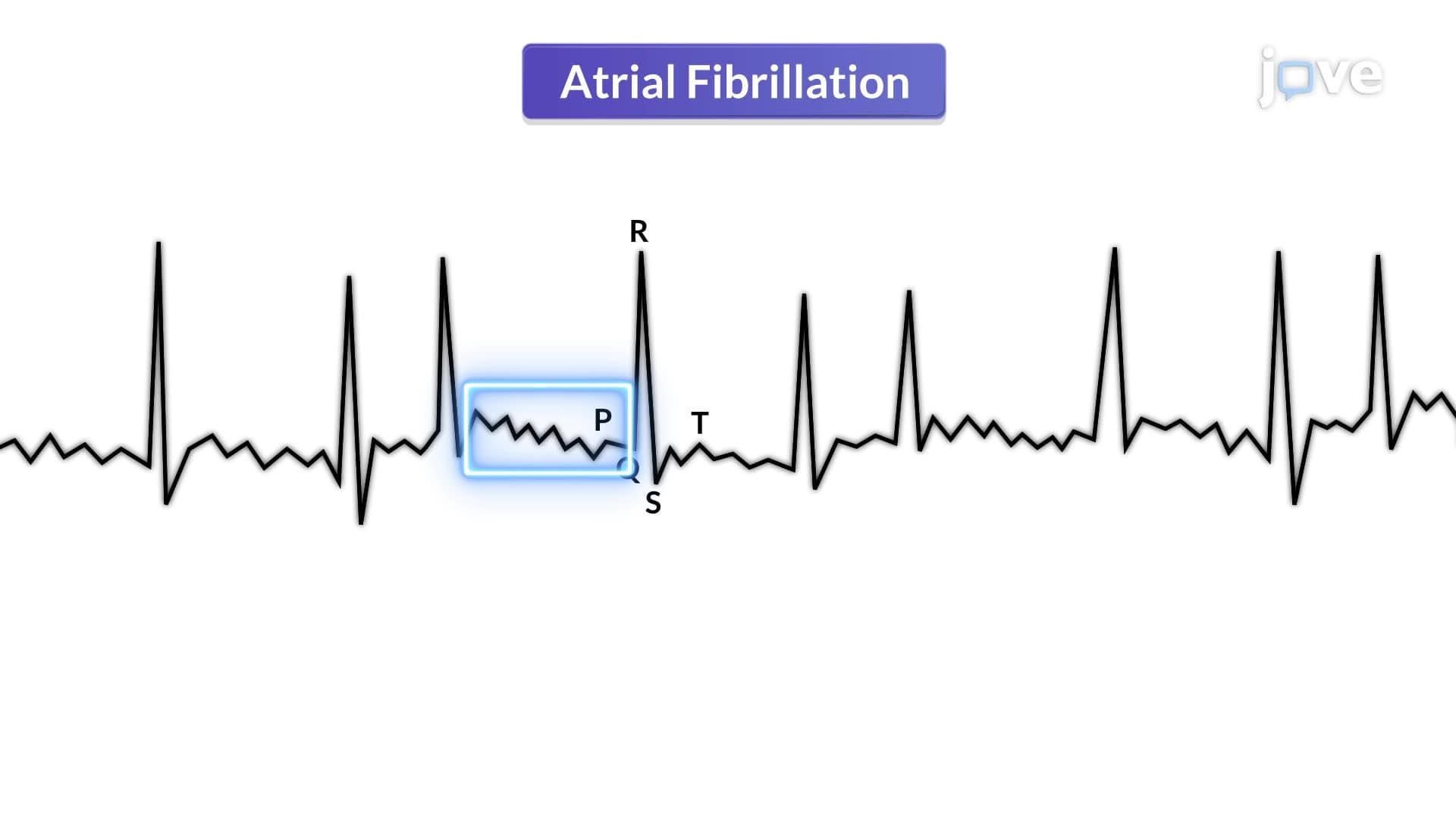
The ECG waveform components represent distinct phases of cardiac electrical activity. The P wave's smooth, rounded morphology indicates coordinated atrial depolarization, while the sharp, narrow QRS complex reflects rapid ventricular depolarization. The PR interval, measuring from P wave onset to QRS beginning, provides critical timing information about atrioventricular conduction-knowledge essential for MCAT preparation and medical school cardiology rotations.
The ST segment and T wave phases represent ventricular repolarization, with abnormalities often indicating ischemia or other pathological conditions. The QT interval encompasses total ventricular electrical activity duration, with prolongation potentially predisposing to dangerous arrhythmias. Understanding these fundamentals proves invaluable for students preparing for healthcare professional exams and clinical rotations at institutions like Johns Hopkins or Cleveland Clinic.
Mastery of electrocardiogram fundamentals extends beyond academic requirements, forming the foundation for clinical decision-making in emergency departments, intensive care units, and outpatient cardiology practices throughout the United States. Students pursuing healthcare careers will encounter these concepts extensively in coursework and licensing examinations, making thorough understanding essential for academic and professional success.
Related Micro-courses