Loading video…


 19 Concepts
 15.5K views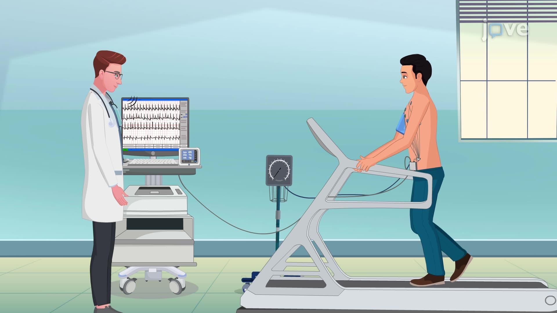

Video Summary: What Is Exercise Stress Test
Every year, over 600,000 Americans die from heart disease, making early detection crucial for survival. An exercise stress test serves as a vital diagnostic tool that reveals hidden cardiovascular problems by monitoring how your heart responds under physical exertion. At major medical centers like the Mayo Clinic, cardiologists use this non-invasive procedure to evaluate chest pain, assess cardiac function, and detect coronary artery disease before symptoms become life-threatening. Watch the full video on JoVE Coach to master this concept with expert-led visuals and step-by-step explanations.
An exercise stress test represents one of the most valuable non-invasive cardiac diagnostic procedures in modern medicine. This sophisticated evaluation method challenges the cardiovascular system through controlled physical exertion, revealing cardiac abnormalities that may remain hidden during rest. Unlike resting electrocardiograms, which only capture baseline cardiac activity, stress testing unmasks coronary artery disease, arrhythmias, and functional cardiac limitations under physiological demand.
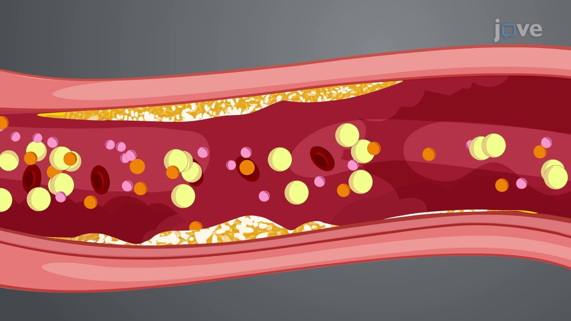
The procedure's diagnostic power stems from a fundamental cardiovascular principle: diseased coronary arteries cannot adequately supply oxygen-rich blood to heart muscle during increased workload. This supply-demand mismatch produces characteristic ECG changes, symptoms, and hemodynamic responses that trained healthcare professionals can interpret to guide treatment decisions.
Healthcare providers across the United States utilize exercise stress tests for multiple diagnostic purposes. Cardiologists at institutions like Cleveland Clinic and Johns Hopkins frequently order these tests to evaluate chest pain of unknown origin, assess exercise tolerance in cardiac rehabilitation patients, and screen high-risk individuals for coronary artery disease. The test proves particularly valuable for detecting multi-vessel coronary disease and determining functional capacity in patients with known cardiac conditions.
Emergency departments often use stress testing to evaluate patients with atypical chest pain who present with normal resting ECGs and cardiac enzymes. This application helps differentiate between cardiac and non-cardiac chest pain, potentially preventing unnecessary hospital admissions while ensuring appropriate cardiac workups for high-risk patients.
The exercise stress test follows a standardized protocol beginning with comprehensive pre-test preparation. Patients must fast for three hours while avoiding stimulants like caffeine, alcohol, and tobacco for 24 hours beforehand. This preparation ensures accurate baseline measurements and prevents interference with cardiac monitoring equipment.
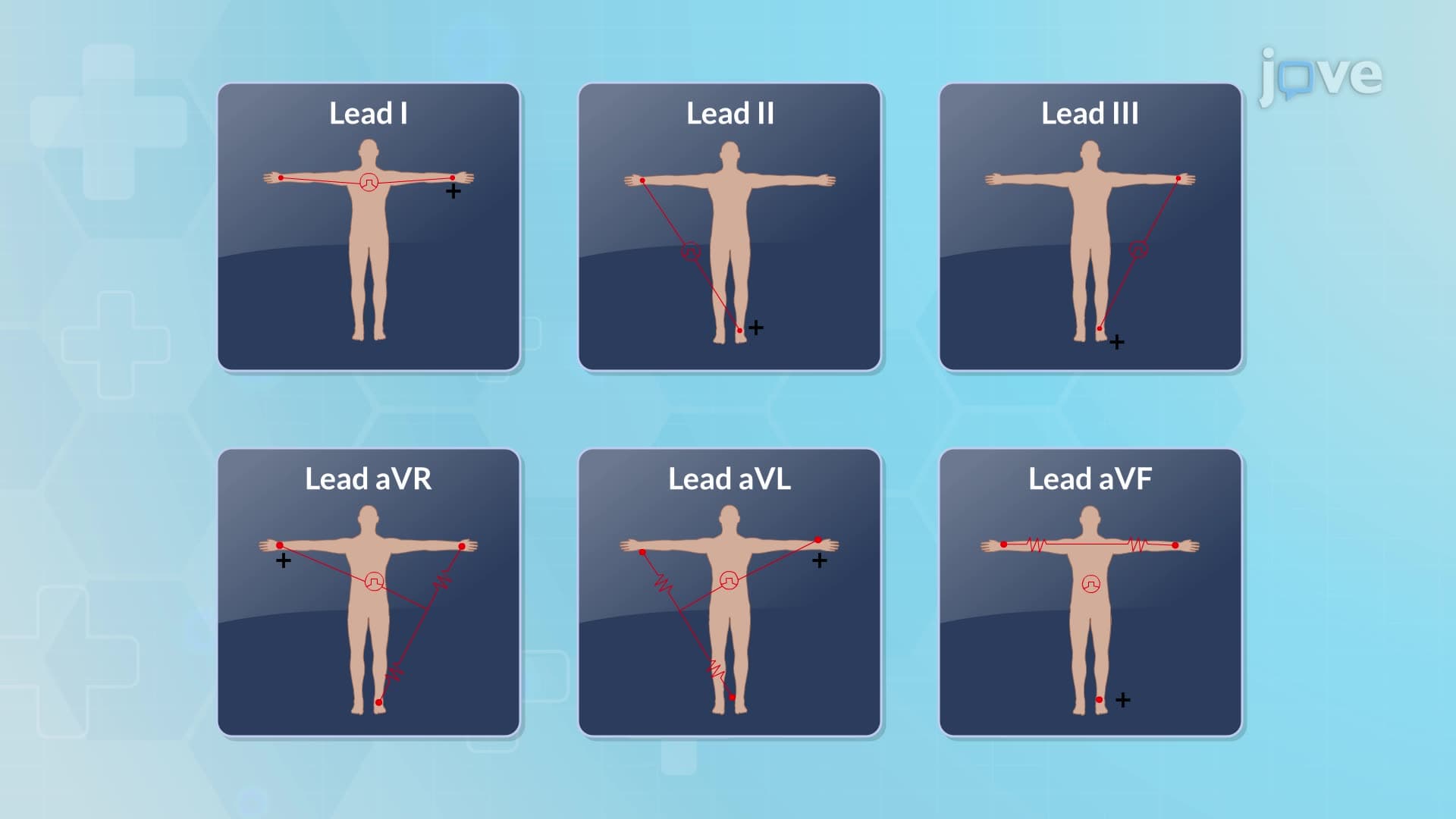
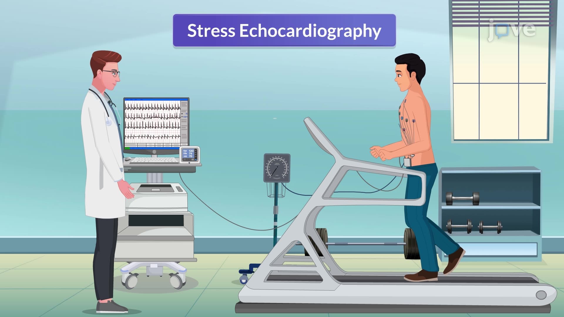
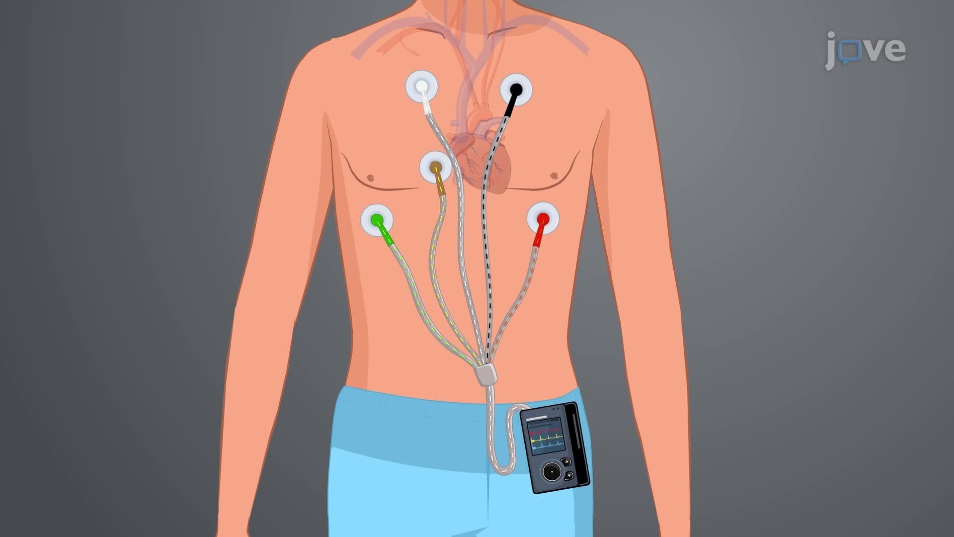
During electrode placement, healthcare professionals position ten ECG leads according to established standards: precordial leads in anatomically correct positions, arm electrodes two centimeters below the clavicles, and leg electrodes below the rib cage. This configuration provides comprehensive cardiac electrical activity monitoring throughout the exercise protocol.
The exercise protocol typically follows the Bruce Protocol, starting with low-intensity activity (approximately 1.7 mph at 10% grade) and progressively increasing speed and incline every three minutes. Continuous monitoring includes 12-lead ECG, heart rate, blood pressure, and symptom assessment. Testing continues until patients reach 85% of their age-predicted maximum heart rate, develop significant symptoms, or demonstrate concerning ECG changes.
Exercise stress test interpretation requires expertise in recognizing both normal and pathological responses to exertion. Negative tests show appropriate heart rate and blood pressure responses without ECG abnormalities or limiting symptoms. These results generally indicate low probability of significant coronary artery disease, though they cannot completely exclude cardiac pathology.
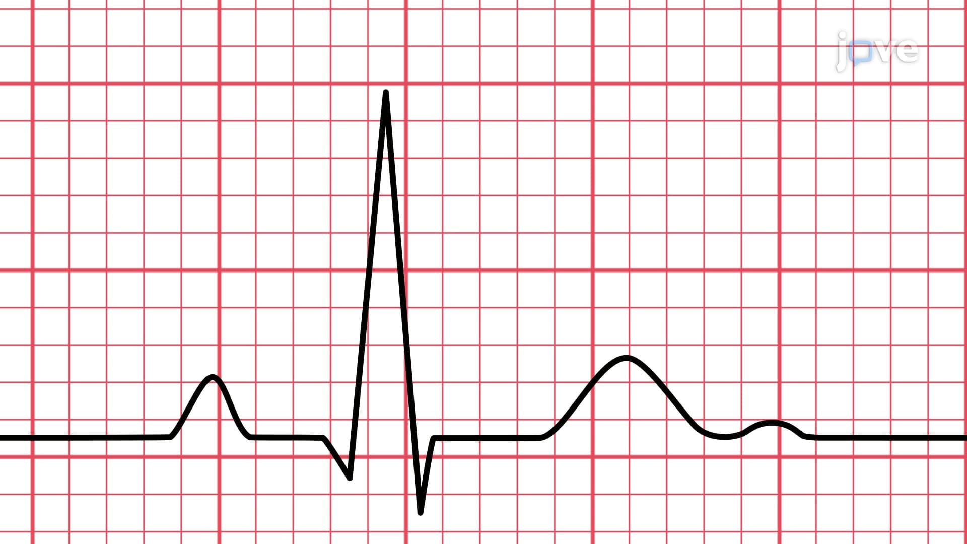
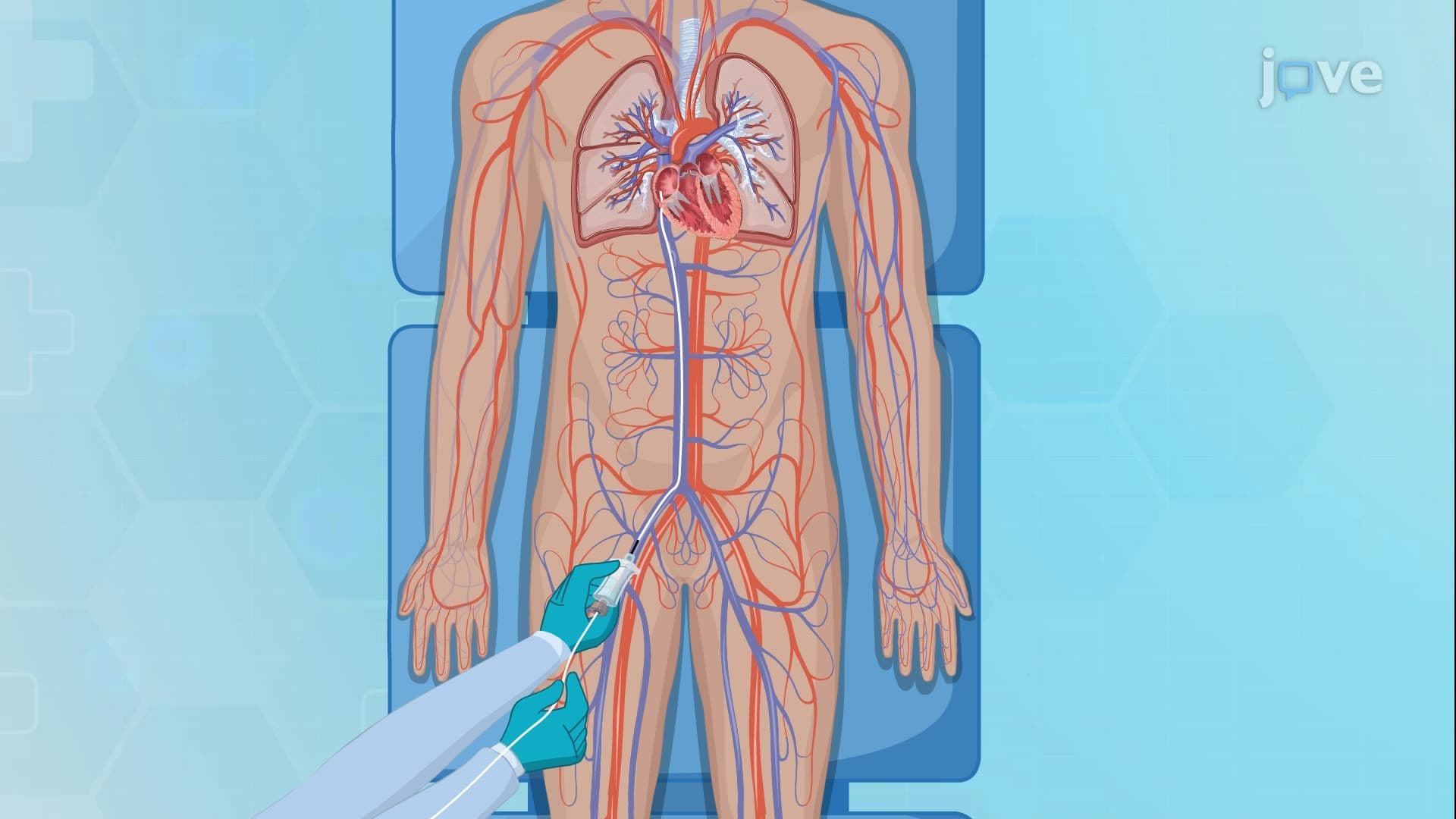
Positive results manifest through various findings including horizontal or downsloping ST-segment depression exceeding 1mm, T-wave inversions, complex arrhythmias, or symptom development with low workloads. These abnormalities suggest myocardial ischemia and warrant further cardiac evaluation, potentially including cardiac catheterization or advanced imaging studies.
Students preparing for healthcare examinations like the MCAT, NCLEX, or college physiology courses should understand that stress test sensitivity ranges from 60-70%, meaning some patients with coronary disease may have false-negative results, particularly those with single-vessel disease or adequate collateral circulation.
Related Micro-courses