Loading subject page…


286,219 views

Video Summary: What Is Heterochromatin
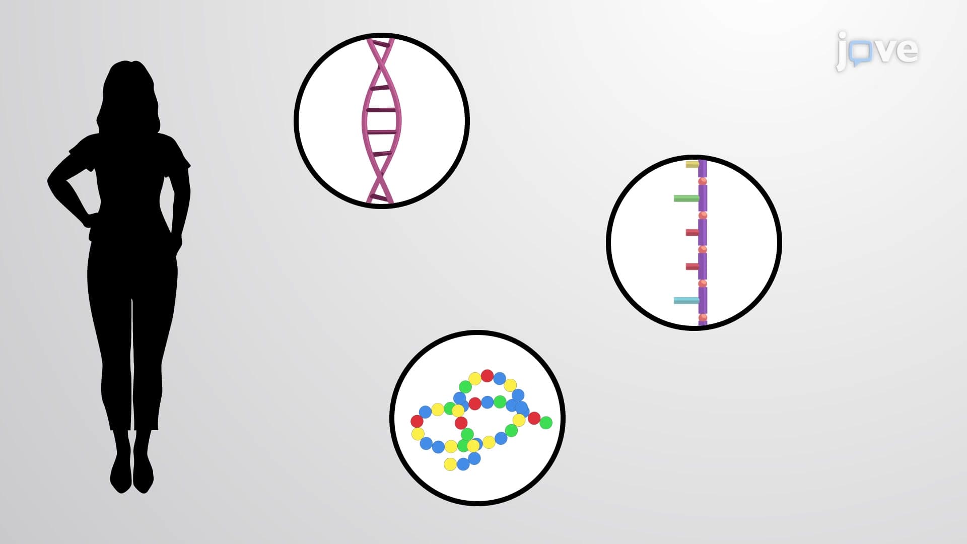
Did you know that in every female human cell, one entire X chromosome is essentially "turned off" and packed away like clothes in a vacuum-sealed bag? This tightly compressed DNA structure is called heterochromatin, a crucial form of chromatin organization that controls gene expression in eukaryotic cells. Understanding what heterochromatin is reveals how cells regulate which genes are active or silenced, from X-chromosome inactivation in women to maintaining chromosome stability during cell division. This concept appears frequently in AP Biology, college genetics courses, and medical school curricula across the United States. Watch the full video on JoVE Coach to master this concept with expert-led visuals and step-by-step explanations.
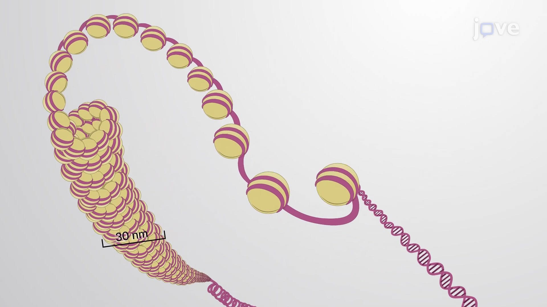
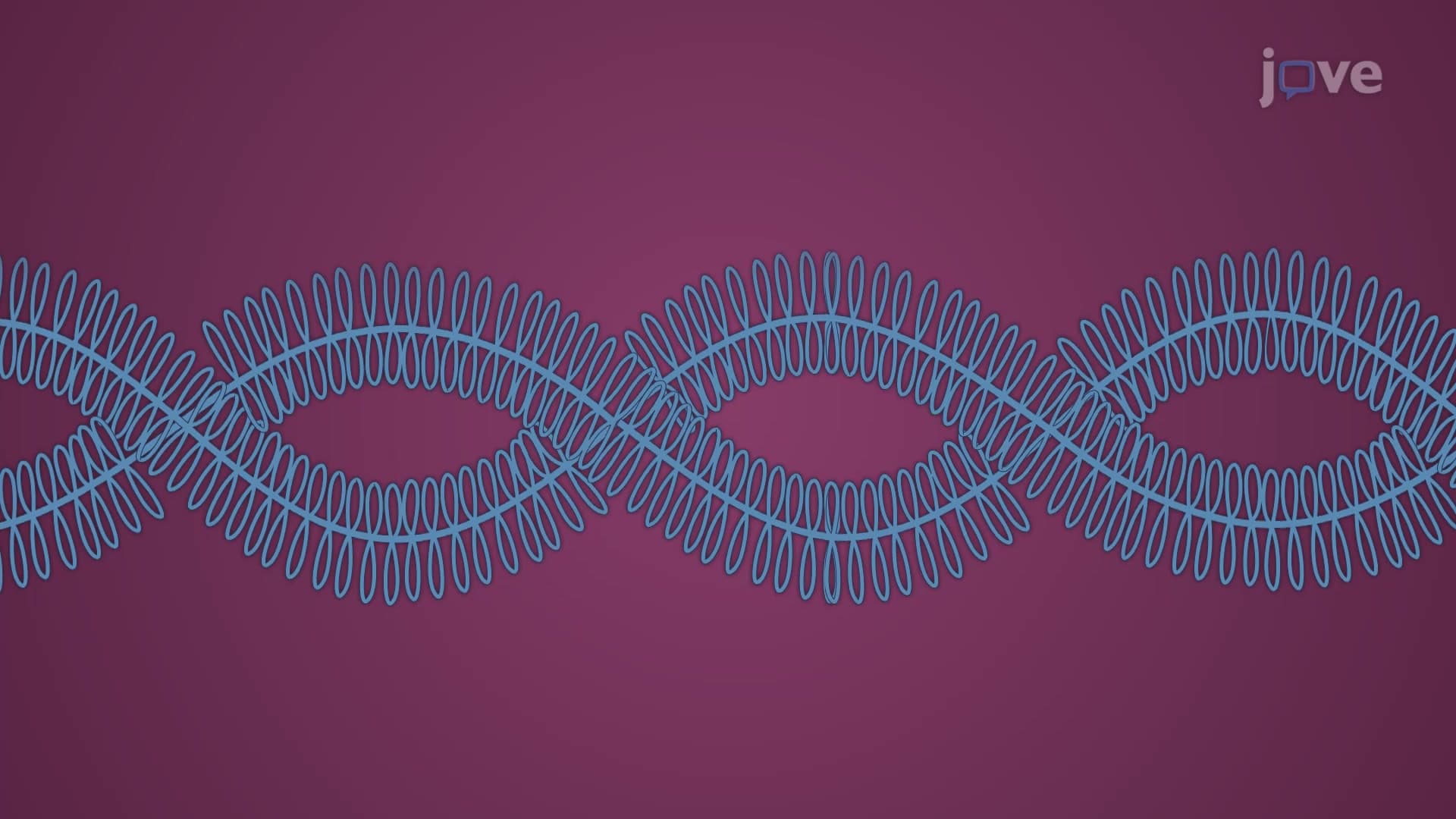
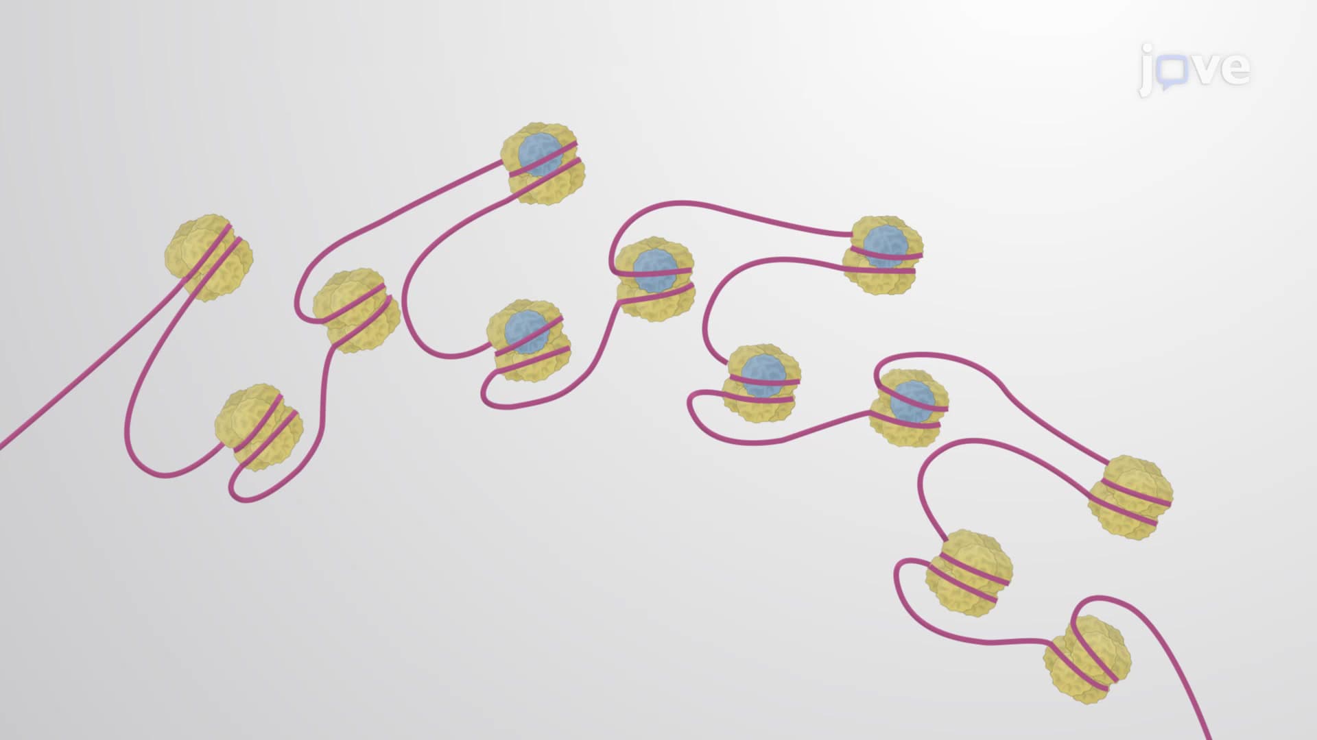
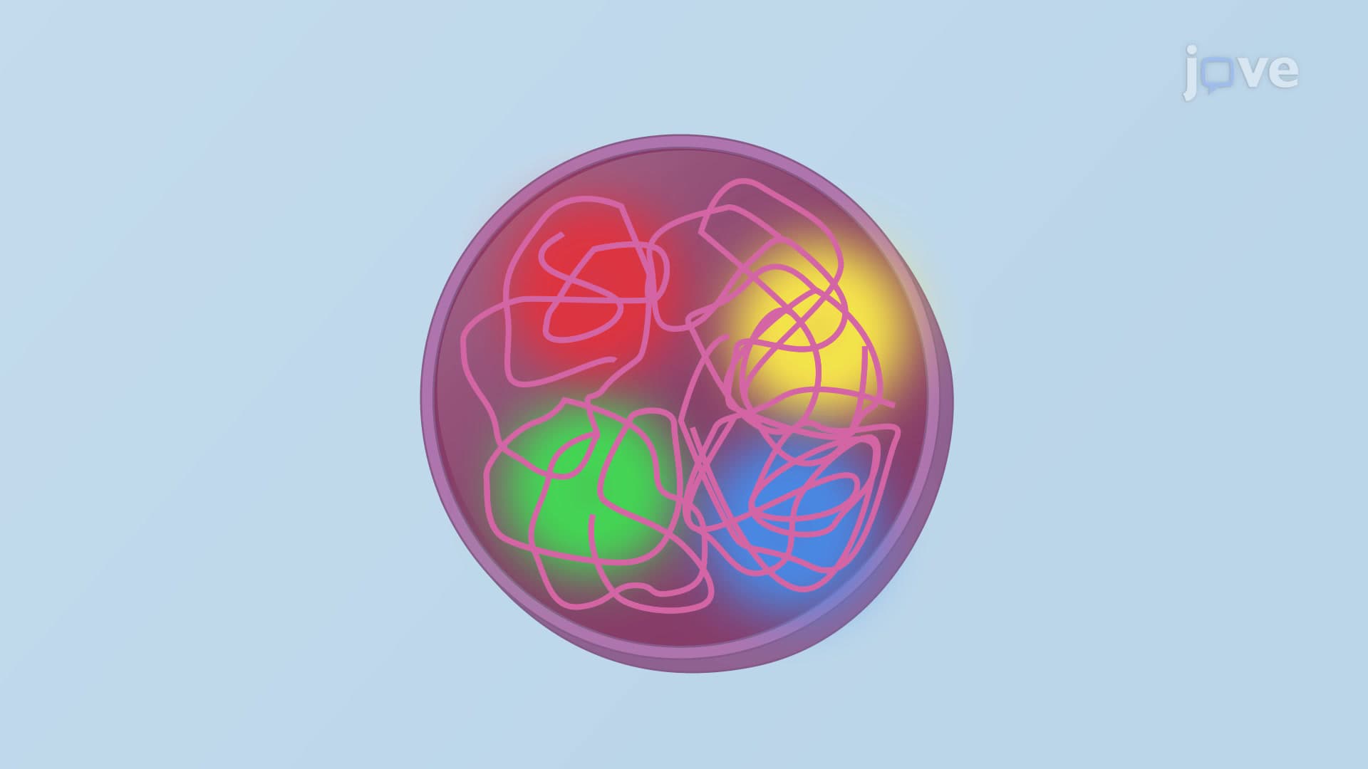
Heterochromatin represents one of the most fascinating examples of how cells organize and control their genetic information. Unlike its loosely-packed counterpart euchromatin, heterochromatin exists in a highly condensed state that severely restricts access to the underlying DNA sequences. This tight packaging serves multiple cellular functions, from permanently silencing repetitive DNA elements to temporarily shutting down entire chromosomes when needed.
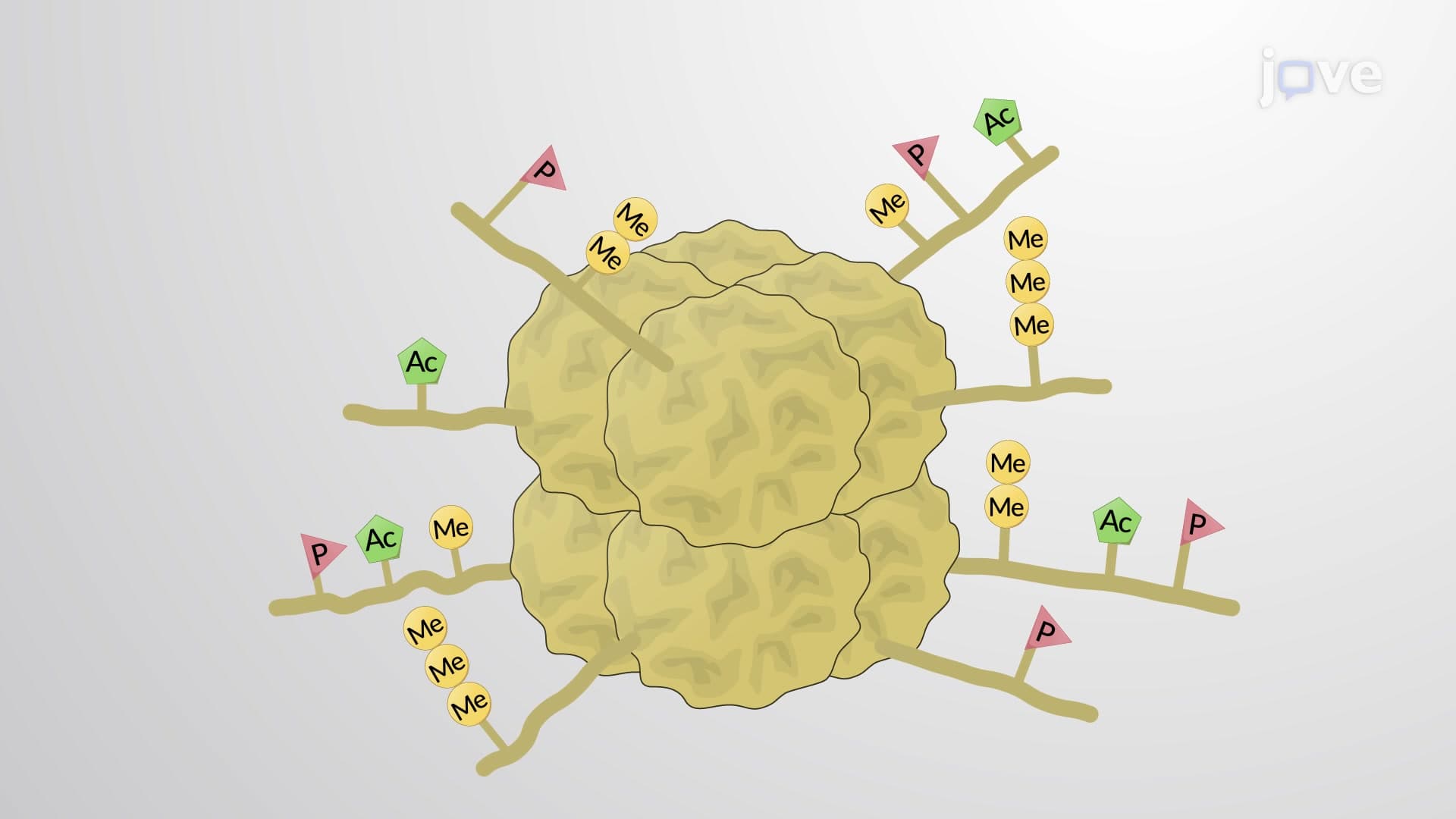
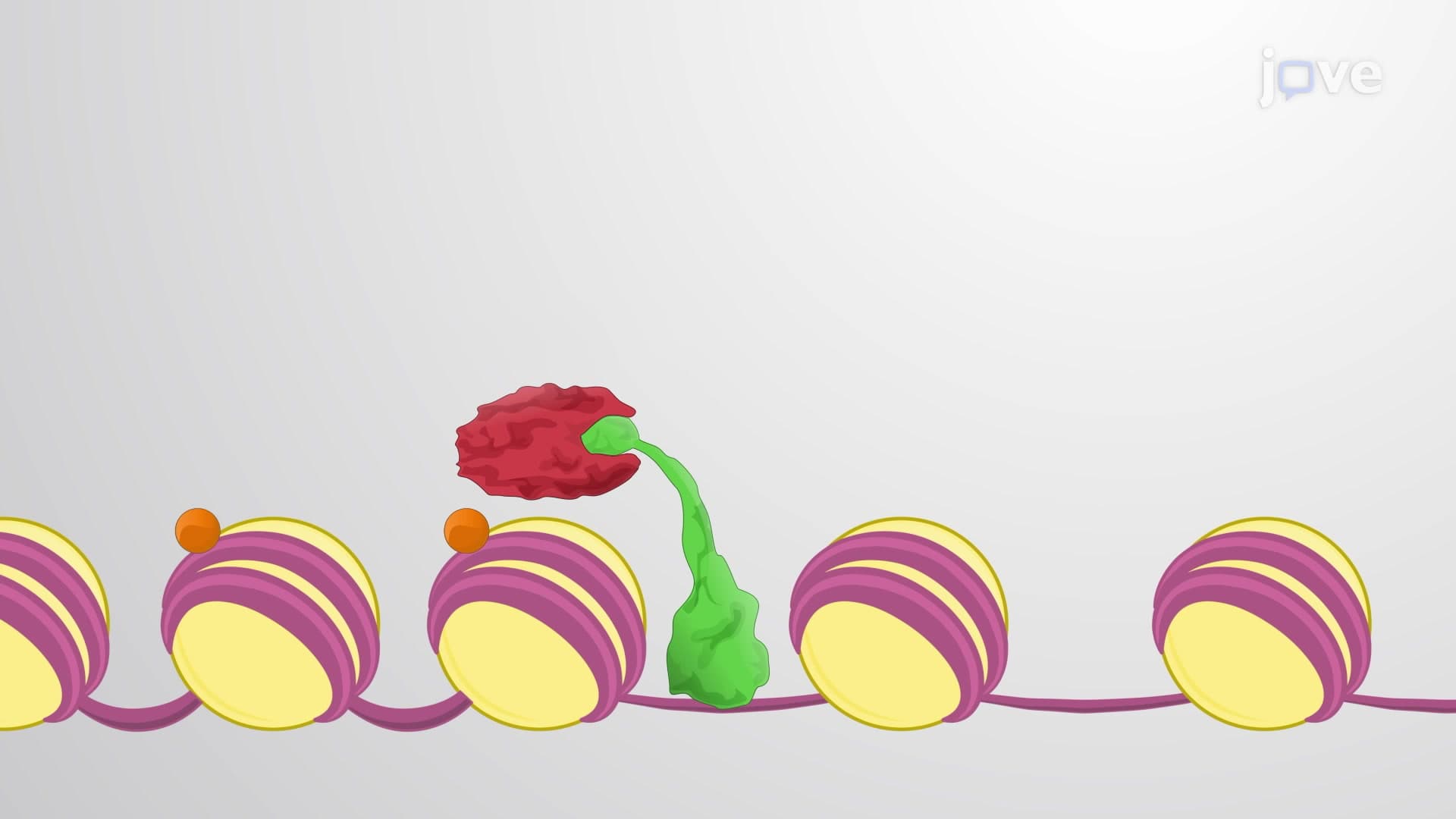
The distinction between constitutive and facultative heterochromatin is crucial for understanding cellular gene regulation. Constitutive heterochromatin acts like a permanent vault, containing repetitive DNA sequences and transposable elements that cells need to keep permanently silenced. These regions, rich in satellite DNA and poor in protein-coding genes, maintain their condensed state throughout the cell cycle. The molecular signature involves specific histone modifications, particularly trimethylation of histone H3 at lysine 9 (H3K9me3), which creates binding sites for Heterochromatin Protein 1 (HP1).
Facultative heterochromatin, in contrast, functions more like a temporary storage system. Genes within these regions can be silenced in some cell types while remaining active in others. This flexibility allows for tissue-specific gene expression patterns and developmental regulation. The Polycomb repressive complex 2 (PRC2) plays a central role here, catalyzing H3K27 trimethylation to establish and maintain facultative heterochromatin domains.
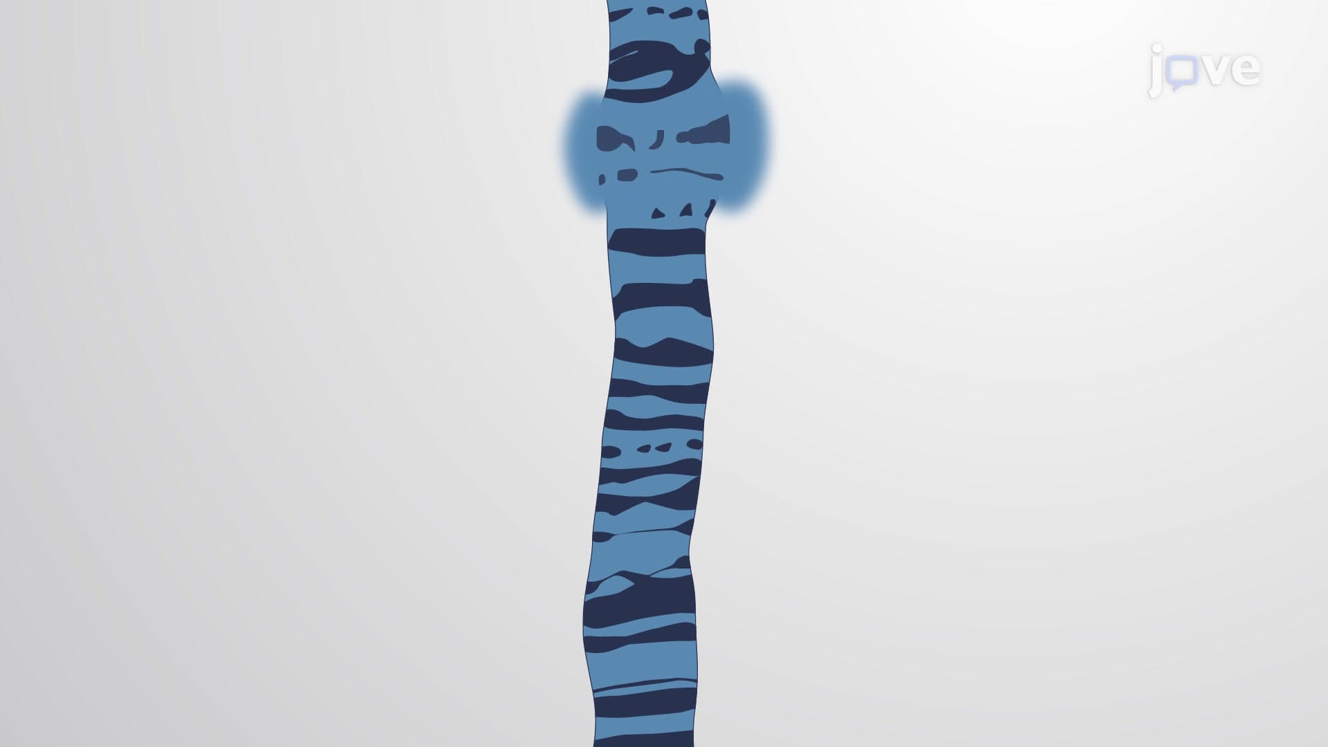
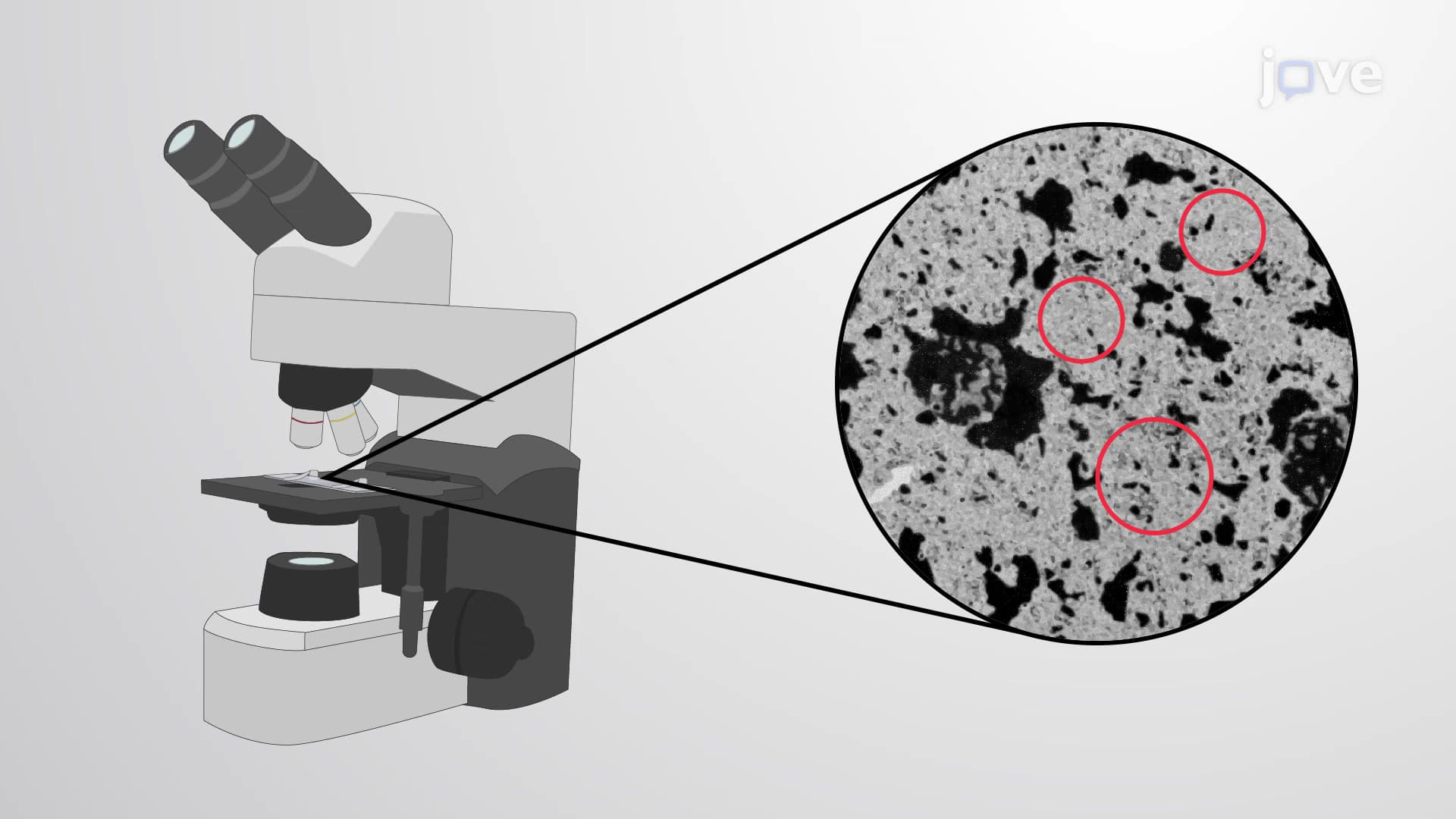
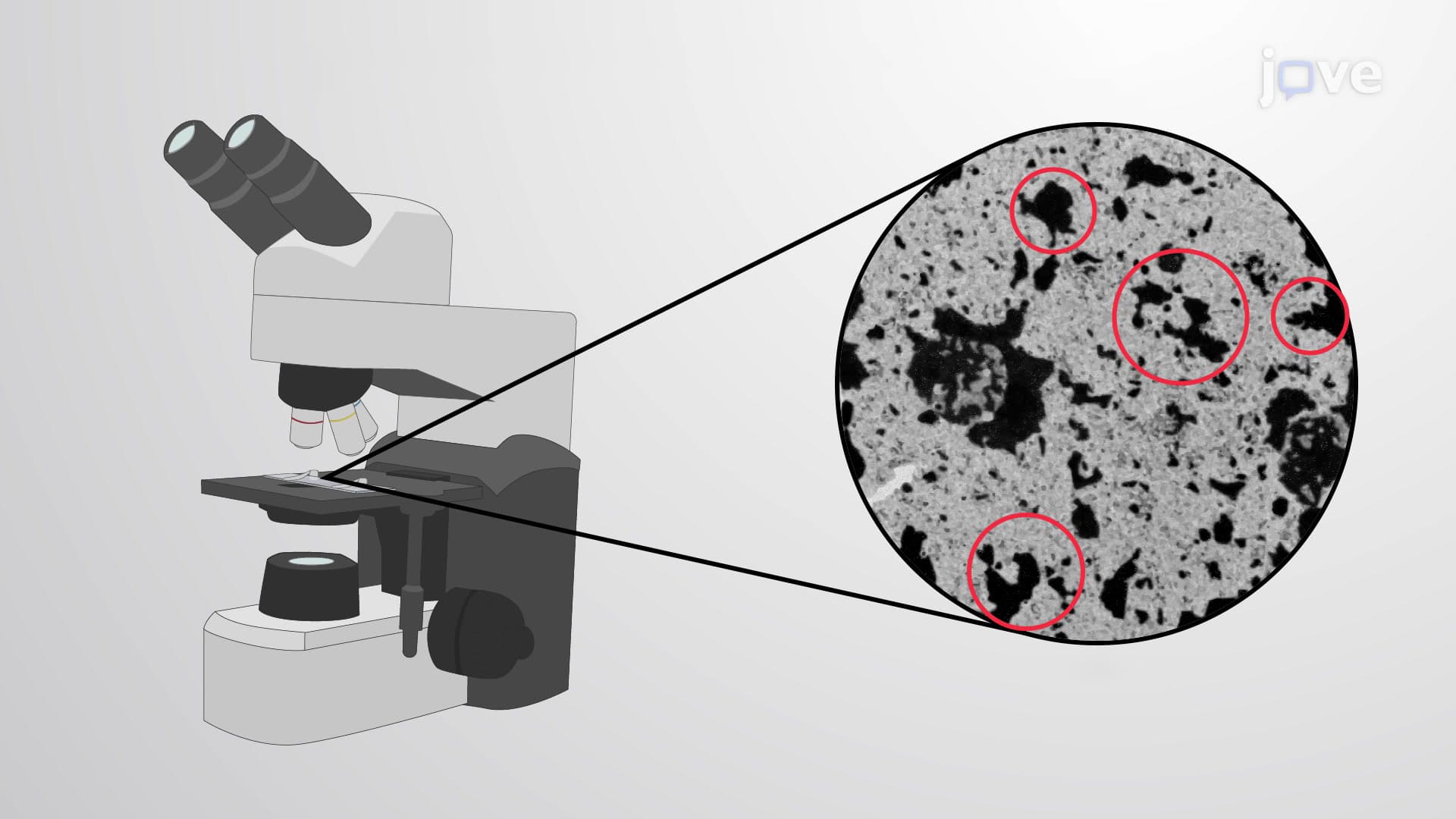
X-chromosome inactivation exemplifies facultative heterochromatin's biological importance. In female mammals, including humans, one X chromosome in each cell becomes heavily heterochromatinized, forming the distinctive Barr body visible under a microscope. This process, discovered by Canadian geneticist Murray Barr, ensures dosage compensation between XX females and XY males. Medical professionals use Barr body analysis in clinical genetics to diagnose certain chromosomal abnormalities and sex chromosome disorders.
Students preparing for the MCAT, AP Biology exam, or college genetics courses should understand that heterochromatin formation involves multiple molecular players working in concert. The process begins with DNA methylation at CpG sites, followed by recruitment of methyl-CpG-binding proteins, histone-modifying enzymes, and finally chromatin compaction factors. This knowledge proves essential for understanding epigenetic inheritance, cancer biology, and developmental disorders.
Heterochromatin dysfunction contributes to various human diseases, including certain cancers where normally silenced oncogenes become inappropriately activated. Pharmaceutical companies are developing drugs that target heterochromatin-modifying enzymes, representing a growing field in precision medicine. Understanding these mechanisms helps explain why identical twins with the same DNA sequence can develop different diseases-epigenetic modifications, including heterochromatin formation, accumulate differently over time.
Related Micro-courses