Loading video…


 20 Concepts
 286.2K views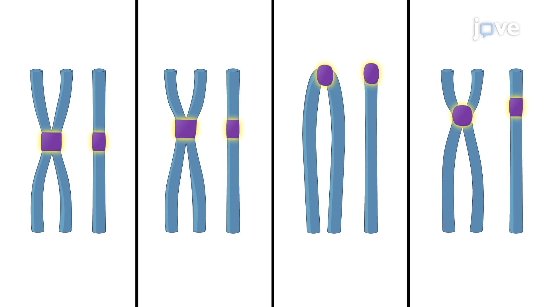

Video Summary: What Is Chromosome Structure
Did you know that if human chromosomes weren't carefully packaged, the DNA in just one cell would stretch over six feet long? Chromosome structure represents one of biology's most elegant organizational systems, transforming miles of genetic material into compact, functional units. During mitosis in cancer research at institutions like MD Anderson Cancer Center, scientists observe how precise chromosome structure enables accurate cell division. Understanding what is chromosome structure reveals the intricate relationship between DNA packaging, centromeres, telomeres, and replication origins that make life possible. Watch the full video on JoVE Coach to master this concept with expert-led visuals and step-by-step explanations.
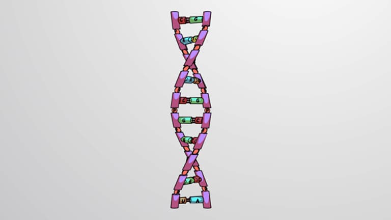
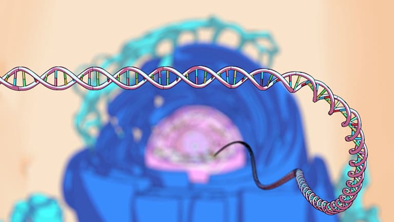
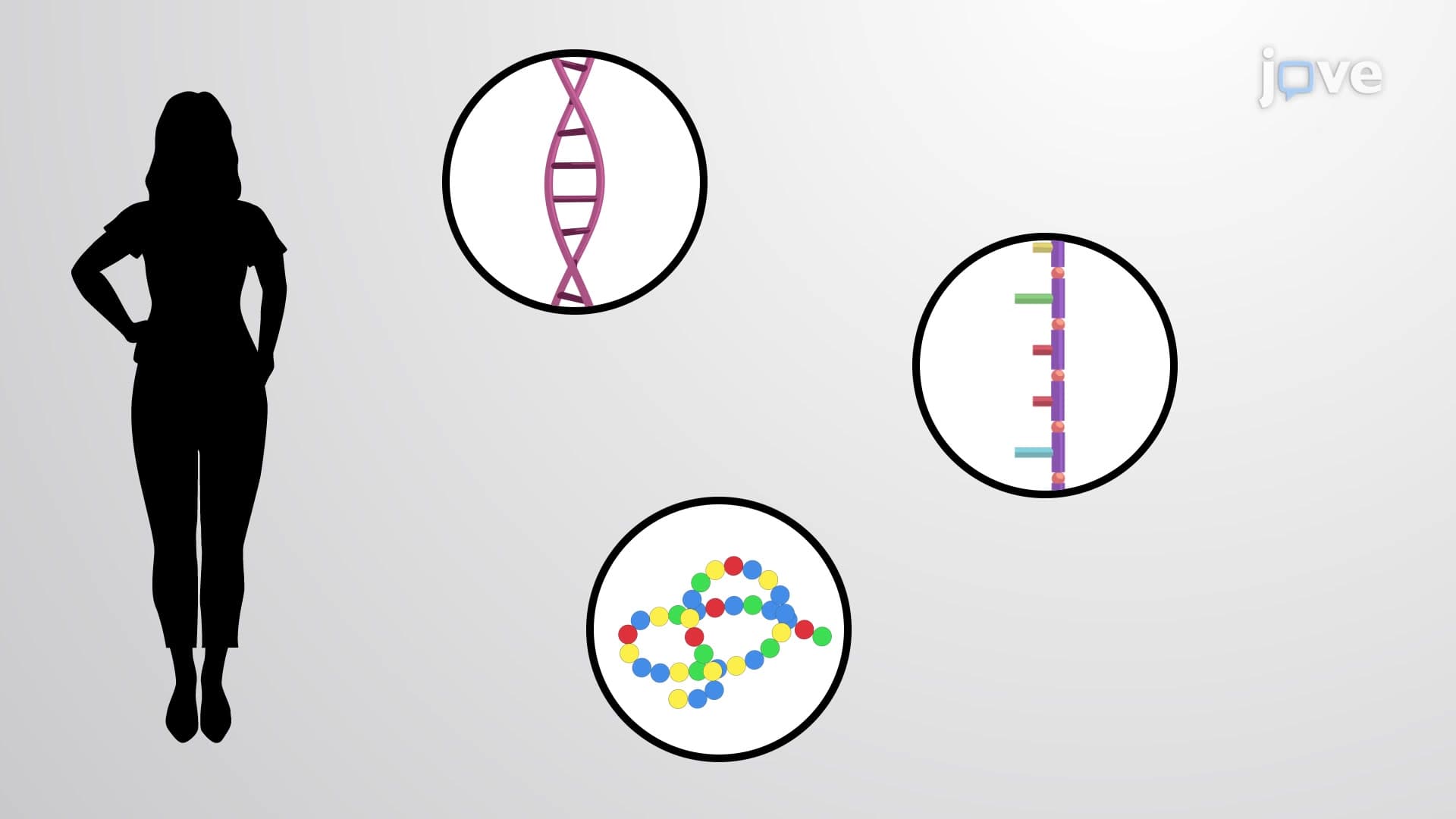
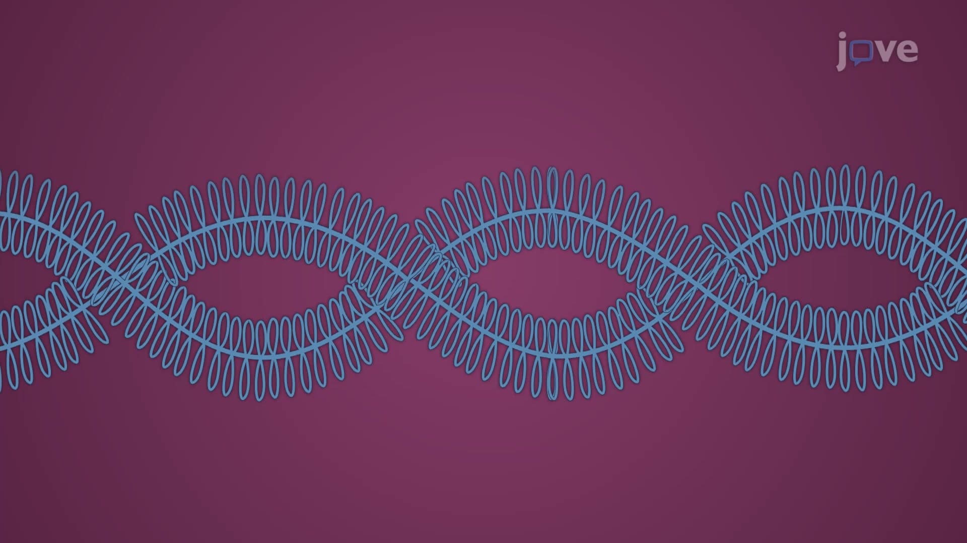
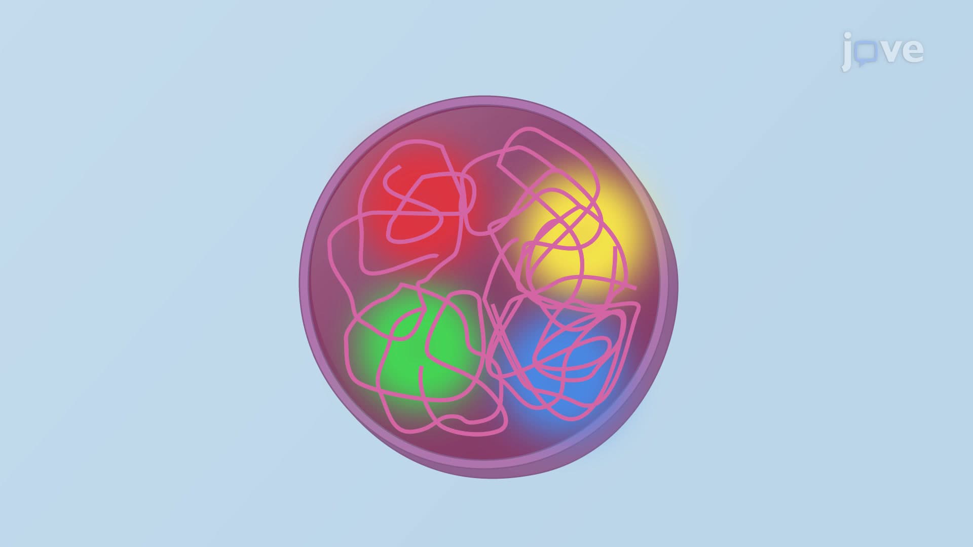
Chromosome structure represents a masterful biological engineering solution to an enormous organizational challenge. Human cells contain approximately 6 billion base pairs of DNA that must be precisely packaged, replicated, and distributed during cell division. The basic building block begins with the DNA-histone complex called chromatin, which undergoes dramatic condensation to form visible chromosomes during cell division.
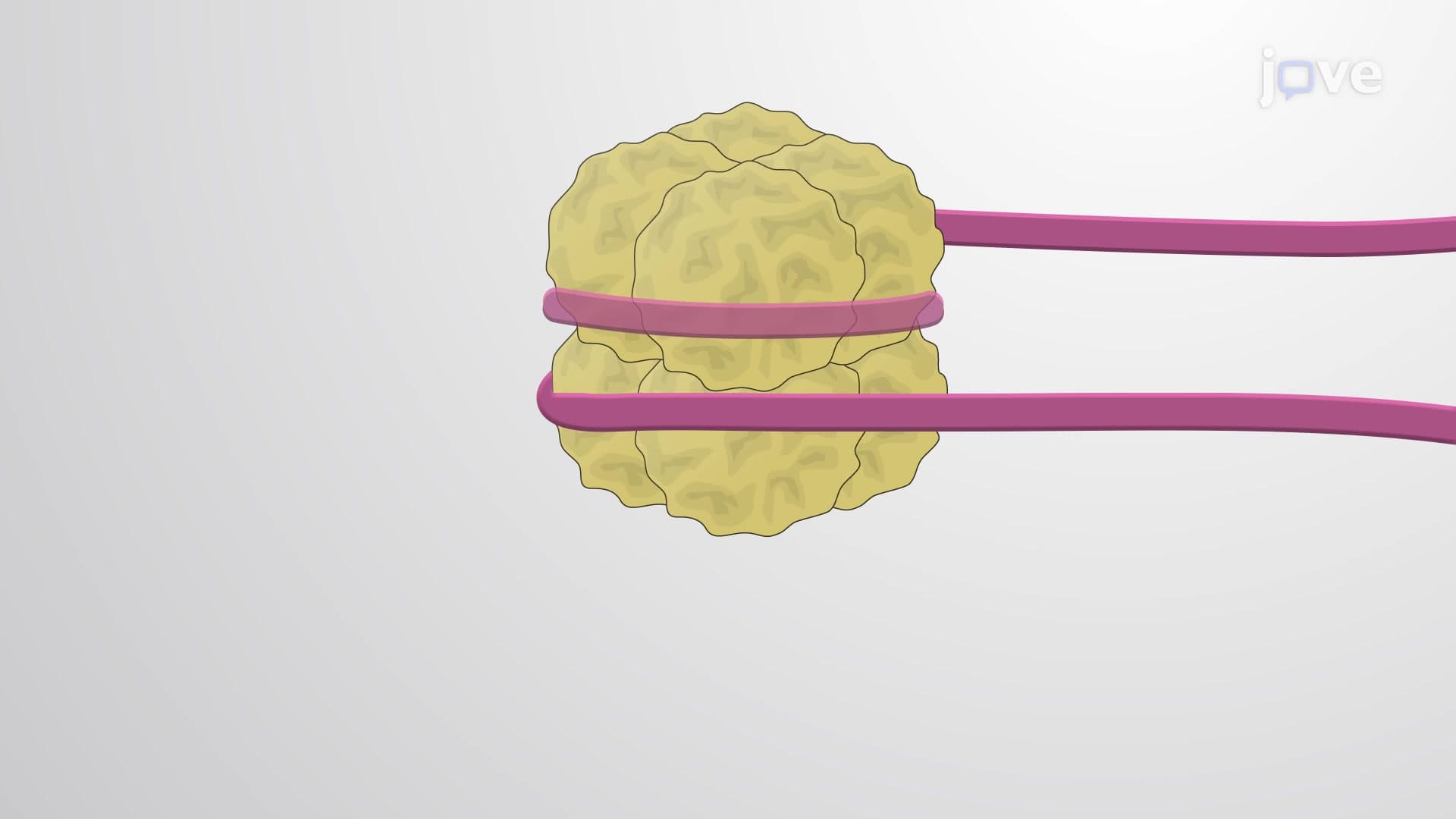
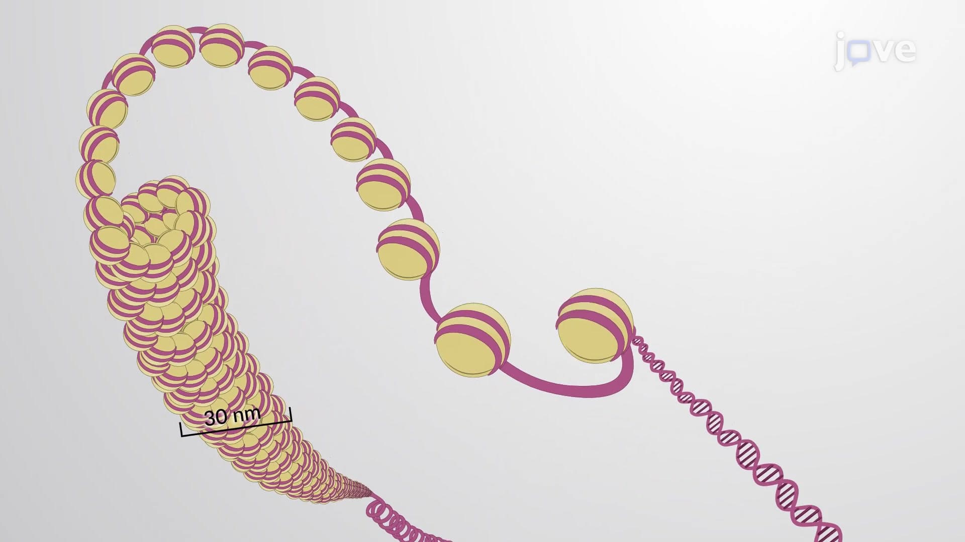
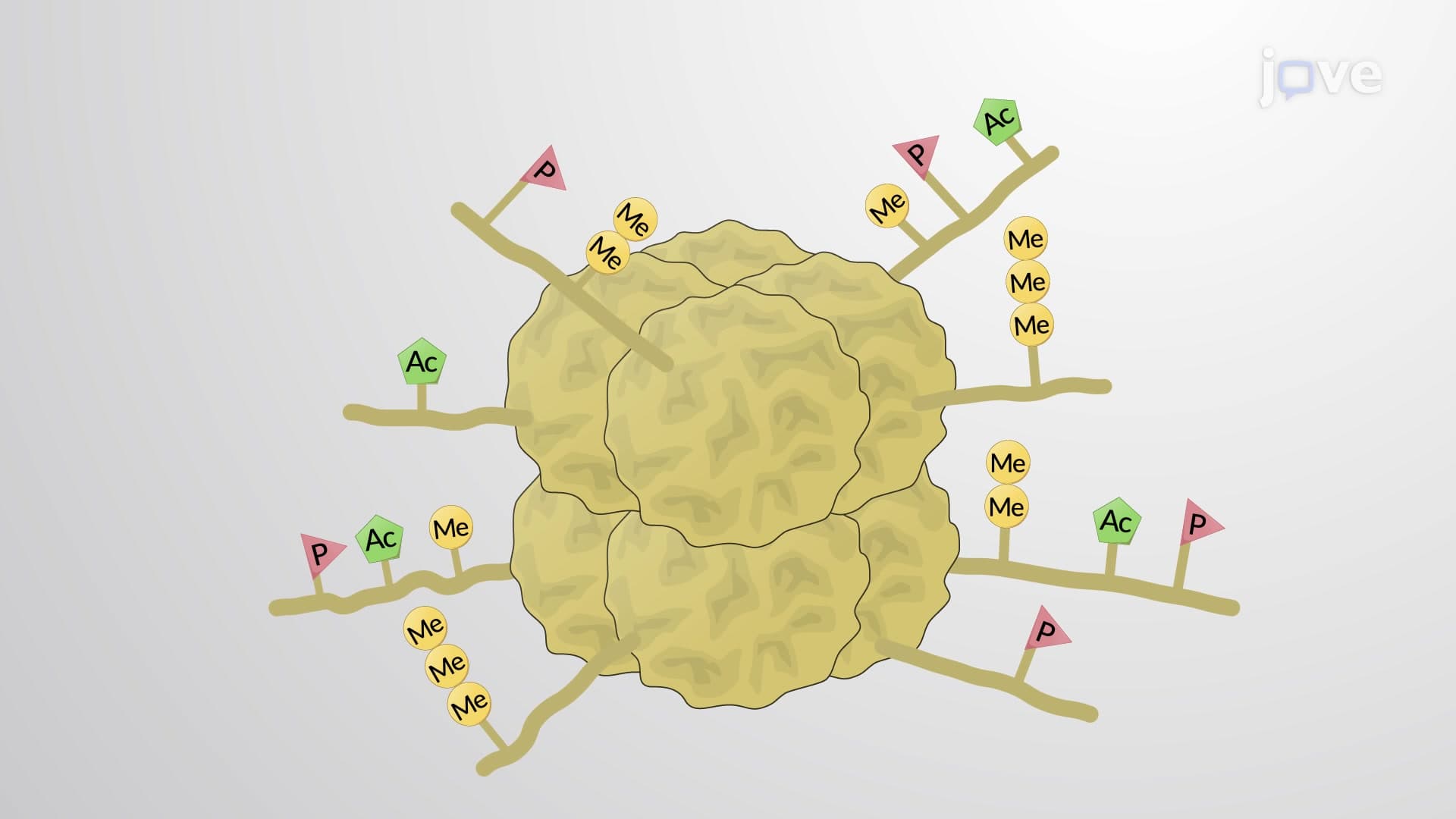
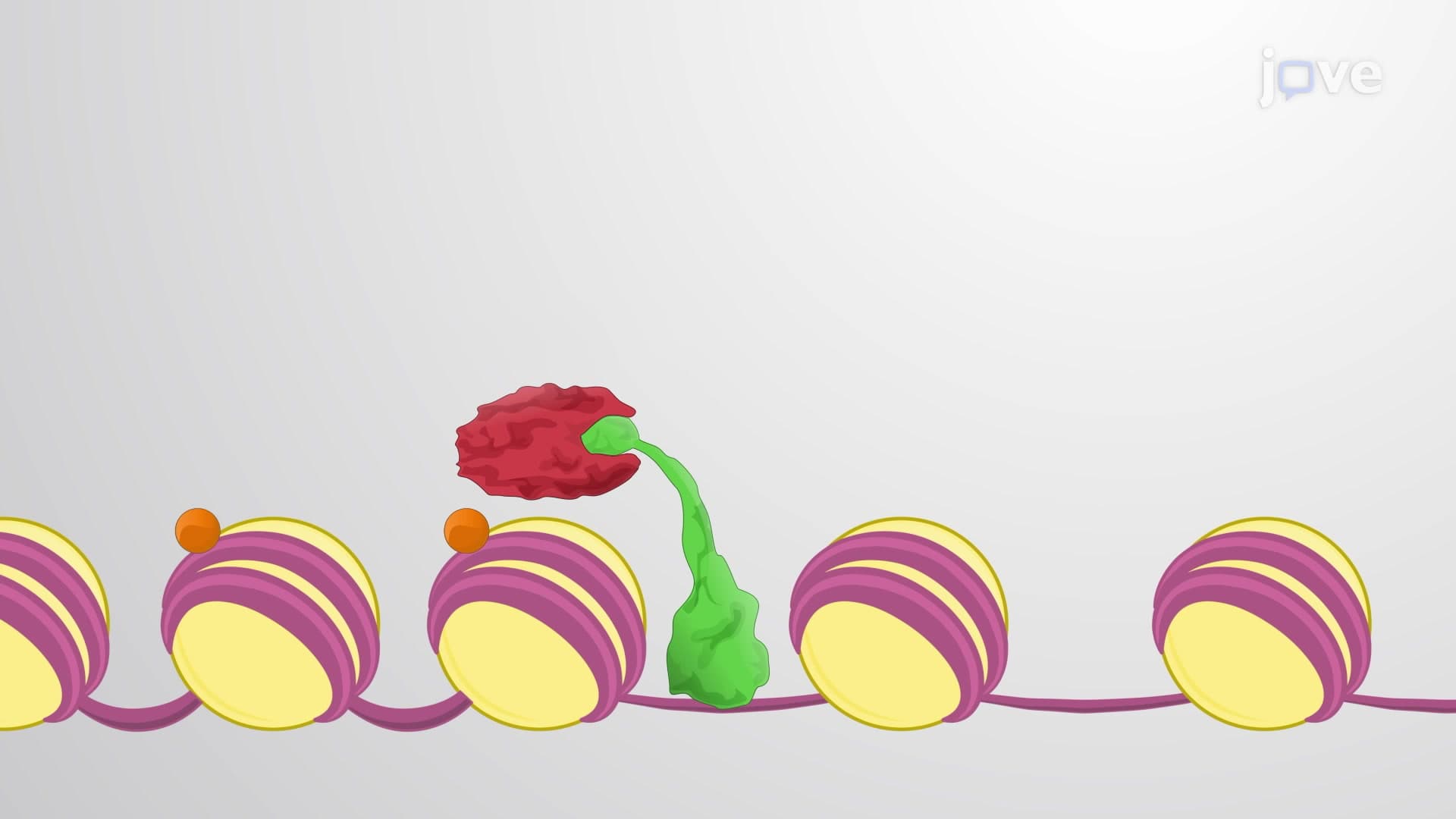
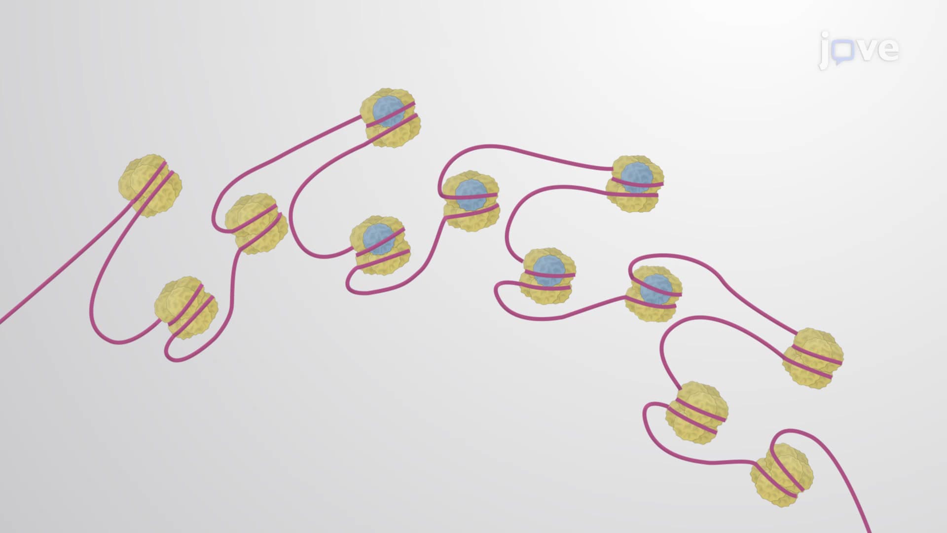
This packaging system operates on multiple levels of organization. At the molecular level, DNA wraps around histone proteins to form nucleosomes, resembling beads on a string. These nucleosomes then coil and fold into increasingly compact structures, ultimately achieving a 10,000-fold compaction during chromosome condensation. This process is essential for fitting massive amounts of genetic information into the microscopic nucleus while maintaining accessibility for cellular processes.
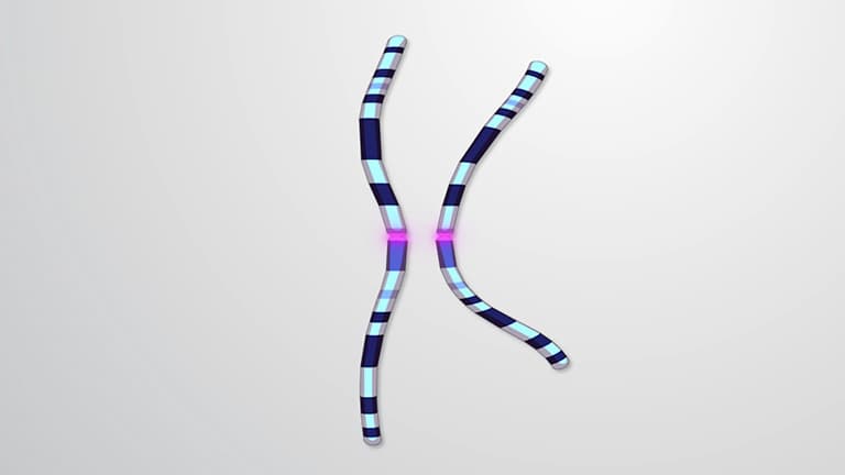
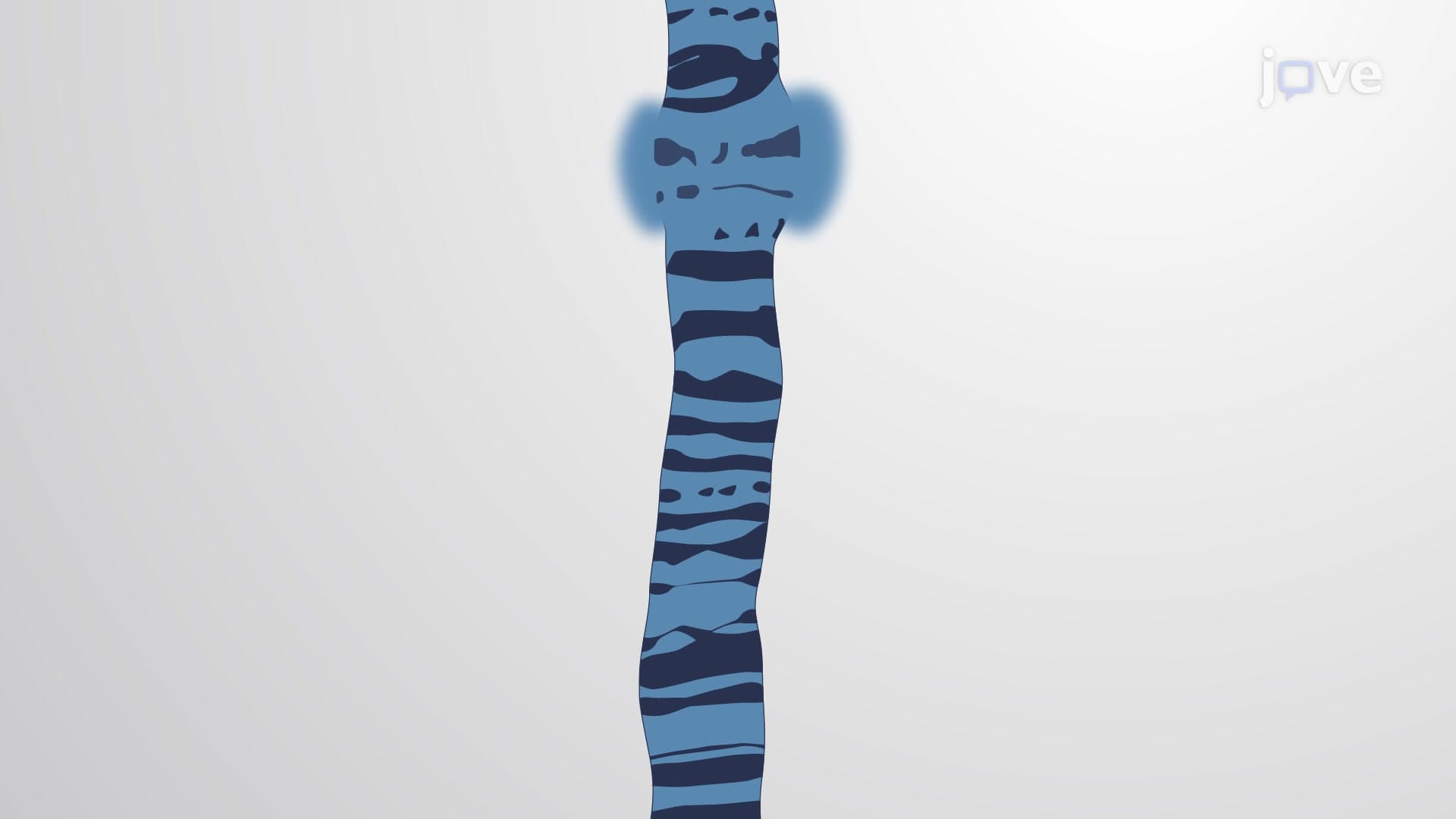
The centromere's position defines four distinct chromosome structure categories that serve as diagnostic tools in clinical genetics. Metacentric chromosomes feature centrally located centromeres, creating arms of equal length-similar to a perfectly balanced scale. Submetacentric chromosomes have off-center centromeres, producing arms of unequal length like an unbalanced scale.
Acrocentric chromosomes, exemplified by human chromosome 21 (involved in Down syndrome), have centromeres positioned near one end, creating a distinctive "stalk and bulb" appearance. The human Y chromosome demonstrates this acrocentric configuration, making it easily identifiable during karyotype analysis in genetics laboratories across the United States. Telocentric chromosomes, with terminal centromeres, create single long arms and are less common in human genetics but important in plant and animal studies.
These configurations aren't merely academic classifications-they're crucial for medical diagnostics. Cytogeneticists at hospitals like Johns Hopkins use these structural features to identify chromosomal abnormalities, guide genetic counseling, and inform treatment decisions for patients with genetic disorders.
Telomeres function as molecular "caps" protecting chromosome ends from degradation and fusion events. These repetitive DNA sequences (TTAGGG in humans) solve the "end replication problem" where standard DNA polymerase cannot completely replicate chromosome tips. Research at institutions like the National Institute on Aging has revealed how telomere shortening relates to cellular aging and cancer development.
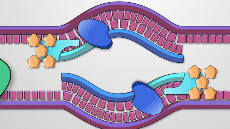
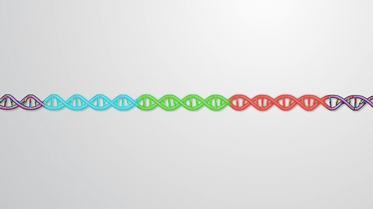
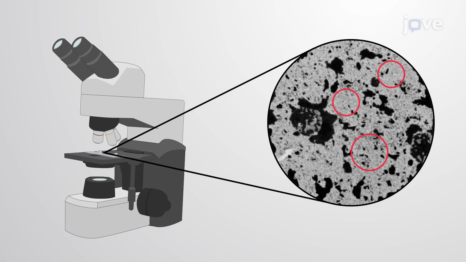
Origins of replication demonstrate another crucial aspect of chromosome structure biology explained. Human chromosomes contain approximately 30,000 origins of replication-specialized DNA sequences where replication machinery assembles. This massive redundancy ensures rapid, accurate DNA copying. Without multiple origins, chromosome replication would require over a month per chromosome, making cell division impossibly slow for complex organisms.
This chromosome structure knowledge directly impacts AP Biology exams, where students analyze cell cycle regulation, and pre-med MCAT preparation, where understanding chromosome dynamics supports genetics and cell biology sections.
Related Micro-courses