Loading subject page…


21,637 views

Video Summary: What Is Pulse Rhythm
Did you know that emergency room physicians at Johns Hopkins Hospital can detect life-threatening heart conditions just by feeling your pulse rhythm? Pulse rhythm represents the pattern of heartbeats within specific time intervals, revealing crucial information about cardiac health. When paramedics check your pulse during emergencies, they're assessing whether your heart maintains a regular, steady pattern or shows dangerous irregularities called dysrhythmias. Watch the full video on JoVE Coach to master this concept with expert-led visuals and step-by-step explanations.
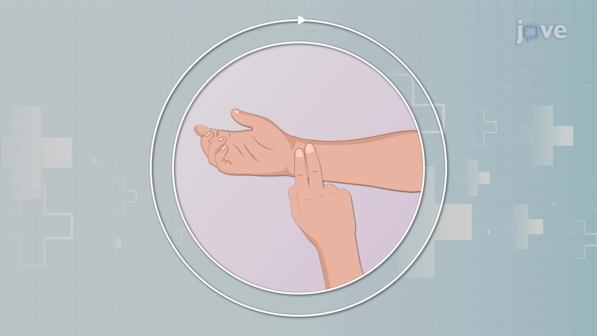
Pulse rhythm serves as a window into cardiovascular health, representing the organized pattern of heartbeats that healthcare providers can detect through physical examination. At its core, pulse rhythm reflects the mechanical manifestation of electrical activity in the heart, where each heartbeat generates a pressure wave that travels through arterial blood vessels. In healthy individuals, this creates a predictable, rhythmic pattern that medical professionals at institutions like the Mayo Clinic and Cleveland Clinic use for initial cardiac assessment.
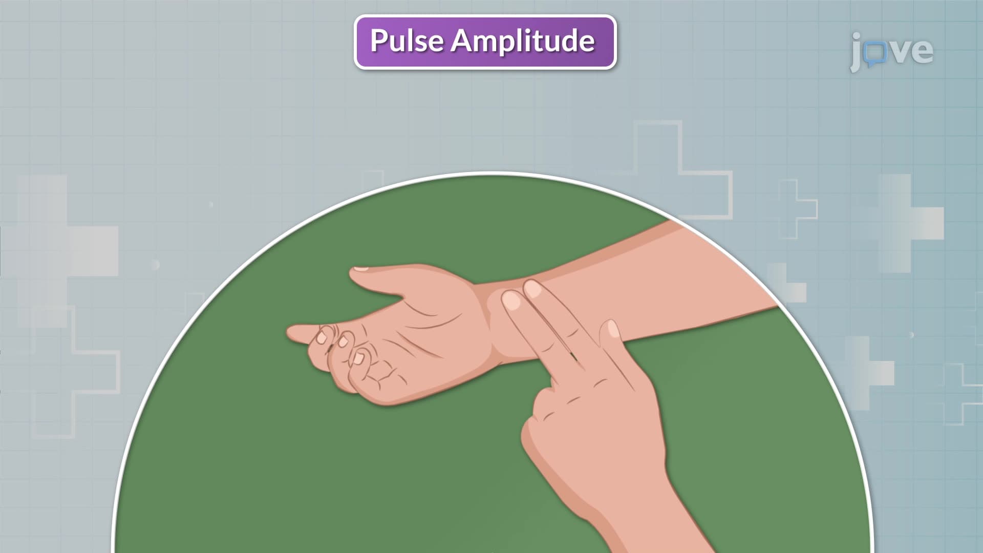
A regular pulse rhythm maintains consistent timing intervals between beats, characterized by uniform waveform patterns and steady pulsation strength. This regularity indicates proper coordination between the heart's electrical conduction system and muscular contractions. However, pulse strength can vary while maintaining regularity-normal pulses provide moderate strength, weak pulses suggest reduced cardiac output or vascular issues, and bounding pulses may indicate conditions like hyperthyroidism or fever.
Irregular pulse rhythms, medically termed dysrhythmias, signal disruptions in normal cardiac function. These irregularities occur when the heart's electrical conduction system experiences interference, leading to abnormal contraction patterns. For students preparing for the MCAT or nursing entrance exams like the NCLEX, understanding that dysrhythmias represent the heart's struggle to maintain coordinated electrical and mechanical activity is crucial for clinical reasoning questions.
Healthcare professionals classify dysrhythmias into two primary categories based on their pattern predictability. Regularly irregular dysrhythmias follow a predictable pattern of irregularity, often involving premature beats that occur at consistent intervals. Examples include pulsus bigeminus (every other beat is premature), coupled extrasystoles (paired abnormal beats), and Wenckebach patterns (progressively lengthening intervals followed by a dropped beat).
Irregularly irregular dysrhythmias present with completely unpredictable timing, making them more concerning clinically. Atrial fibrillation, commonly seen in emergency departments across the United States, exemplifies this pattern where chaotic electrical activity in the atria creates an entirely random ventricular response. Premature ventricular contractions and various ectopic beats also fall into this category, requiring immediate medical attention in many cases.
Modern medicine employs sophisticated tools for pulse rhythm evaluation beyond traditional palpation. Electrocardiograms (ECGs) provide the gold standard for cardiac rhythm assessment, recording electrical activity that healthcare providers at institutions like Johns Hopkins and Massachusetts General Hospital interpret daily. For AP Biology students, understanding that ECGs translate ionic movements across cardiac cell membranes into readable waveforms connects cellular physiology to clinical practice.
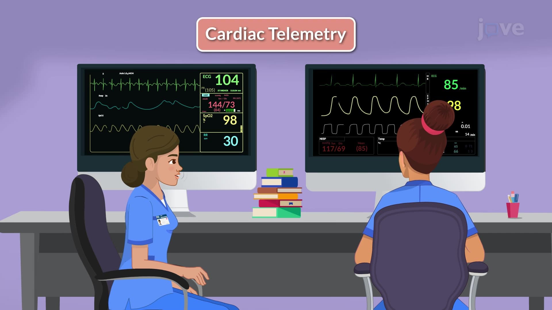
Holter monitoring extends ECG capability by providing 24-hour continuous recording, capturing intermittent rhythm disturbances that brief office visits might miss. Cardiac telemetry takes monitoring further, offering real-time observation crucial in intensive care settings where immediate intervention may be necessary for life-threatening rhythm changes.
Related Micro-courses