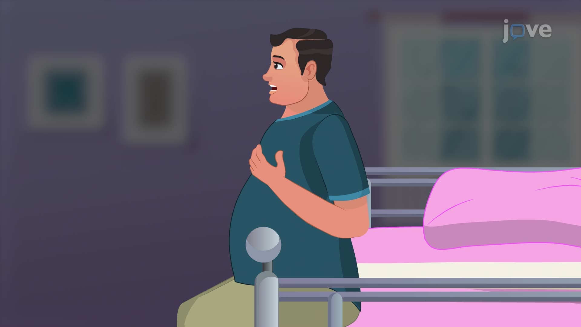
Vital signs: respiration assessment is fundamental to healthcare practice, encompassing the measurement and interpretation of breathing patterns, respiratory rate, depth, and rhythm. This comprehensive guide explores respiratory physiology, assessment techniques, and clinical applications essential for nursing students and healthcare professionals. Master respiratory rate assessment skills through JoVE Coach's systematic approach to vital signs evaluation.
Understand the physiological mechanisms controlling respiration and gas exchange processes
Learn proper techniques for measuring respiratory rate and assessing breathing patterns
Identify normal versus abnormal respiratory findings including tachypnea and bradypnea
Explore factors affecting respiration including age, medications, and medical conditions
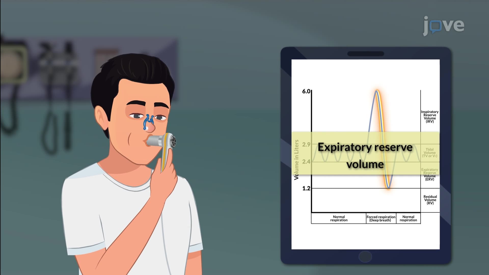
Analyze respiratory volumes, capacities, and their clinical significance
Apply systematic respiratory assessment methods in clinical settings
Recognize respiratory distress signs and accessory muscle usage
Interpret pulse oximetry readings and oxygen saturation values
1. Respiratory Physiology and Gas Exchange
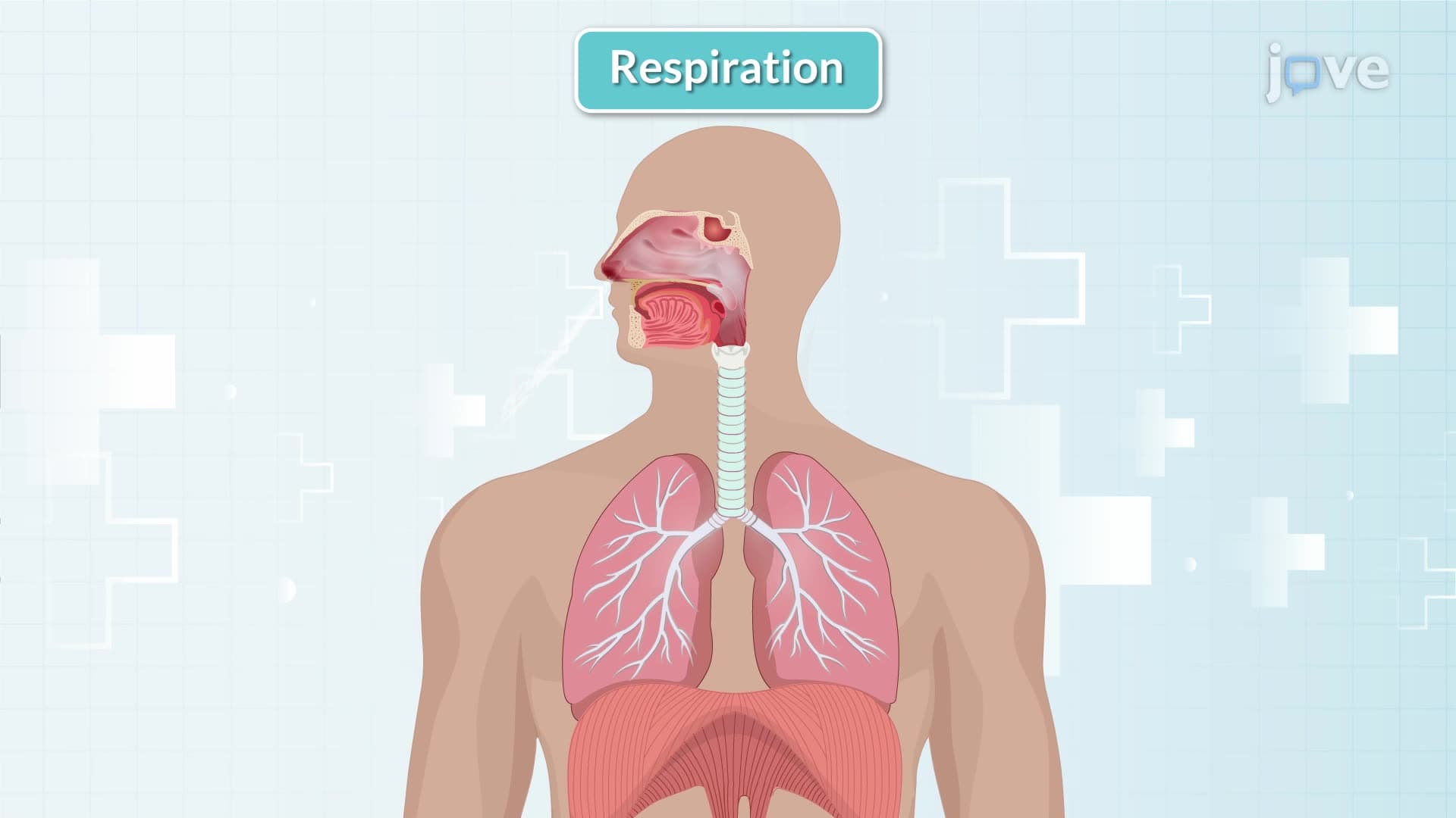
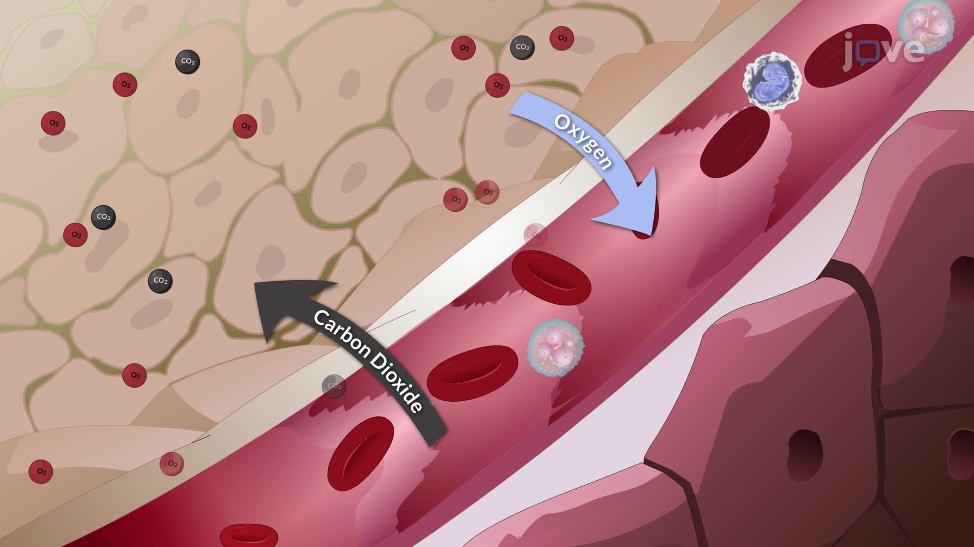
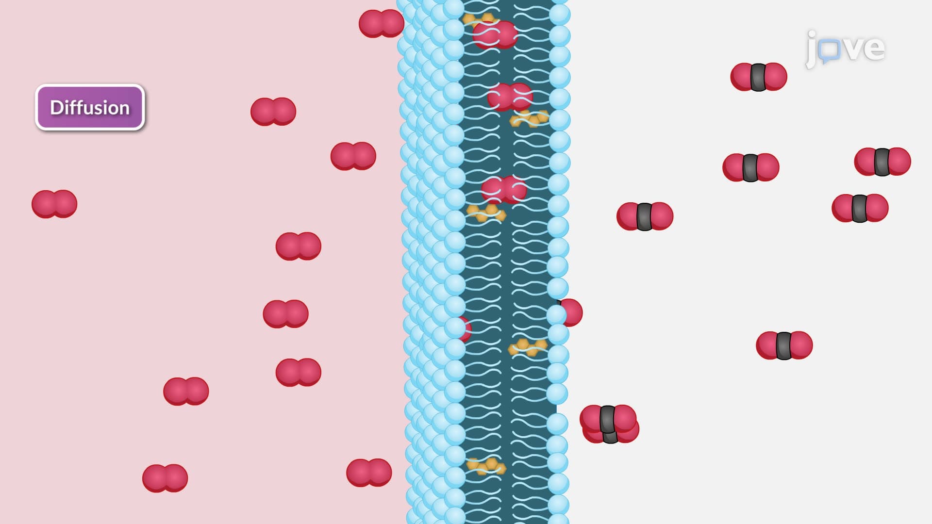
Respiration involves pulmonary ventilation, diffusion, and perfusion working together to maintain cellular metabolism. During inspiration, the diaphragm contracts and intercostal muscles elevate the ribcage, creating negative pressure that draws air into the lungs. Gas exchange occurs through diffusion, where oxygen moves from alveoli to pulmonary capillaries while carbon dioxide transfers from blood to alveolar air. The medulla oblongata and pons regulate breathing through chemoreceptors that monitor blood pH and carbon dioxide levels, adjusting respiratory rate and depth accordingly.
2. Respiratory Rate Assessment Techniques
Accurate respiratory rate assessment requires counting complete breath cycles (inspiration plus expiration) for one full minute while the patient remains unaware of monitoring. Normal adult respiratory rates range from 12-18 breaths per minute. Healthcare providers should observe chest rise and fall, noting any irregularities in rhythm or depth. Tachypnea (>20 breaths/minute) may indicate anxiety, pain, or metabolic acidosis, while bradypnea (<12 breaths/minute) suggests respiratory depression or neurological impairment. Patients with rates above 24 require close monitoring, and rates exceeding 27 indicate potential cardiac arrest risk.
3. Breathing Patterns and Abnormalities
Various breathing patterns indicate specific pathophysiological conditions requiring immediate recognition. Cheyne-Stokes breathing shows gradual increase and decrease in depth followed by apneic periods, commonly seen in heart failure patients. Kussmaul breathing presents as deep, rapid respirations associated with diabetic ketoacidosis. Biot's breathing displays irregular rate and depth with sudden apneic episodes, often indicating brainstem damage. Healthcare providers must distinguish between normal eupnea and pathological patterns to ensure appropriate interventions and prevent patient deterioration.
4. Factors Influencing Respiratory Function
Multiple variables affect respiratory rate assessment and breathing patterns in clinical practice. Age significantly impacts normal values, with newborns breathing 30-60 times per minute compared to adults. Exercise increases both rate and depth to meet oxygen demands, while pain typically decreases inspiratory effort, resulting in shallow, rapid breathing. Medications like opioids reduce respiratory drive, whereas stimulants increase breathing rate. Body position affects lung expansion, with upright postures enabling optimal chest movement compared to supine positions that may restrict respiratory mechanics.
5. Clinical Assessment and Documentation
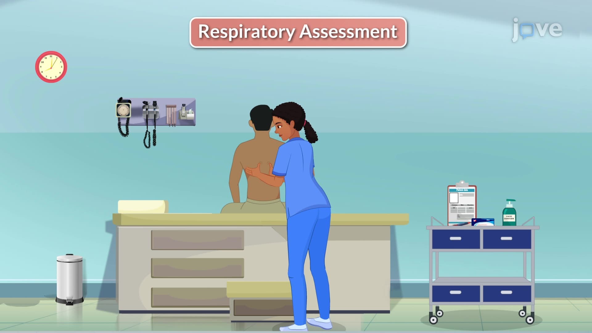
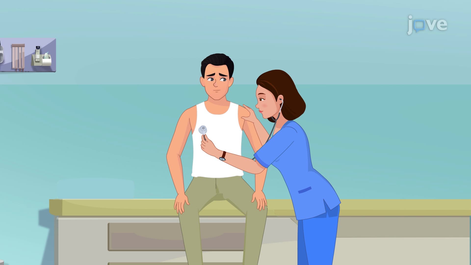
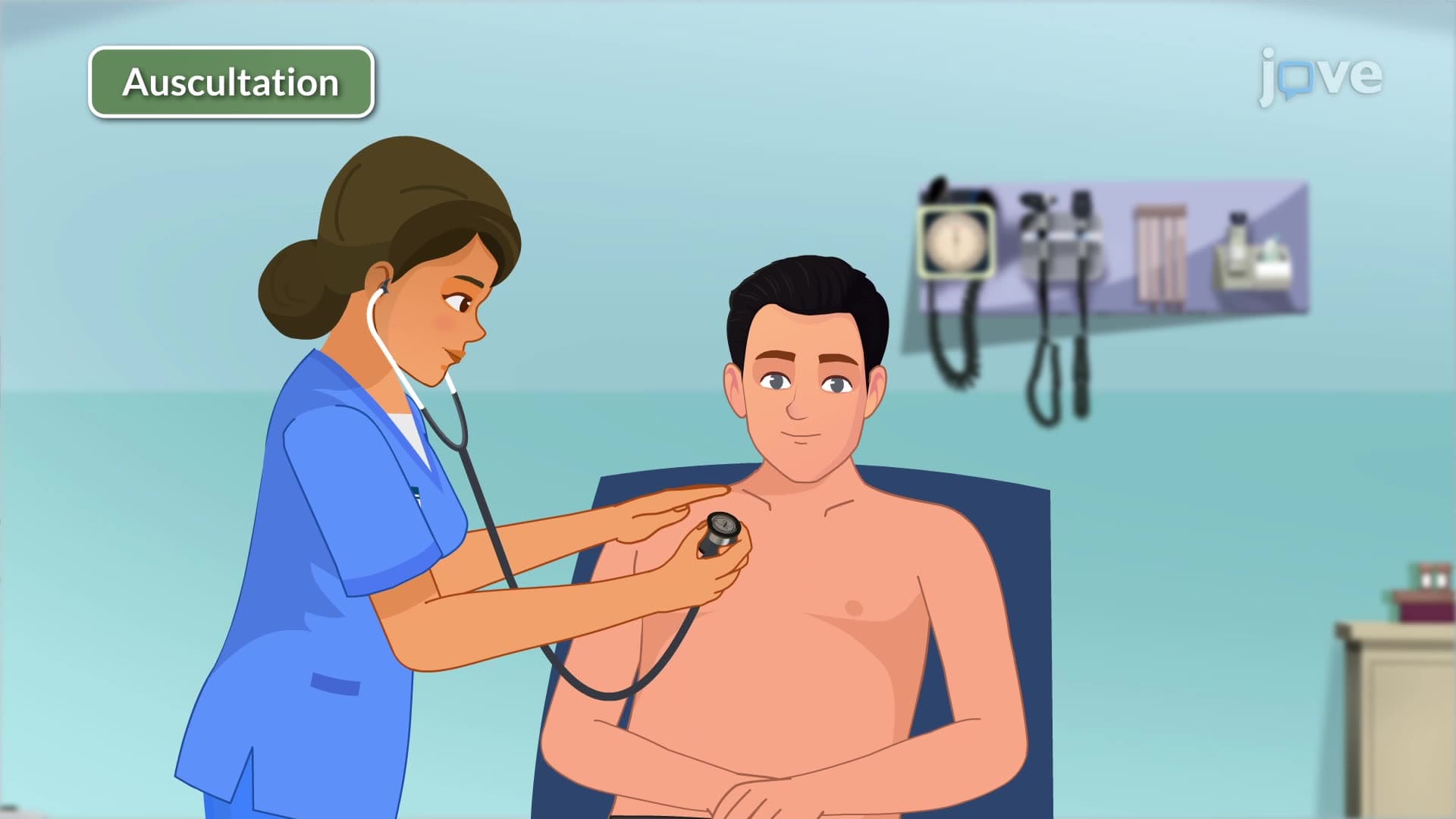
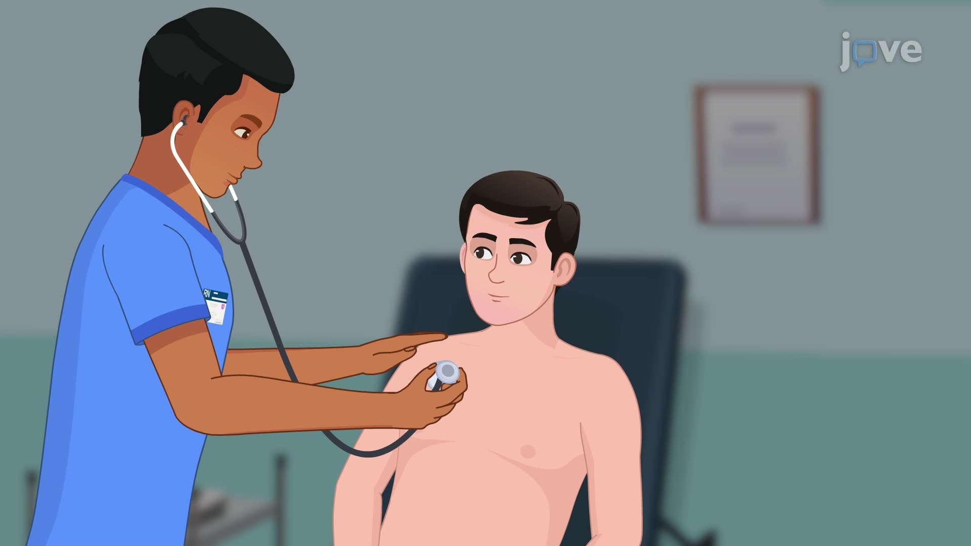
Comprehensive respiratory assessment involves systematic evaluation of airway patency, breathing effort, and oxygenation status. Healthcare providers assess skin color for cyanosis, observe accessory muscle usage indicating respiratory distress, and evaluate chest expansion symmetry. Pulse oximetry provides objective oxygen saturation measurements, with normal values ranging 95-100%. Documentation must include respiratory rate, depth, rhythm, oxygen saturation, and any abnormal findings such as adventitious breath sounds. Proper assessment techniques ensure early detection of respiratory compromise and guide appropriate therapeutic interventions in healthcare settings.
Frequently Asked Questions
Normal adult respiratory rates range from 12-18 breaths per minute, while newborns typically breathe 30-60 times per minute. Children's rates gradually decrease with age, reaching adult values by adolescence. These age-related differences reflect metabolic demands and lung development patterns.
NCLEX questions often focus on identifying abnormal respiratory patterns, recognizing signs of respiratory distress, and understanding when to report critical findings. Expect scenarios requiring interpretation of vital signs trends and appropriate nursing interventions for respiratory emergencies.
Tachypnea (>20 breaths/minute) may indicate anxiety, pain, fever, or underlying respiratory/cardiac conditions. Rates above 24 require close monitoring, while rates exceeding 27 suggest imminent cardiac arrest risk within 72 hours, necessitating immediate medical intervention.
Conscious awareness of breathing assessment can alter both rate and depth through voluntary control, leading to inaccurate measurements. Healthcare providers should count respirations while appearing to assess pulse to maintain assessment accuracy.
Pulse oximetry measures oxygen saturation non-invasively, with normal values ranging 95-100%. Values below 90% indicate hypoxemia requiring immediate attention. However, pulse oximetry may be unreliable in cases of carbon monoxide poisoning, severe anemia, or poor peripheral circulation.
Practice counting respirations on classmates, memorize normal values for different age groups, and understand pathophysiology behind abnormal patterns. Create flashcards linking breathing patterns to specific conditions, and practice systematic assessment techniques regularly.
Observe for sternocleidomastoid muscle contraction in the neck, intercostal retractions between ribs, and use of abdominal muscles during expiration. These findings suggest increased work of breathing and potential respiratory compromise requiring immediate evaluation.
Upright positions enable optimal chest expansion and accurate rate counting, while supine positions may restrict lung movement and alter breathing patterns. Consider positional effects when interpreting respiratory findings and ensure consistent positioning for serial assessments.
This microcourse includes 18 concept videos that walk you through the building blocks of Nursing. Each video is short, about 1 minute, so you can cover a full topic during a coffee break or between classes. The full sequence starts with Respiration and ends with Assessment of Respiration.
The natural next step is Vital Signs: Blood Pressure. Once you finish those, the full Nursing curriculum of 14 microcourses on JoVE Coach opens up, taking you from foundational concepts to advanced systems.