Loading subject page…


21,353 views
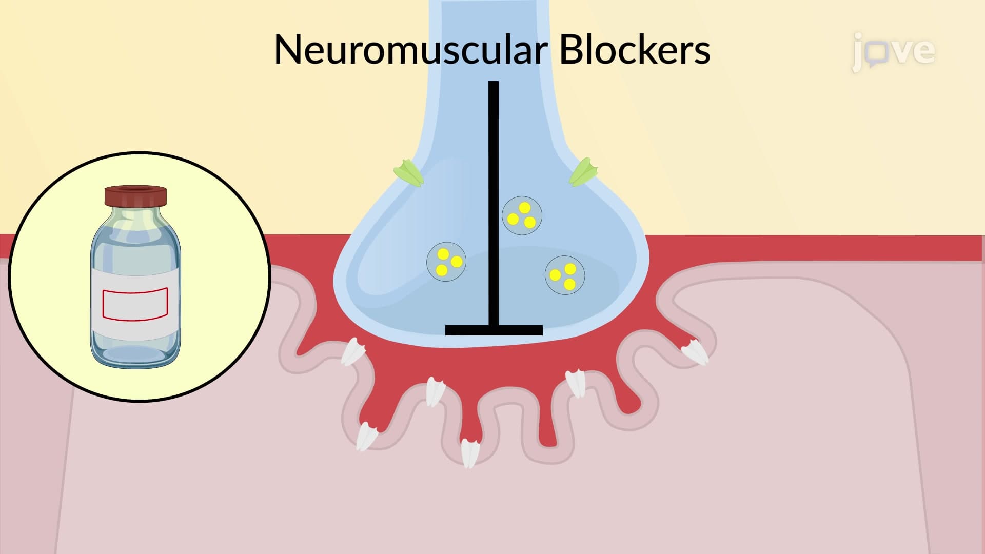

Video Summary: What Is Neuromuscular Junction and Blockade
Ever wonder how anesthesiologists ensure patients remain completely still during delicate brain surgery at Mayo Clinic? Neuromuscular junction blockade is the precise mechanism that allows medical professionals to temporarily paralyze muscles by interrupting communication between nerves and muscle fibers. This critical process involves blocking acetylcholine receptors at the neuromuscular junction, preventing the normal signals that cause muscle contraction. Understanding what is neuromuscular junction and blockade reveals how drugs like succinylcholine enable safe, controlled muscle relaxation during surgical procedures. Watch the full video on JoVE Coach to master this concept with expert-led visuals and step-by-step explanations.
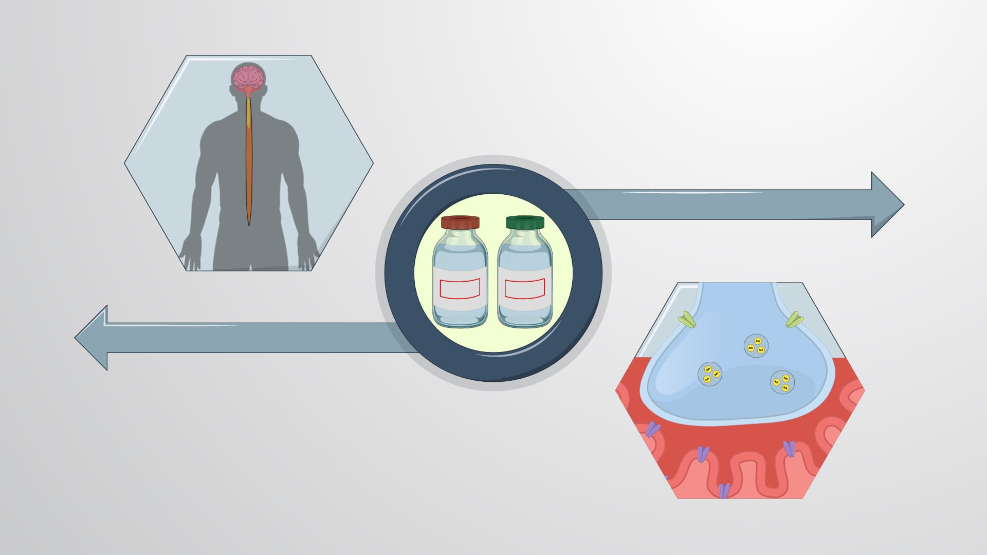
The neuromuscular junction represents one of the most precisely engineered communication systems in human physiology. This specialized synapse connects motor neurons from the spinal cord to individual skeletal muscle fibers, enabling voluntary movement from lifting a pencil to running a marathon. At Johns Hopkins School of Medicine, medical students learn that disrupting this communication through neuromuscular junction blockade forms the foundation of modern surgical anesthesia.
The neuromuscular junction consists of three critical components working in perfect synchronization. The presynaptic motor nerve terminal contains thousands of synaptic vesicles packed with acetylcholine molecules. The synaptic cleft, measuring only 50-100 nanometers wide, separates the nerve from the muscle fiber. The postsynaptic motor end plate contains densely packed nicotinic acetylcholine receptors, each capable of opening ion channels when activated.
This intricate structure becomes clinically relevant when anesthesiologists at institutions like Cleveland Clinic administer neuromuscular blocking agents. Students preparing for the MCAT often encounter questions about how these drugs exploit the junction's normal physiology to achieve therapeutic muscle paralysis.
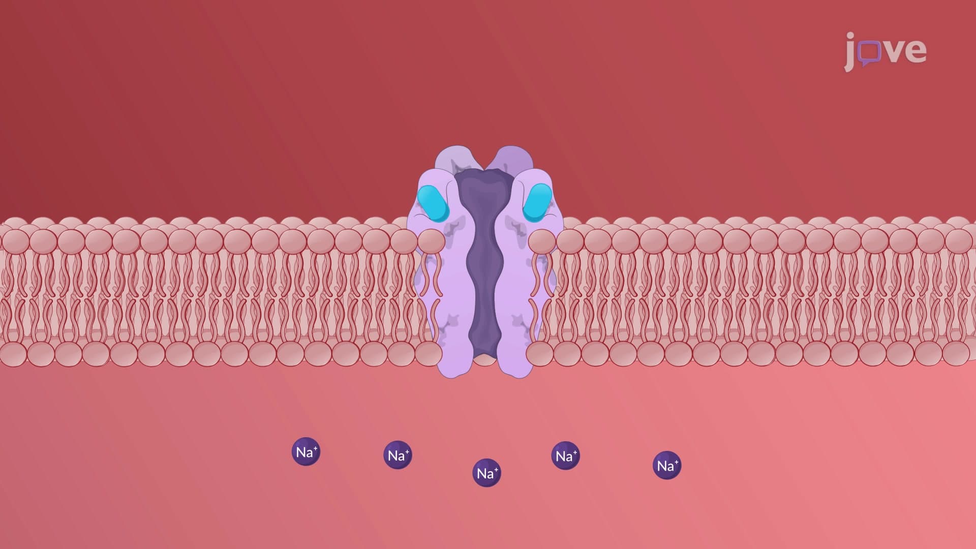
Normal neuromuscular transmission begins when an action potential reaches the nerve terminal, triggering calcium-dependent exocytosis of acetylcholine. These neurotransmitter molecules diffuse across the synaptic cleft and bind to nicotinic receptors, causing conformational changes that open sodium channels. The resulting depolarization spreads across the muscle membrane, ultimately triggering contraction through calcium release from the sarcoplasmic reticulum.
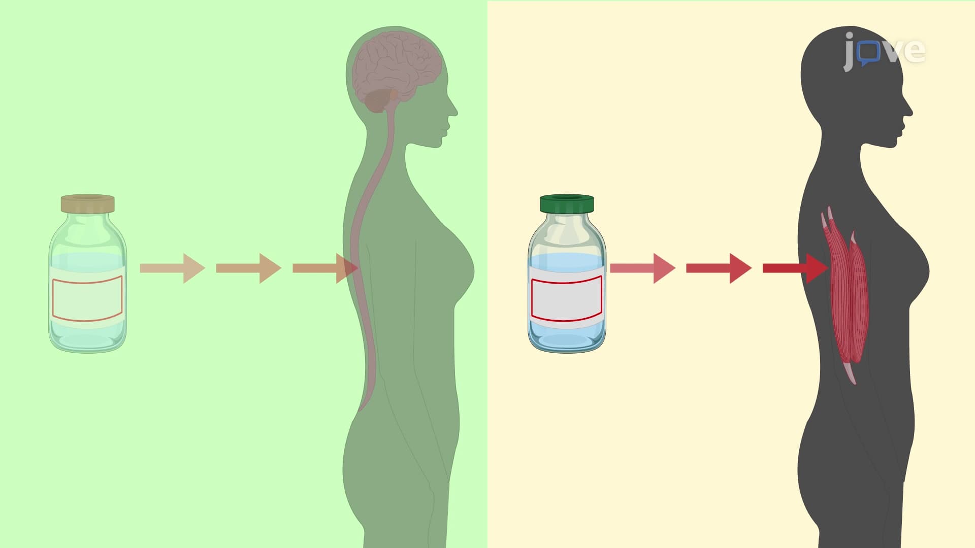
Neuromuscular blocking agents interrupt this process through competitive or non-competitive inhibition. Succinylcholine, commonly used in emergency departments across the United States, mimics acetylcholine's structure closely enough to bind nicotinic receptors but lacks the ability to properly open ion channels. This competitive blockade prevents normal muscle activation while maintaining the patient's consciousness and vital functions.
Understanding neuromuscular junction blockade proves essential for students pursuing healthcare careers. AP Biology students encounter this topic when studying cell signaling and neurotransmission. Pre-med students face detailed questions about receptor pharmacology on the MCAT, while nursing students preparing for the NCLEX must understand the monitoring and safety protocols surrounding these medications.
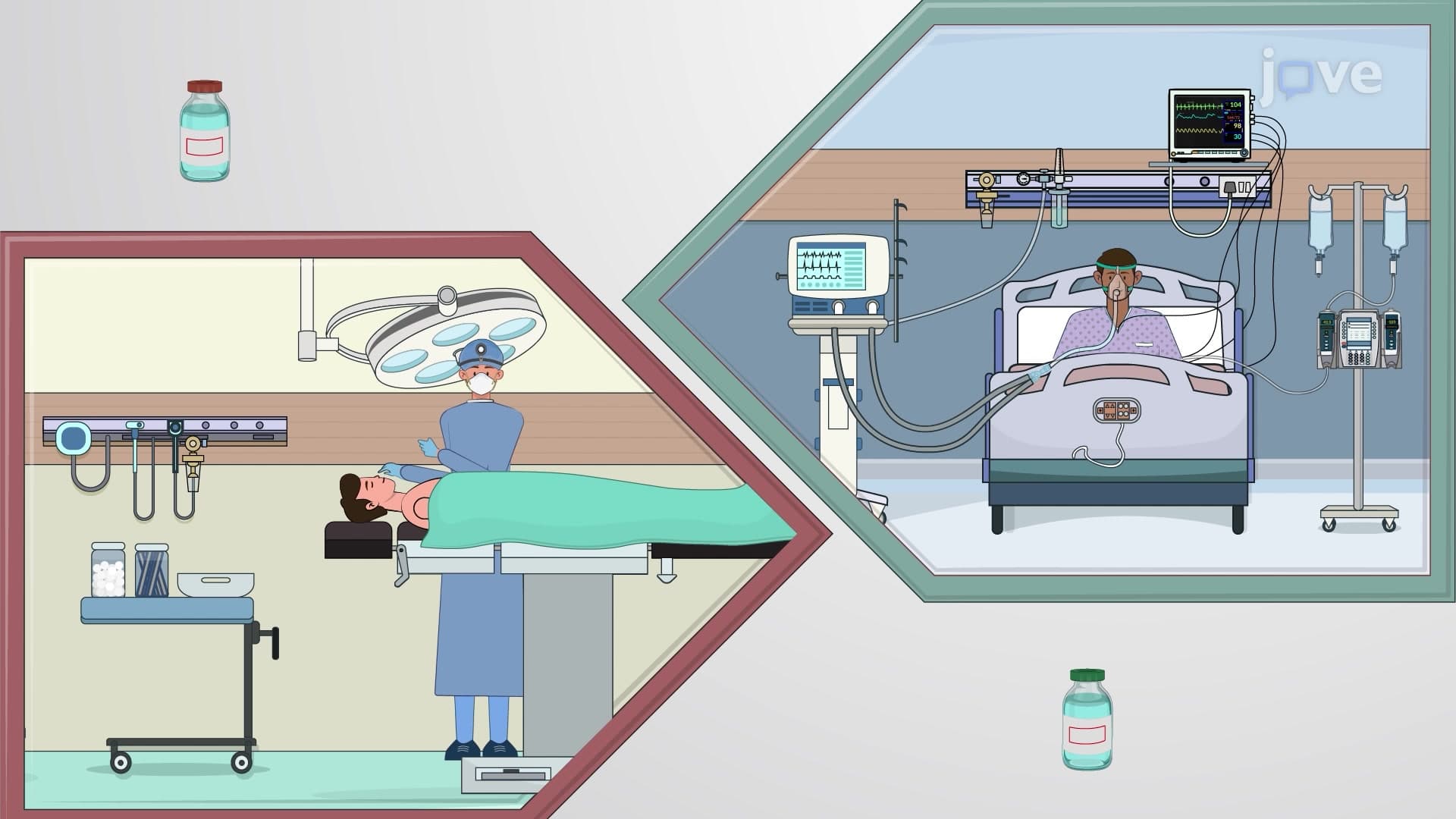
In clinical practice, anesthesiologists at major medical centers like Massachusetts General Hospital rely on precise neuromuscular blockade to ensure patient safety during procedures ranging from appendectomies to cardiac surgery. The ability to temporarily eliminate muscle movement allows surgeons to work with unprecedented precision while maintaining patient comfort and safety.
Related Micro-courses