Loading video…


 25 Concepts
 84.7K views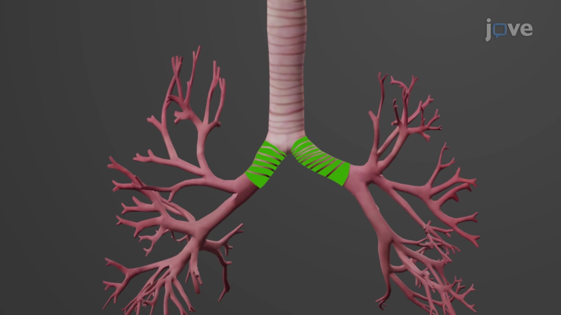

Video Summary: What Is the Bronchial Tree
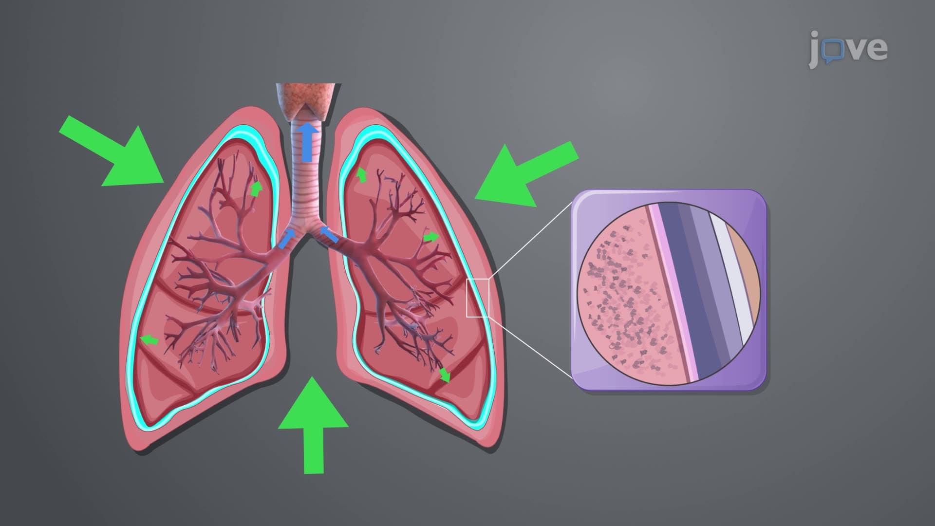
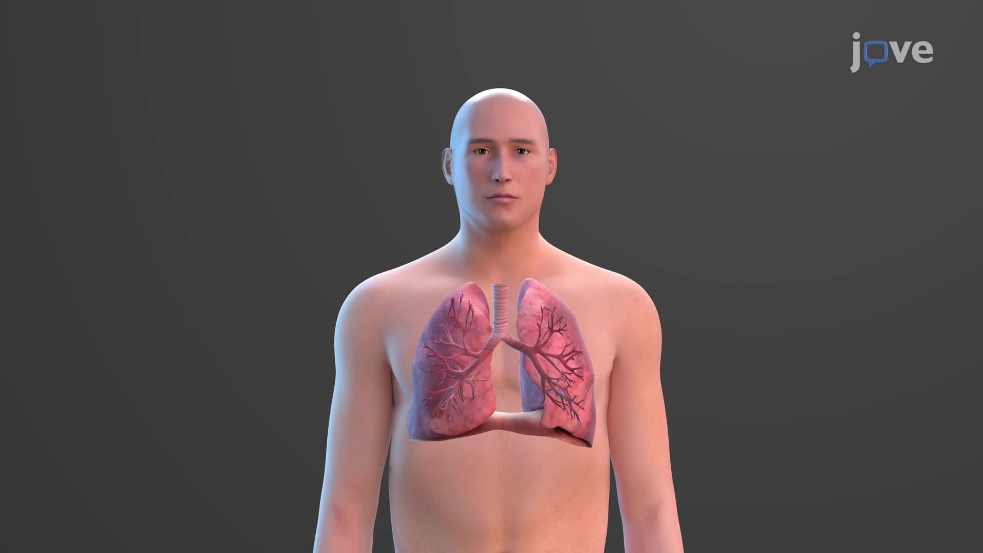
Ever wonder how air travels from your nose to the tiniest air sacs in your lungs? The bronchial tree anatomy biology reveals an intricate highway system that branches like an upside-down tree throughout your chest. When you take a deep breath during a high school track meet, air flows through the trachea to the carina, where it splits into primary bronchi before branching into progressively smaller airways. What is The Bronchial Tree becomes clear when you understand this systematic branching pattern that ensures oxygen reaches every corner of your lungs. Watch the full video on JoVE Coach to master this concept with expert-led visuals and step-by-step explanations.
The bronchial tree anatomy biology represents one of nature's most efficient distribution systems, designed to deliver air to approximately 300 million alveoli in human lungs. This airway bronchial tree explained begins at the carina-a critical anatomical landmark located at the T5 vertebral level where emergency physicians insert endotracheal tubes during CPR procedures.
The structural design follows engineering principles seen in river deltas or electrical circuits. The right primary bronchus measures approximately 2.5 cm long with a 1.4 cm diameter, while the left primary bronchus extends 5 cm with a 1.1 cm diameter. This asymmetry explains why aspirated objects more commonly lodge in the right lung-a crucial concept for MCAT respiratory physiology questions.
What is the bronchial tree in respiratory anatomy becomes apparent when examining its systematic branching. Each lung lobe receives its dedicated lobar bronchus: three on the right (upper, middle, lower) and two on the left (upper, lower). This anatomical organization allows thoracic surgeons at institutions like Johns Hopkins to perform precise lobectomies when treating lung cancer.
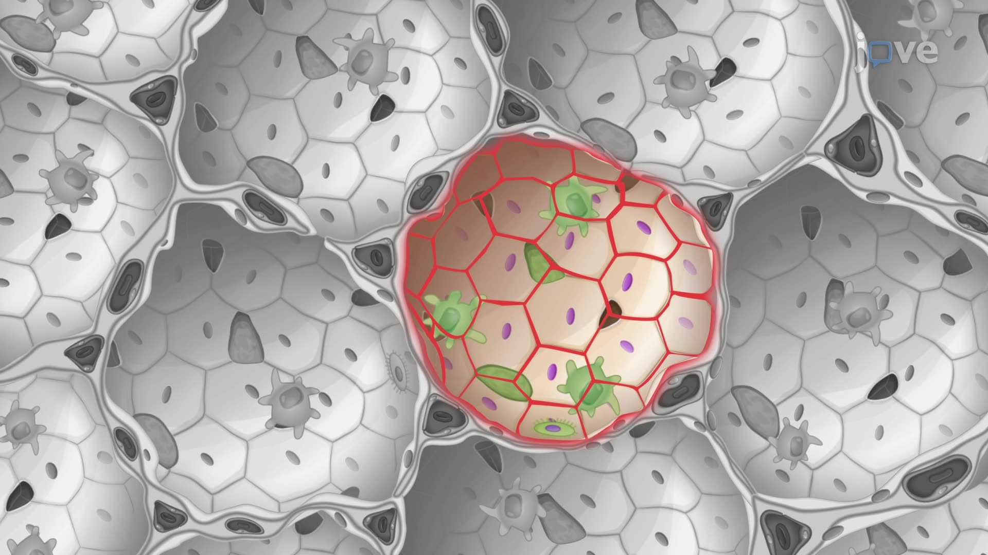
The segmental bronchi create ten distinct bronchopulmonary segments in the right lung and eight to ten in the left lung. Radiology students at medical schools across the United States learn to identify these segments on CT scans, as each segment has independent blood supply and drainage-essential for surgical planning and understanding pneumonia patterns.
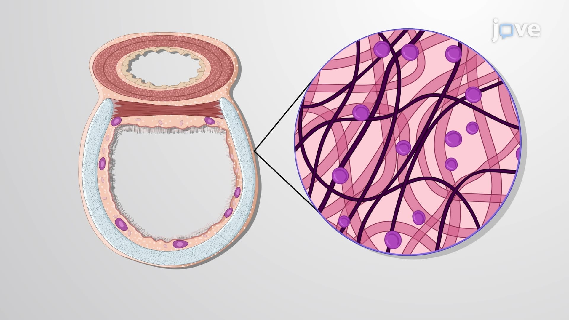
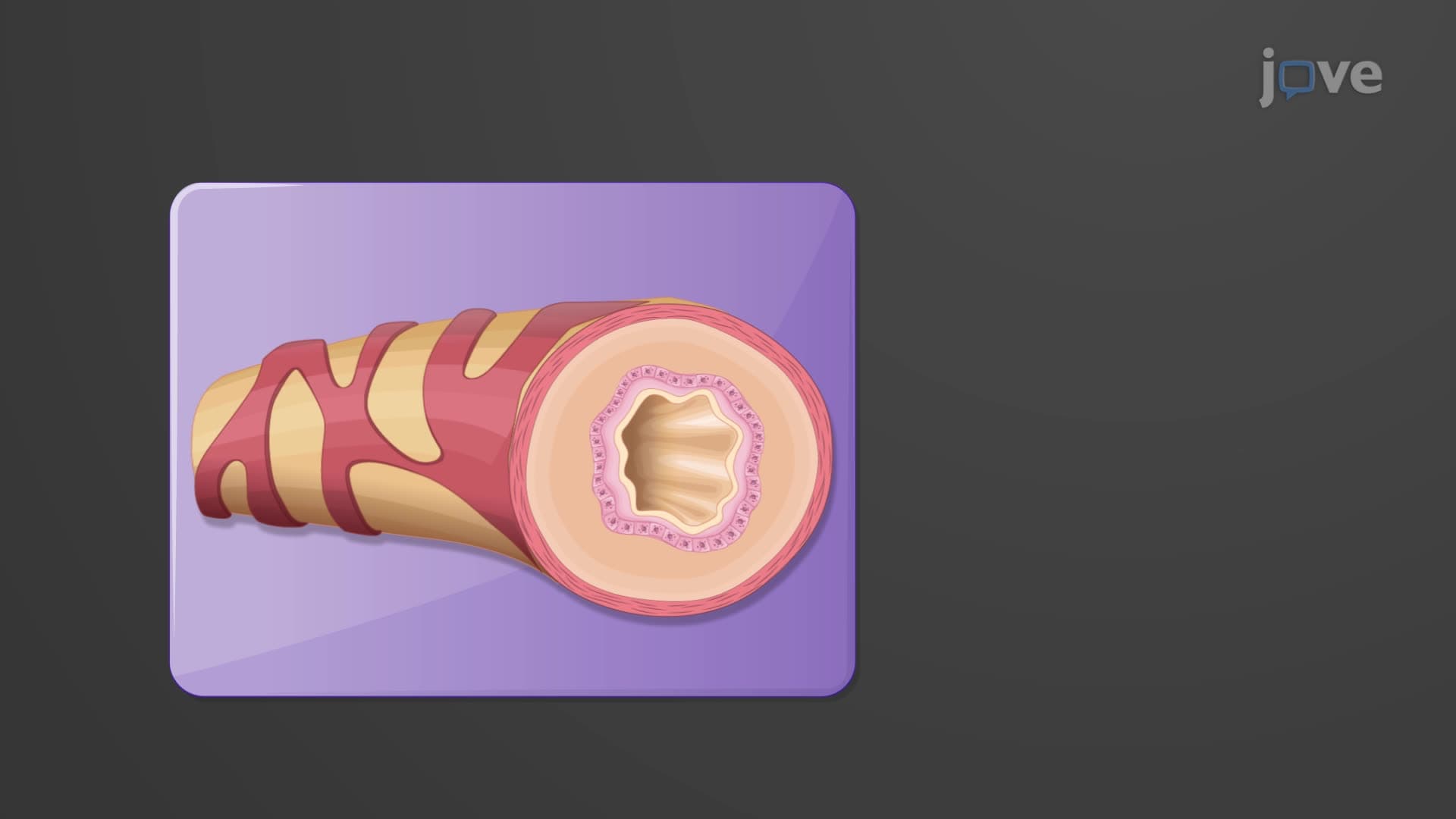
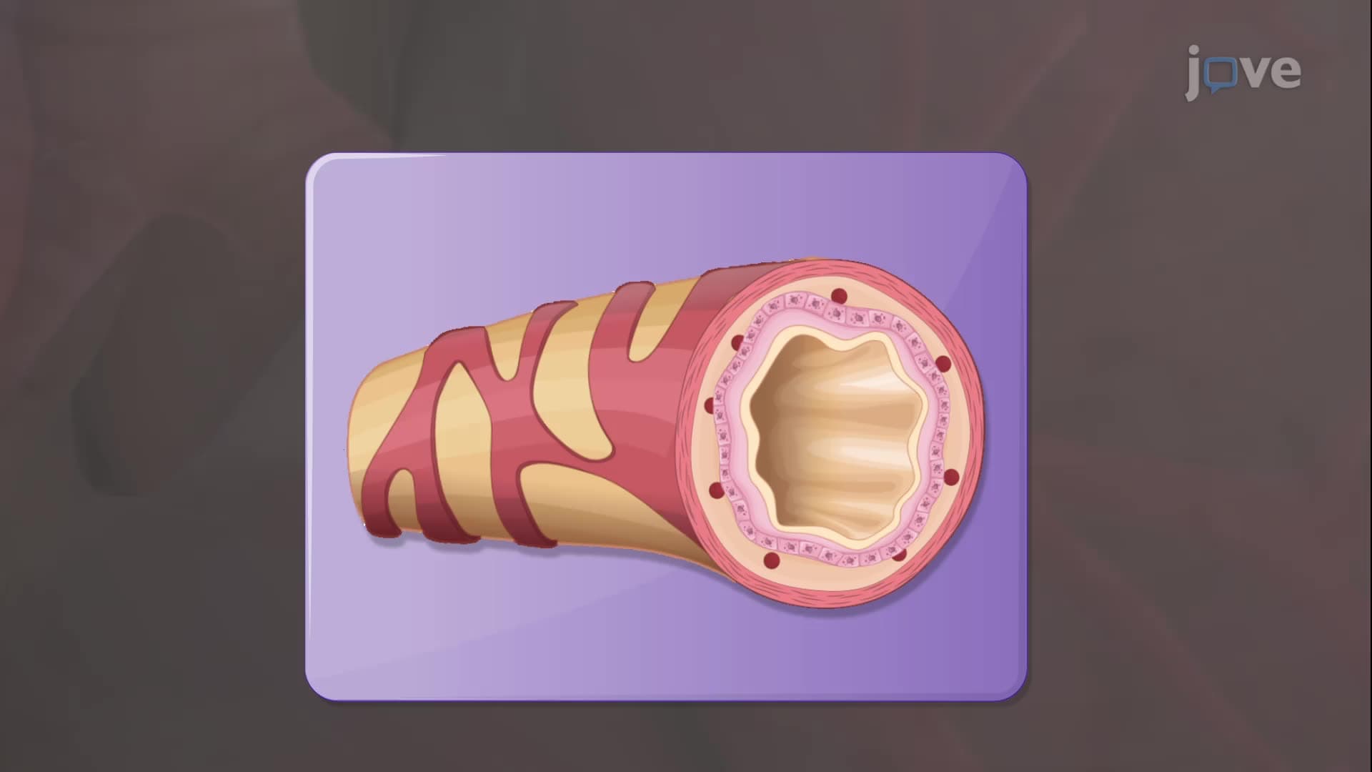
The bronchial tree lung anatomy reveals remarkable cellular specialization. Primary and secondary bronchi maintain incomplete cartilage rings similar to the trachea, providing structural support while allowing esophageal expansion during swallowing. As airways narrow, cartilage gradually disappears, replaced by increased smooth muscle-the target of bronchodilator medications used in asthma treatment.
Terminal bronchioles mark the conducting zone bronchial tree's endpoint, containing specialized club cells that perform multiple functions. These non-ciliated cells, discovered by Clara in 1937, produce surfactant proteins and serve as stem cells for bronchiolar repair. Understanding club cell function appears frequently on USMLE Step 1 examinations and explains why certain environmental toxins specifically damage small airways.
Bronchial tree anatomy directly impacts patient care in American hospitals. Pulmonologists use bronchoscopy to navigate these pathways, reaching specific segments for biopsy or treatment. The branching pattern determines how inhaled medications distribute, influencing treatment strategies for conditions like cystic fibrosis-a genetic disorder affecting approximately 30,000 Americans.
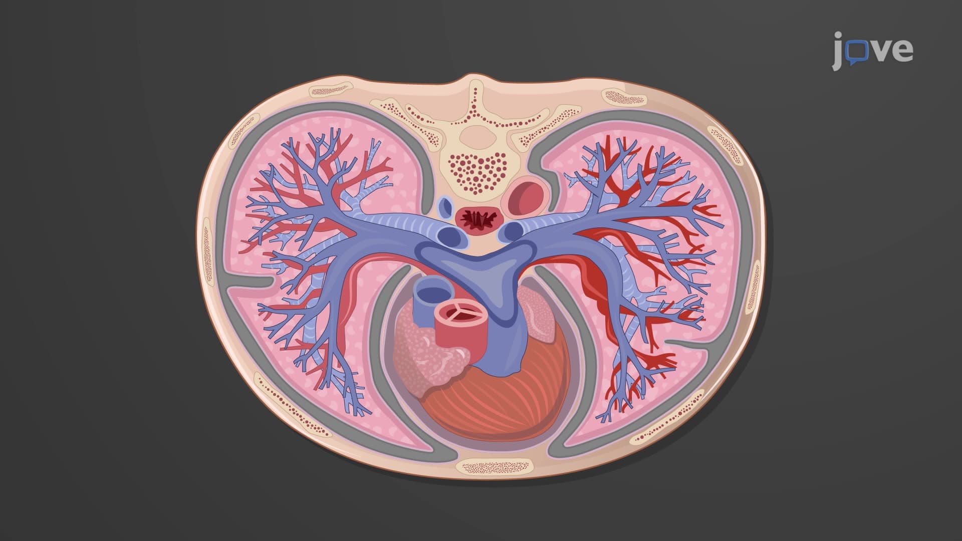
For students preparing for AP Biology or college anatomy courses, understanding bronchial tree organization provides the foundation for studying respiratory mechanics, gas exchange, and pathophysiology. This knowledge integrates with cardiovascular anatomy, as pulmonary arteries follow bronchial branching patterns, creating the lung's dual circulation system essential for efficient oxygenation.
Related Micro-courses