Loading video…


 12 Concepts
 78.9K views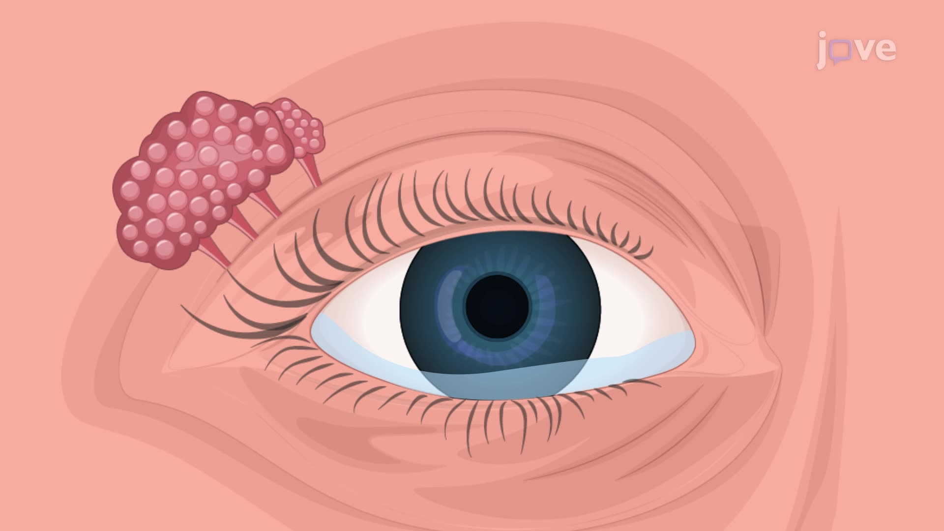

Video Summary: What Is Accessory Structures Eye
Did you know that without your eyebrows, a single drop of sweat could blur your vision while taking the SAT? Eye accessory structures anatomy encompasses the protective components surrounding your eyes that work tirelessly to maintain clear vision. From the conjunctiva that keeps your eyes moist during long study sessions to the lacrimal apparatus that produces tears when you're stressed about finals, understanding what is accessory structures eye reveals how your body protects one of its most vital sensory organs. Watch the full video on JoVE Coach to master this concept with expert-led visuals and step-by-step explanations.
The accessory structures of the eye form a sophisticated protective system that ensures optimal visual function while safeguarding this delicate sensory organ. Unlike the eye's internal components responsible for vision, these accessory structures primarily serve protective, supportive, and maintenance roles. For students preparing for the MCAT or AP Biology exams, understanding these structures is crucial as they frequently appear in questions about sensory organ anatomy and physiology.
The eyelids (palpebrae) represent the most visible accessory eye protection mechanism. The upper and lower eyelids are separated by the palpebral fissure and meet at two points called commissures-the medial commissure (near the nose) and lateral commissure (toward the temple). These structures contain specialized muscles: the orbicularis oculi, which closes the eyelids, and the levator palpebrae superioris, which lifts the upper eyelid. This muscular coordination becomes particularly relevant when studying neurological conditions affecting facial nerve function, commonly tested on the USMLE Step 1.
The conjunctiva, a transparent mucous membrane, lines the inner eyelids and covers the sclera (but notably not the cornea). This structure produces mucin, which helps maintain the eye's tear film stability. Medical students often encounter conjunctivitis cases during clinical rotations, making this anatomy practically relevant for future healthcare professionals.
The lacrimal apparatus represents one of the most clinically significant accessory eye structures. Located in the upper lateral region of each orbit, the lacrimal gland continuously produces tears containing water, electrolytes, proteins, and antimicrobial substances. These tears spread across the eye surface during blinking, then drain through the lacrimal puncta (small openings at the inner corners of each eyelid) into the lacrimal canaliculi and finally into the lacrimal sac.
This drainage system becomes particularly important in clinical settings. For instance, blocked lacrimal ducts can cause excessive tearing, a condition frequently seen in pediatric ophthalmology practices across the United States. Students studying for the NCLEX or HESI A2 exams should understand this pathway as it relates to eye infections and tear duct disorders.
Though often overlooked, eyebrows serve critical protective functions beyond aesthetics. These dense, short hairs create a physical barrier that redirects sweat, rain, and debris away from the eyes. They also provide some protection from UV radiation, particularly important given increasing skin cancer rates in the United States. Understanding eyebrow anatomy becomes relevant when studying facial trauma cases or surgical procedures affecting the forehead region.
Related Micro-courses