Loading video…


 20 Concepts
 222.1K views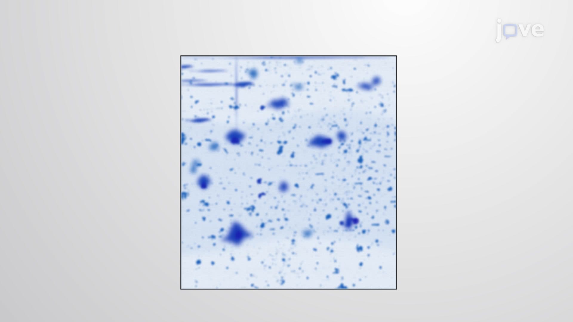

Video Summary: What Is Two Dimensional Gel Electrophoresis
Did you know that researchers at Harvard Medical School can distinguish between thousands of proteins in a single cell using a technique that separates molecules like sorting books by both author and publication date? Two dimensional gel electrophoresis achieves this remarkable precision by combining two separation methods: first by electrical charge, then by molecular weight. This powerful laboratory technique has revolutionized protein analysis at institutions like Johns Hopkins University, enabling scientists to detect even single amino acid differences between similar proteins. Watch the full video on JoVE Coach to master this concept with expert-led visuals and step-by-step explanations.
Two dimensional gel electrophoresis represents one of the most sophisticated protein separation techniques available in modern biochemistry laboratories. Unlike traditional single-dimension methods that separate proteins based on only one property, this advanced approach utilizes two perpendicular separation mechanisms to achieve unprecedented resolution. The technique has become indispensable in proteomics research at major US institutions, from Stanford University's biochemistry programs to clinical laboratories at the Mayo Clinic.
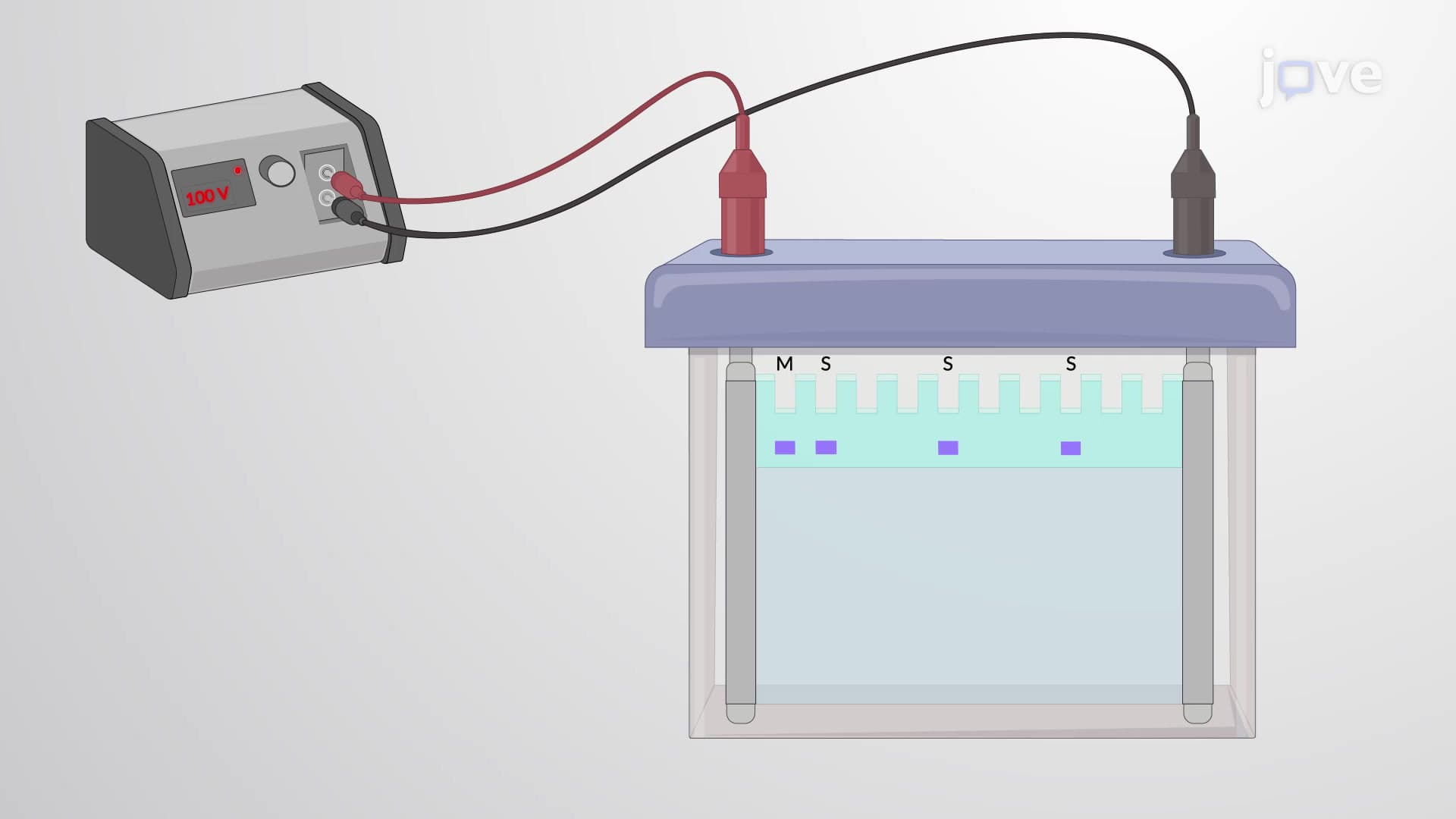
The first dimension employs isoelectric focusing (IEF) using immobilized pH gradient (IPG) strips. These specialized strips contain a stable pH gradient that ranges from acidic to basic conditions. When proteins are loaded onto the strip and an electric field is applied, each protein migrates until it reaches its isoelectric point-the specific pH where the protein carries zero net charge. At this point, the protein becomes immobilized and stops moving.
This charge-based separation is remarkably sensitive. Students preparing for the MCAT or advanced placement biology exams should understand that proteins differing by even a single charged amino acid residue will migrate to different positions. For example, if a lysine residue (positively charged) is substituted for a glutamate residue (negatively charged), the resulting protein will have a different isoelectric point and migrate to a distinct location on the IPG strip.
Following isoelectric focusing, the IPG strip undergoes treatment with sodium dodecyl sulfate (SDS), a powerful denaturing detergent. This treatment serves multiple purposes: it disrupts protein secondary and tertiary structures, coats proteins with negative charges proportional to their length, and prepares them for the second dimension of separation.
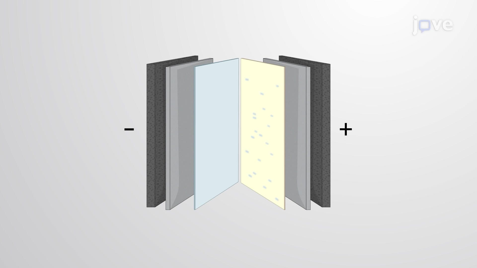
The treated IPG strip is then placed atop a polyacrylamide gel for SDS-PAGE separation. In this perpendicular direction to the first separation, proteins migrate based solely on their molecular weight. Smaller proteins navigate through the gel matrix more easily and travel farther, while larger proteins encounter greater resistance and migrate shorter distances.
Two dimensional gel electrophoresis has found extensive applications in US medical research and clinical diagnostics. At research institutions like the National Institutes of Health, scientists use this technique to study disease-related protein changes. For instance, researchers investigating Alzheimer's disease can compare brain protein profiles from affected and healthy individuals, identifying specific proteins that undergo modification during disease progression.
College students studying biochemistry will encounter this technique in advanced laboratory courses, where it serves as a powerful tool for understanding protein complexity and cellular responses to different conditions. The technique's ability to detect post-translational modifications makes it particularly valuable for studying how cellular stress, drug treatments, or developmental changes affect protein expression patterns.
Related Micro-courses