Loading subject page…


51,001 views
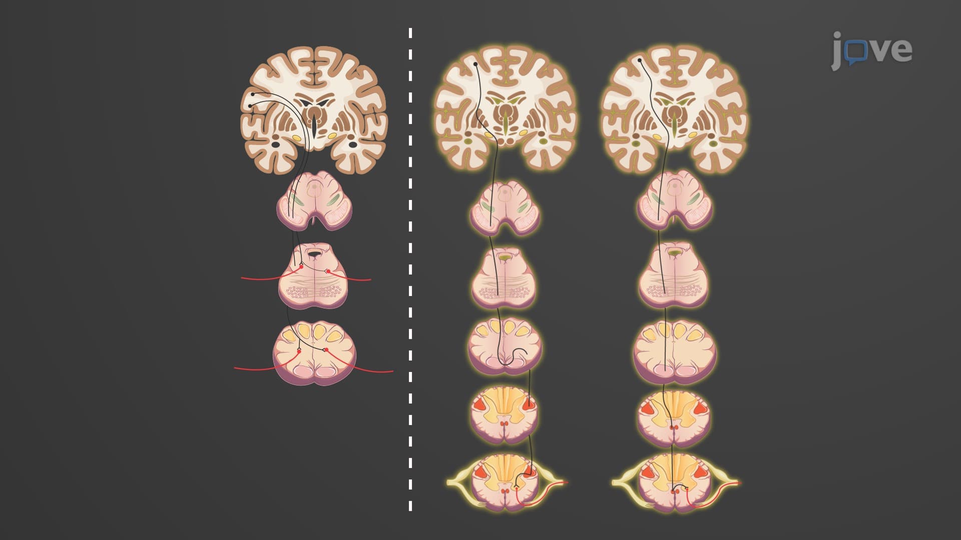

Video Summary: What are Direct Motor Pathways
Ever wonder how a professional pianist's fingers move with such precision, or how a surgeon performs delicate operations? The direct motor pathways anatomy controls these voluntary, conscious movements through specialized neural circuits. In the US, medical students studying for the MCAT learn that these pyramidal pathways connect the brain's motor cortex directly to spinal cord neurons, enabling everything from writing your name to throwing a baseball. Understanding what are direct motor pathways reveals how your brain transforms thoughts into precise muscle actions. Watch the full video on JoVE Coach to master this concept with expert-led visuals and step-by-step explanations.
The direct motor pathways anatomy represents one of the most elegant control systems in human neuroscience. These pathways, also called pyramidal tracts, form the primary route for voluntary motor commands from your brain to reach skeletal muscles. Unlike reflexive movements that occur automatically, the voluntary motor pathway explained through these circuits allows for conscious, deliberate actions-from a baseball pitcher's precise throw to a violinist's intricate fingering.
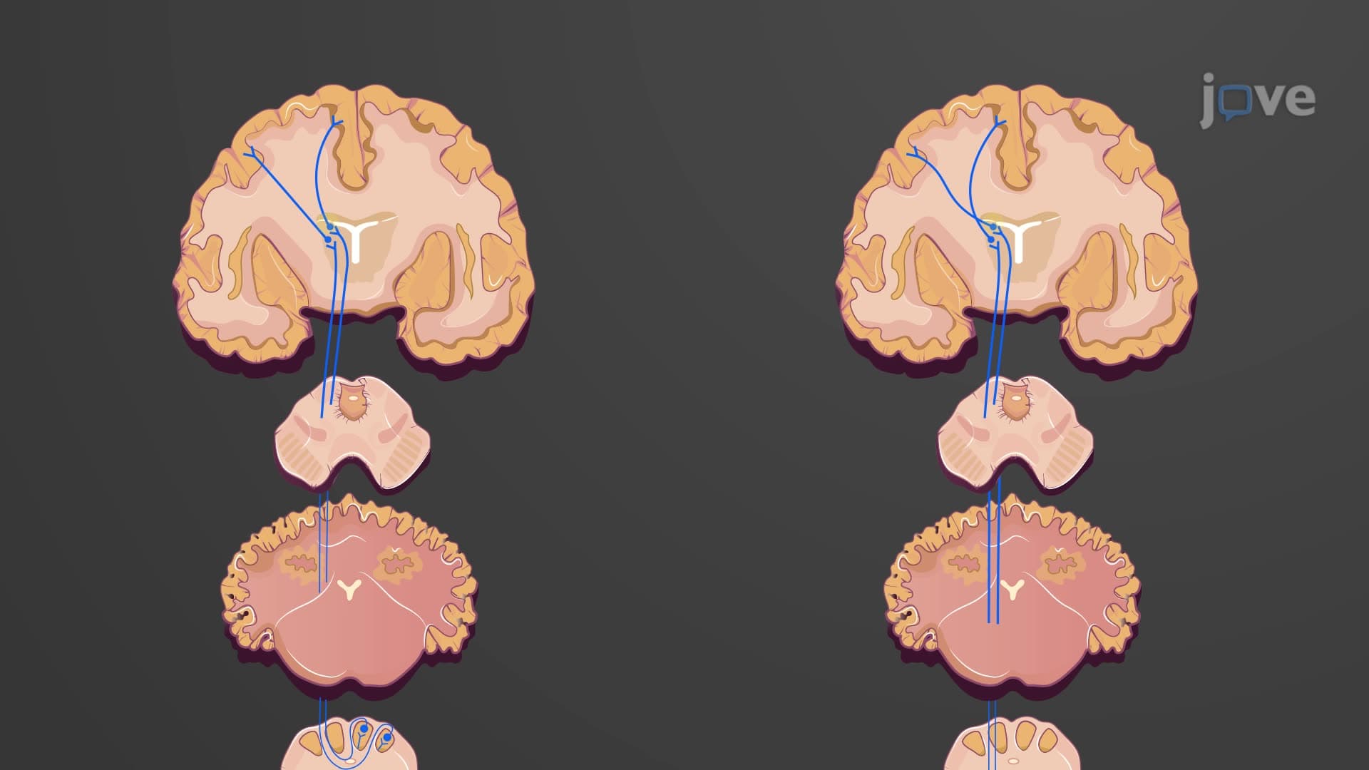
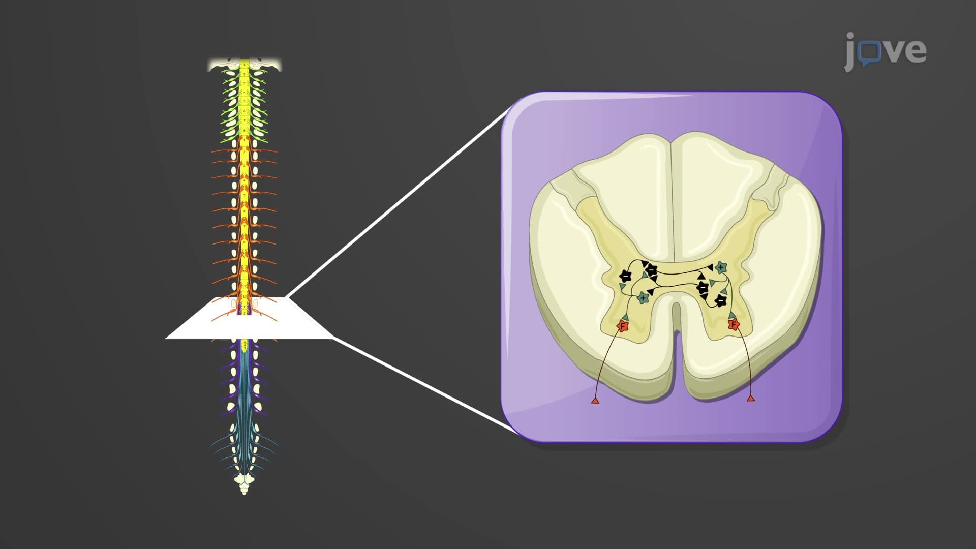
The architecture of what are the direct motor pathways in the CNS involves a two-neuron system. Upper motor neurons originate in the primary motor cortex (Brodmann area 4) and supplementary motor areas. These neurons project their axons downward through the internal capsule, cerebral peduncles, and into the brainstem or spinal cord. Lower motor neurons, located in cranial nerve nuclei or spinal cord anterior horns, receive these cortical inputs and directly innervate skeletal muscles.
This organization proves crucial for US medical students preparing for USMLE Step 1, where distinguishing upper versus lower motor neuron lesions appears frequently in clinical vignettes. Upper motor neuron damage typically causes spasticity and hyperreflexia, while lower motor neuron damage results in flaccid paralysis and muscle atrophy.
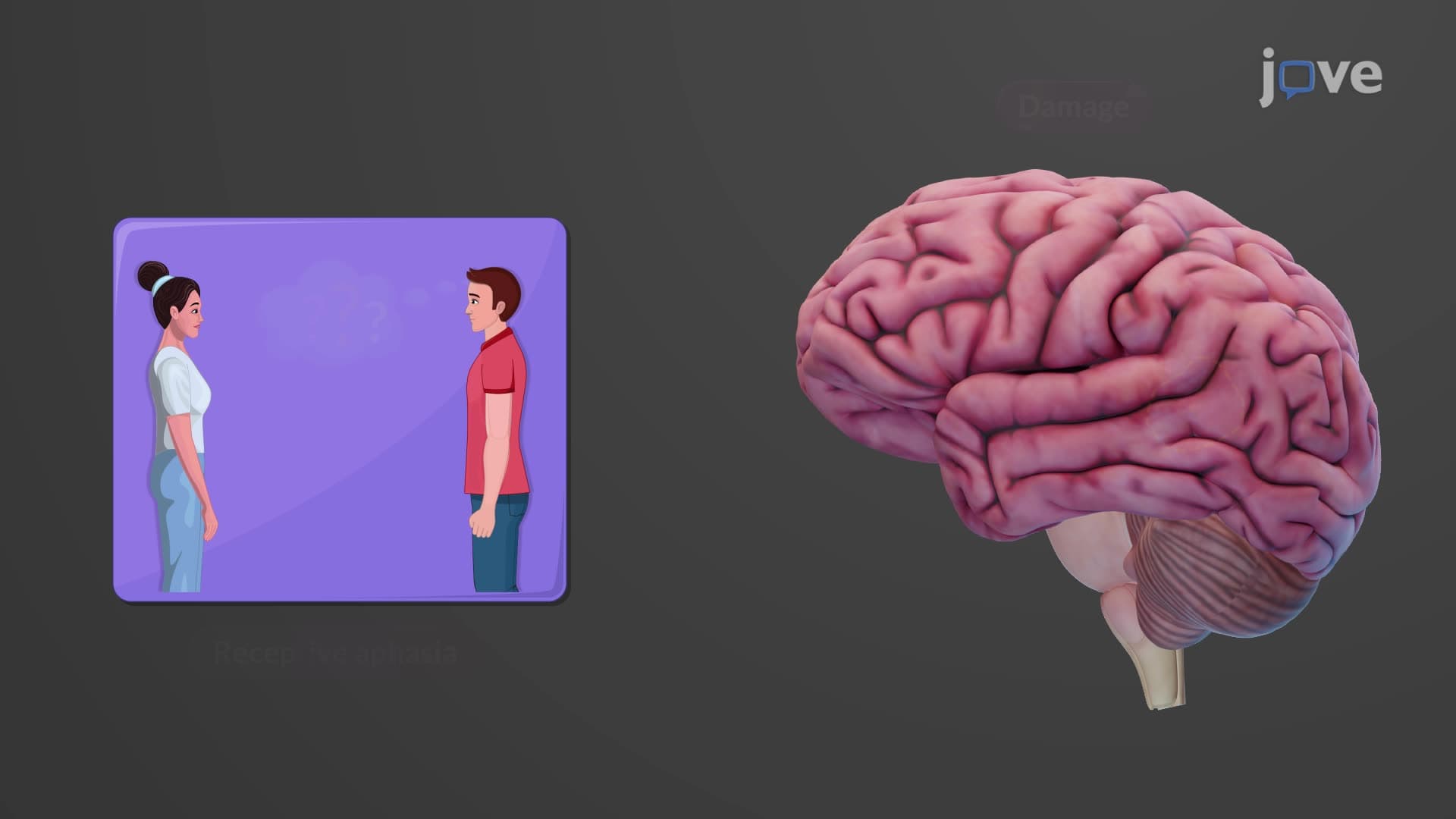
The corticobulbar tract manages voluntary movements of cranial muscles. These upper motor neuron direct pathways control facial expressions, eye movements, chewing, swallowing, and speech articulation. For instance, when a patient suffers a stroke affecting the corticobulbar tract, they might develop dysarthria (difficulty speaking) or dysphagia (difficulty swallowing)-conditions commonly encountered in US hospitals and nursing programs.
The corticospinal tract voluntary system divides into lateral and anterior components. The lateral corticospinal tract handles precise, skilled movements of distal limb muscles. Approximately 85-90% of these fibers decussate (cross) at the pyramidal decussation in the medulla oblongata. This crossing explains why left brain damage affects right-side movement and vice versa-a fundamental concept in AP Biology and college anatomy courses.
The anterior corticospinal tract comprises the remaining 10-15% of fibers that don't cross in the medulla. Instead, these fibers decussate at spinal cord levels and primarily control axial muscles of the trunk and proximal limb muscles responsible for posture and gross movements.
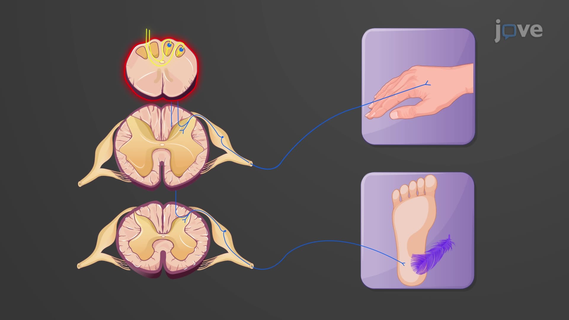
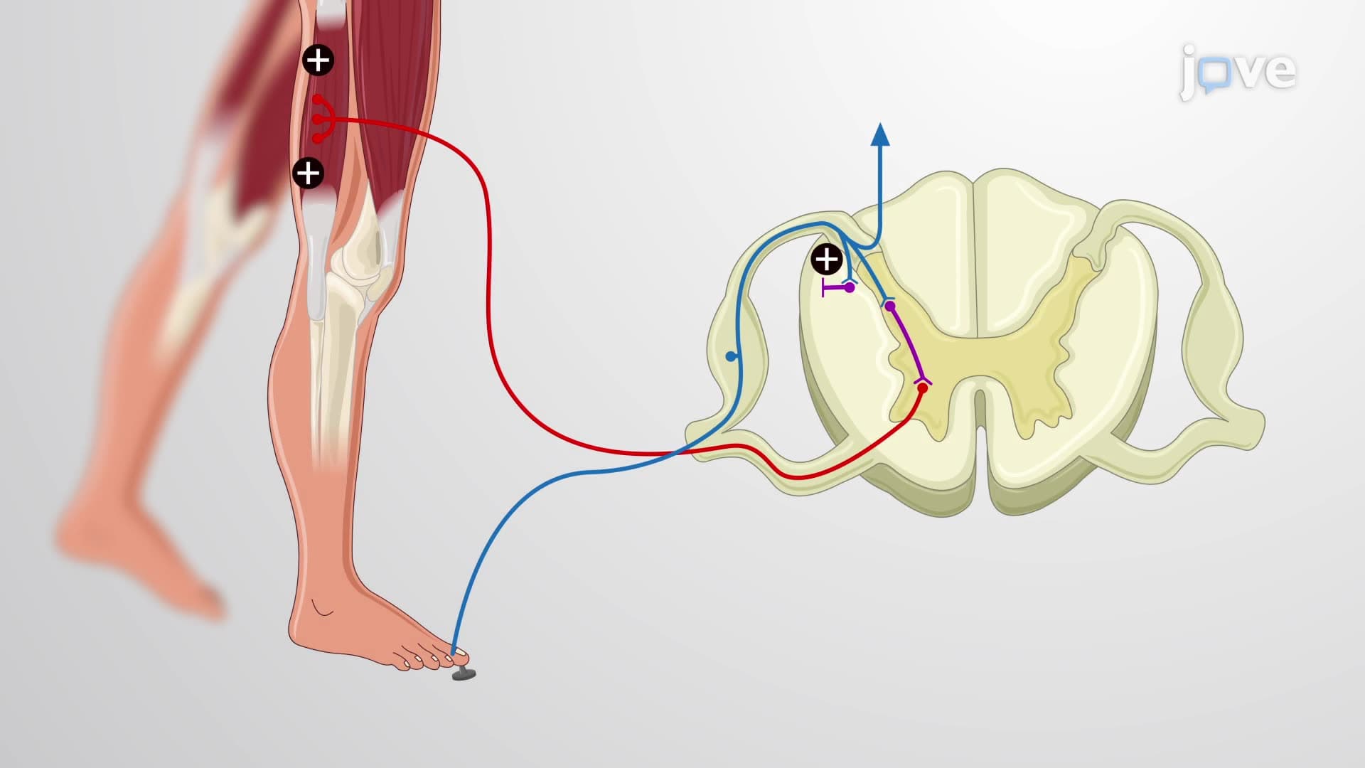
Understanding pyramidal tract motor pathway dysfunction helps explain conditions like cerebral palsy, stroke, and spinal cord injuries. In US clinical settings, the Babinski sign-where the big toe extends upward when the sole is stroked-indicates pyramidal tract damage. This assessment appears regularly on NCLEX-RN examinations and medical school practical exams.
For students taking the AP Biology exam, remember that these pathways exemplify how nervous system organization enables complex behaviors. The hierarchical control from cortex to muscle demonstrates principles of biological organization that extend beyond just human anatomy.
Related Micro-courses