Loading subject page…


84,702 views

Video Summary: What Is Gross Anatomy Lungs
Did you know your right lung is actually larger than your left lung because your heart needs space? Understanding lung gross anatomy biology reveals how these cone-shaped organs fit perfectly within your ribcage, with the right lung containing three distinct lobes while the left has only two. Medical students at Johns Hopkins University spend hours studying these structural differences to prepare for clinical rotations. Watch the full video on JoVE Coach to master this concept with expert-led visuals and step-by-step explanations.
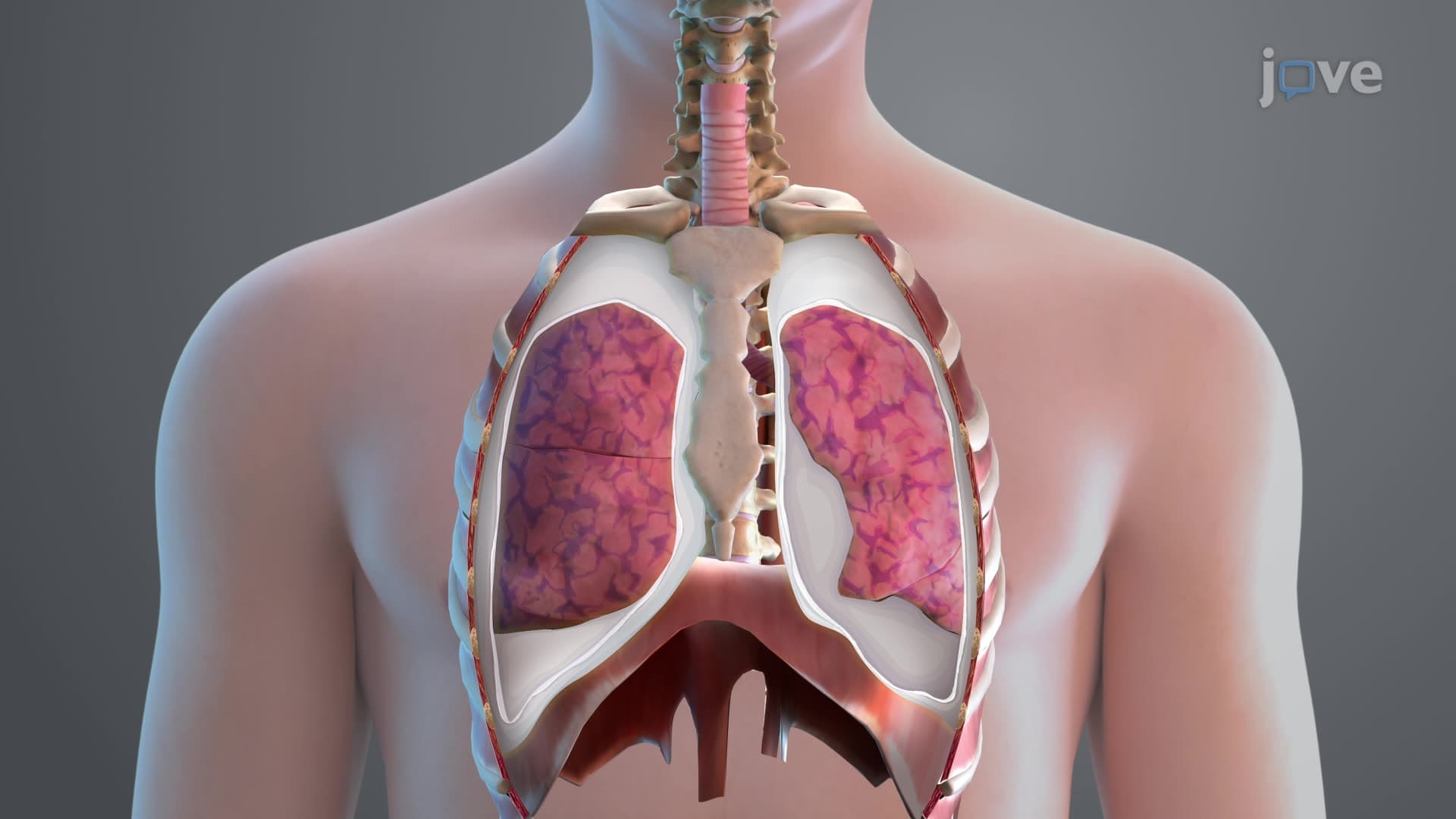
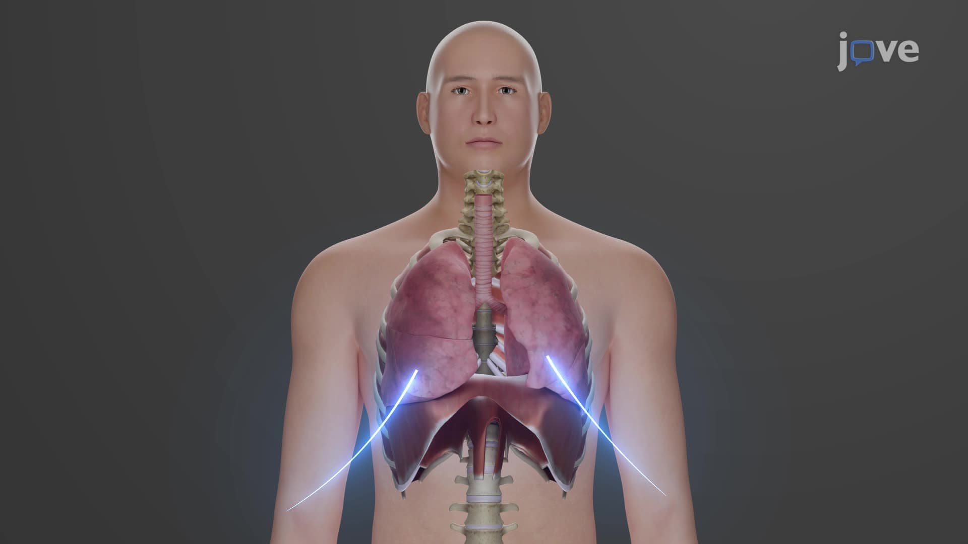
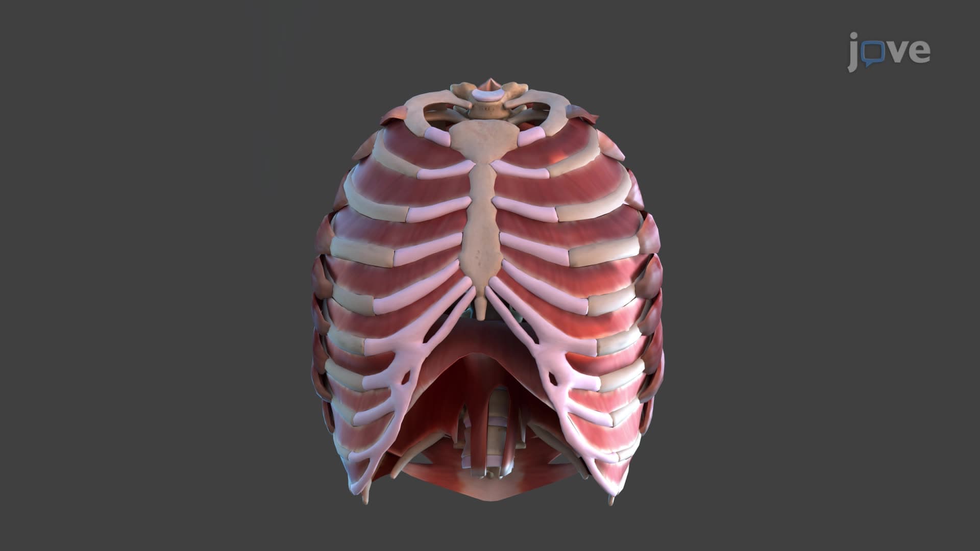
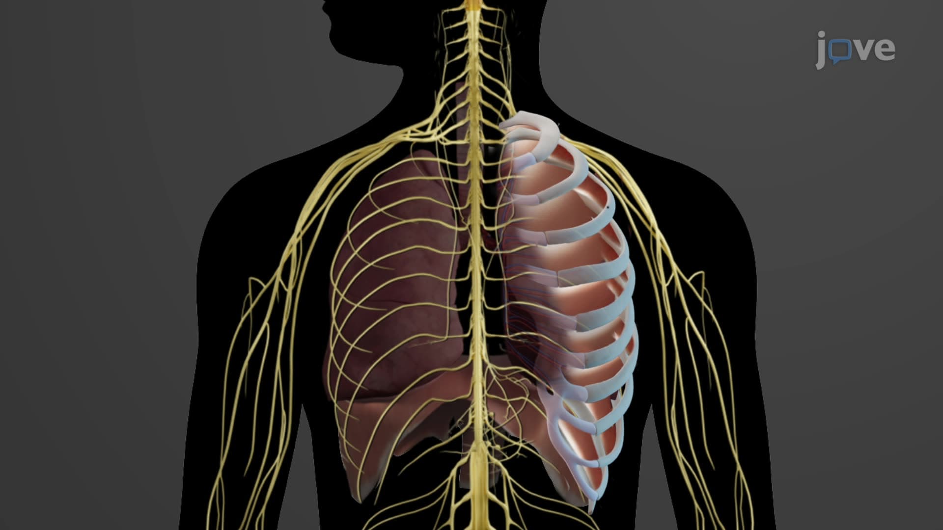
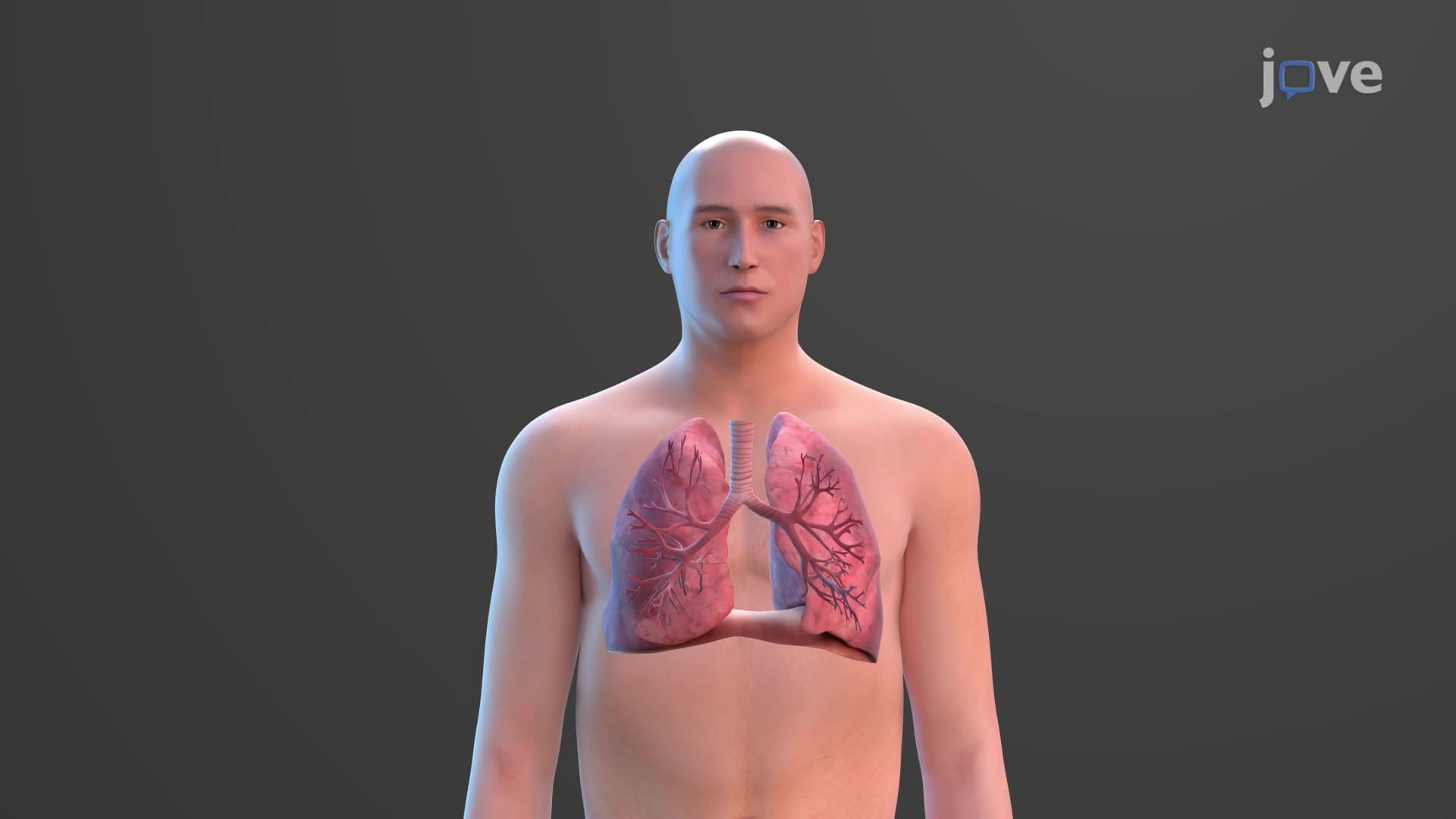
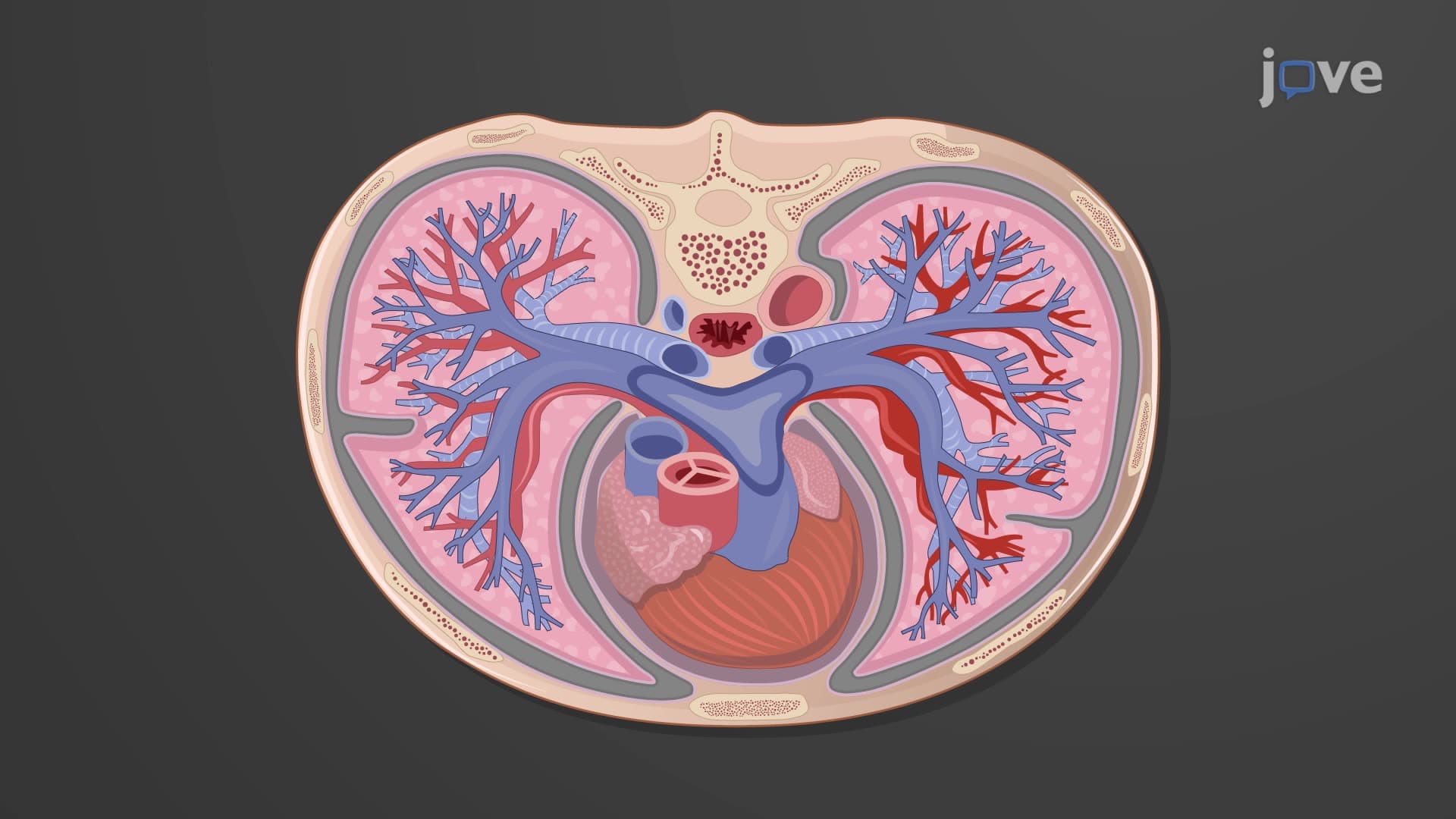
Lung gross anatomy biology encompasses the macroscopic structure and spatial relationships of these vital respiratory organs. The lungs occupy most of the thoracic cavity, positioned laterally to the heart and major blood vessels of the mediastinum. Each lung displays a characteristic cone shape, with the narrow apex extending beneath the clavicle and the broad base resting on the diaphragm. This positioning allows for maximum respiratory capacity while accommodating other thoracic structures.
The asymmetrical design of human lungs reflects evolutionary adaptation to cardiac positioning. The right lung contains three distinct lobes-superior, middle, and inferior-separated by horizontal and oblique fissures. In contrast, the left lung features only two lobes (superior and inferior) divided by a single oblique fissure. The left lung's unique cardiac notch creates space for the heart's left border, demonstrating how lung fissure lobe anatomy accommodates neighboring organs.
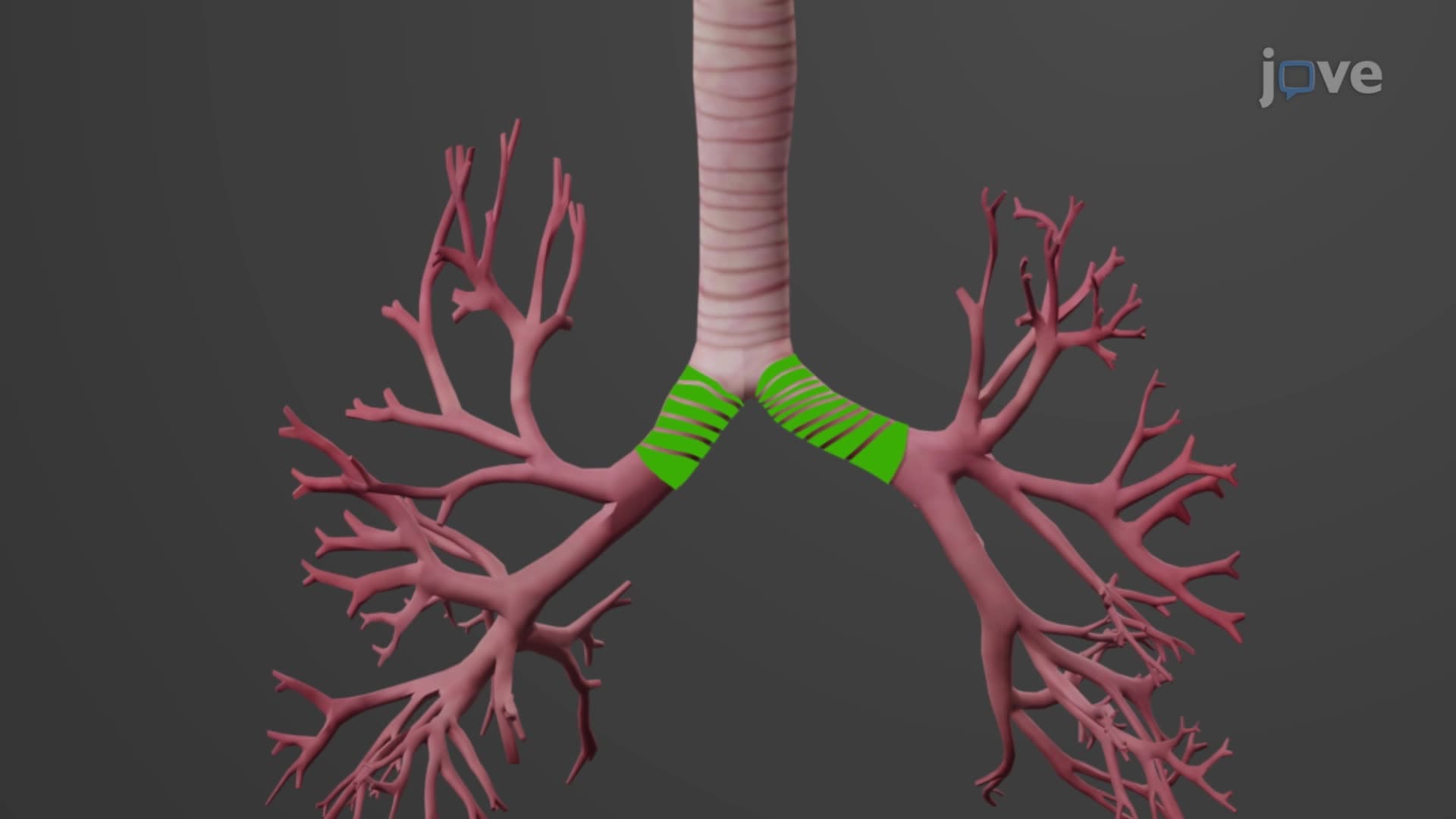
The lung hilum hilus anatomy serves as the crucial entry and exit point for bronchi, blood vessels, lymphatics, and nerves. Located on each lung's mediastinal surface, the hilum connects to the lung root-a collection of structures that anchor the lungs to the mediastinum. This arrangement allows for essential gas exchange while maintaining structural stability during respiratory movements. Medical students studying for the MCAT must understand these relationships to answer questions about pulmonary circulation and innervation.
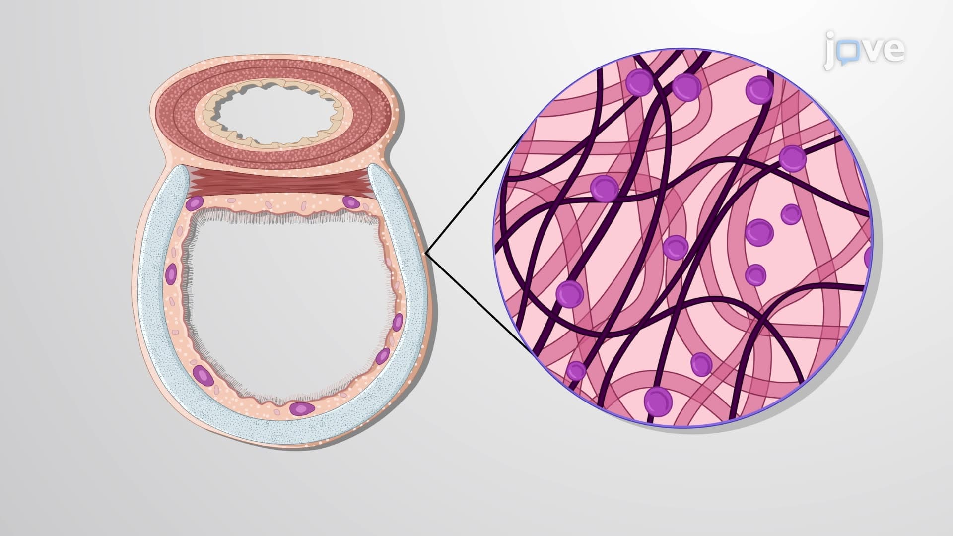
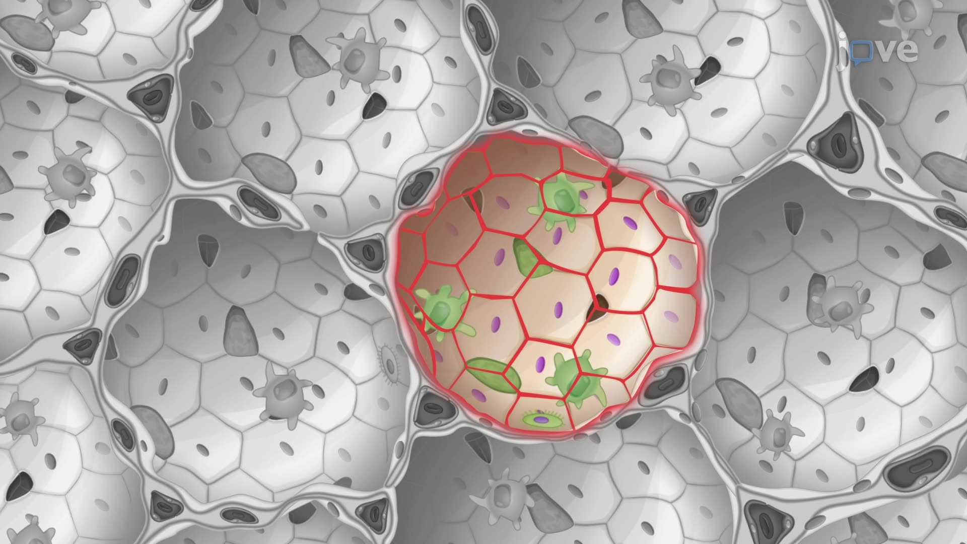
Beyond lobar divisions, each lung contains functionally independent bronchopulmonary segments-10 in the right lung and 8-10 in the left. These segments, separated by connective tissue septa, represent the smallest surgically resectable units of lung tissue. Understanding this lung gross structure proves essential for radiologists interpreting chest imaging and surgeons planning lobectomies. Each segment further subdivides into lobules, visible as hexagonal patterns on the lung surface, reflecting the organized branching of bronchioles and blood vessels.
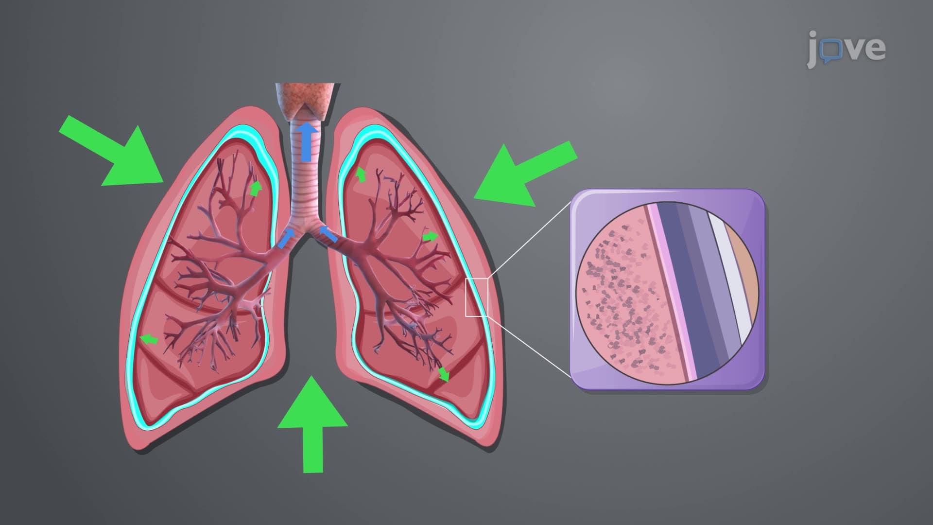
The pleural membrane lung relationship creates a sealed environment essential for breathing mechanics, with visceral pleura covering lung surfaces and parietal pleura lining the thoracic cavity. This arrangement facilitates smooth lung expansion during inspiration while preventing lung collapse.
Related Micro-courses