Loading video…


 25 Concepts
 84.7K views
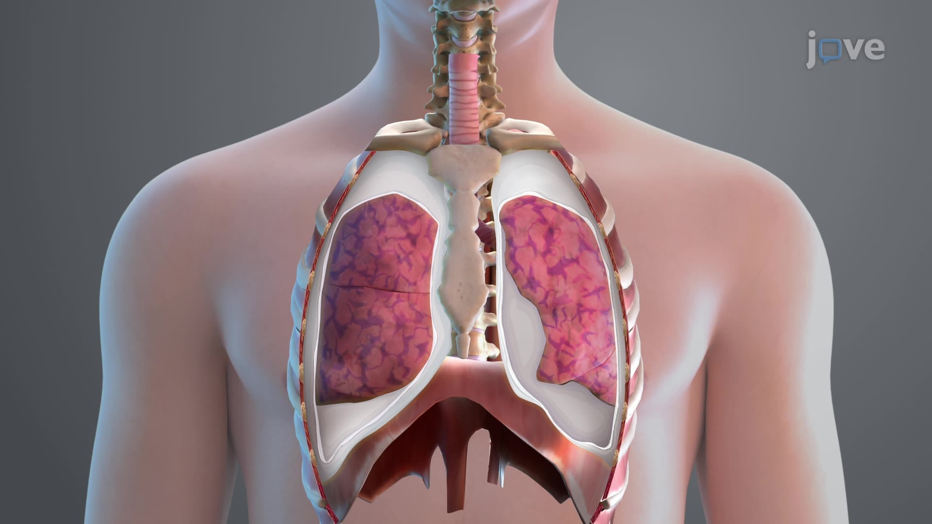

Video Summary: What Is Pleura of the Lungs
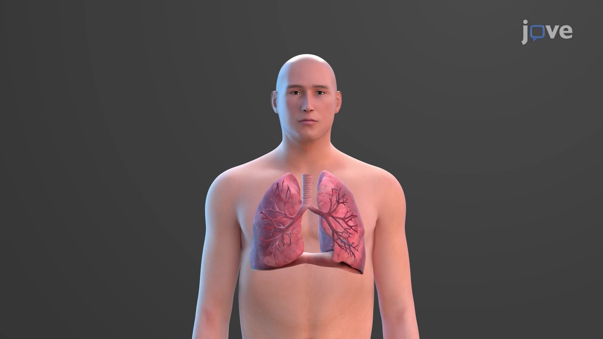
Ever wonder why your lungs don't collapse every time you exhale? The lung pleura anatomy biology reveals a remarkable double-membrane system that keeps your respiratory organs functioning seamlessly. This pleural membrane creates a vacuum-like seal similar to how suction cups work, preventing lung collapse-a critical factor in conditions like pneumothorax that emergency rooms across American hospitals treat daily. Understanding what is pleura of the lungs transforms how students grasp respiratory mechanics. Watch the full video on JoVE Coach to master this concept with expert-led visuals and step-by-step explanations.
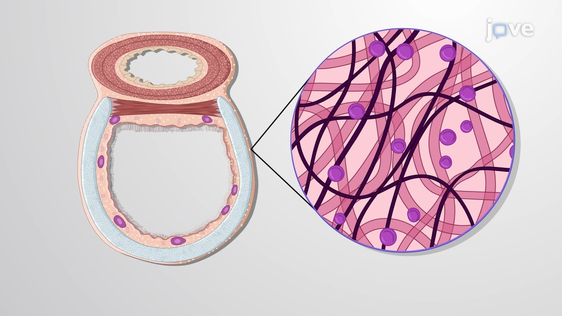
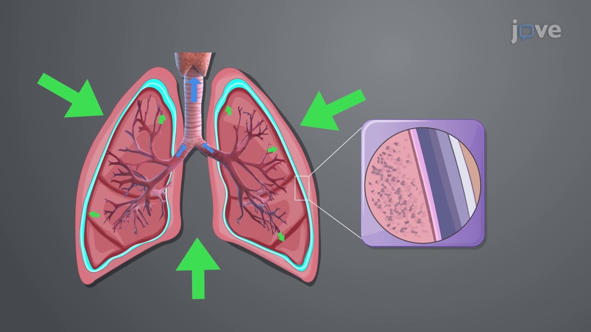
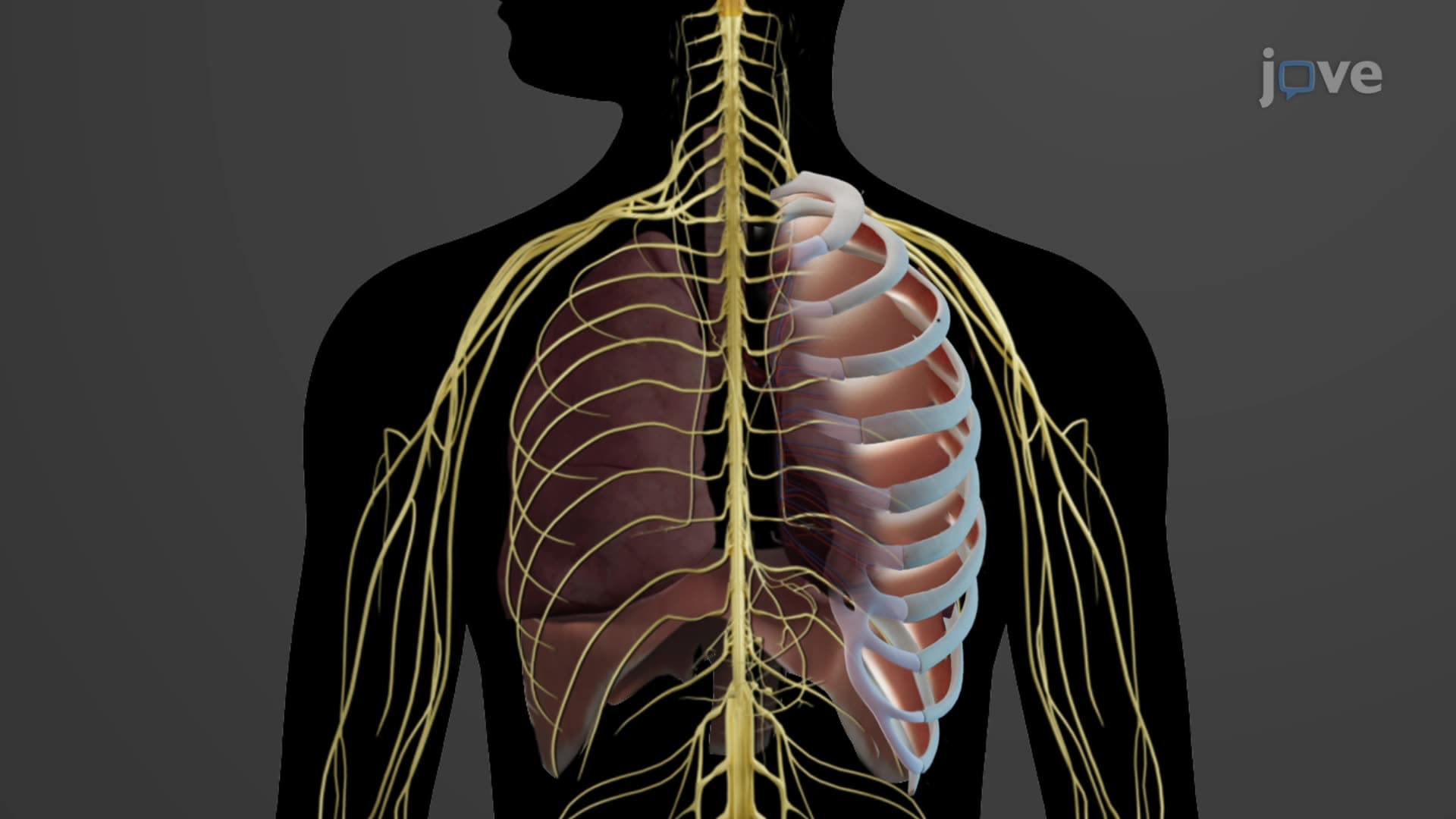
The pleural membrane explained anatomy centers on a sophisticated double-layered serous membrane system that exemplifies biological engineering at its finest. Unlike simple protective coverings, the pleura creates a dynamic interface between your lungs and chest cavity, functioning as both lubricant and anchor system.
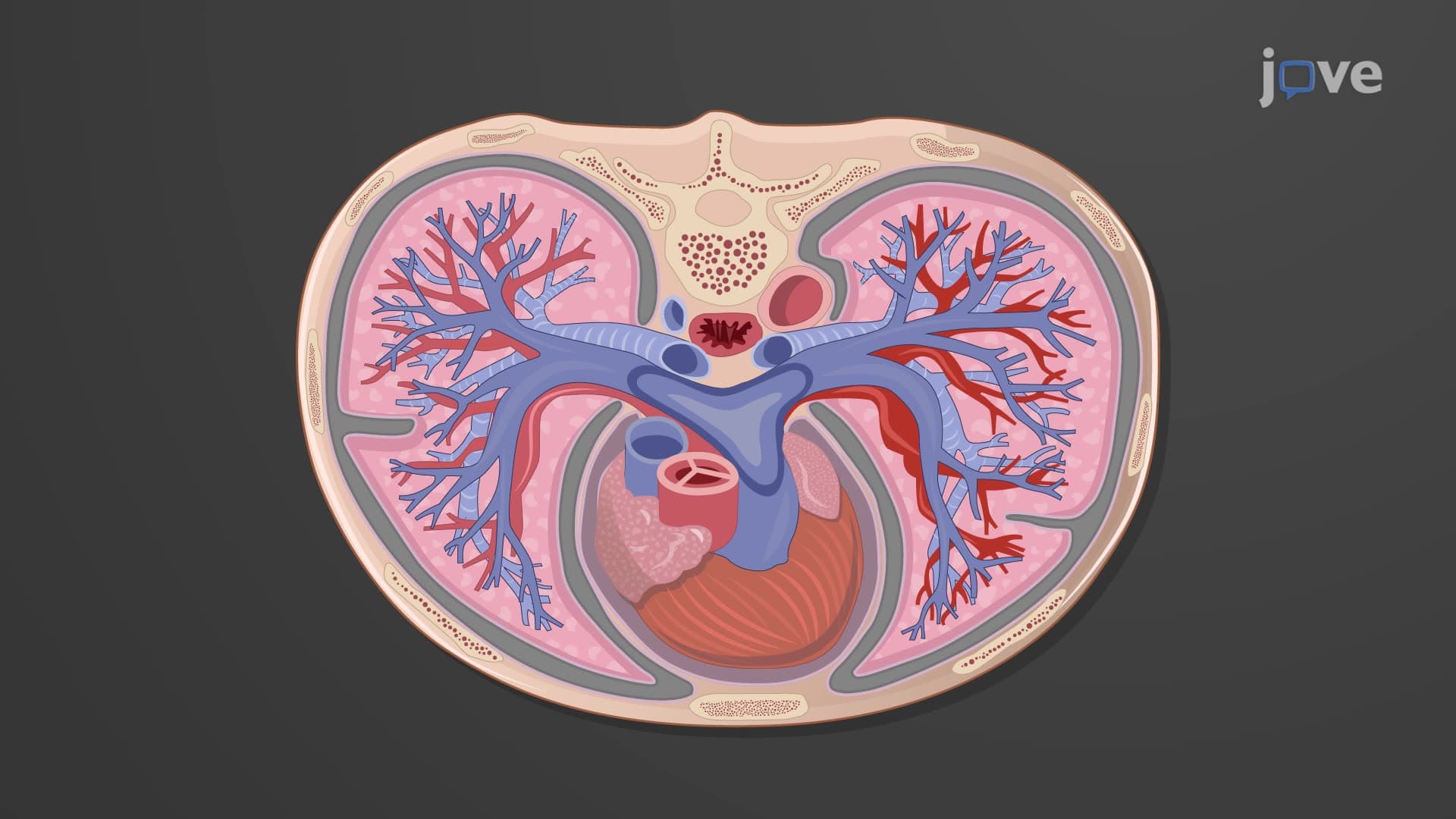
This membrane system consists of two specialized layers working in perfect coordination. The visceral pleura intimately covers each lung's surface, following every contour and fissure. Meanwhile, the parietal pleura lines the inner chest wall, diaphragm, and mediastinum, creating a sealed compartment around each lung.
The visceral parietal pleura creates a narrow space called the pleural cavity, containing approximately 10-15 milliliters of pleural fluid in healthy adults. This fluid serves multiple critical functions that AP Biology students must understand for exam success.
The fluid's surface tension creates a cohesive force stronger than typical adhesives, ensuring lungs expand with chest wall movement during inspiration. Without this mechanism, conditions like pneumothorax occur, where air enters the pleural space and lung collapse follows-a medical emergency treated in over 74,000 cases annually across US emergency departments.
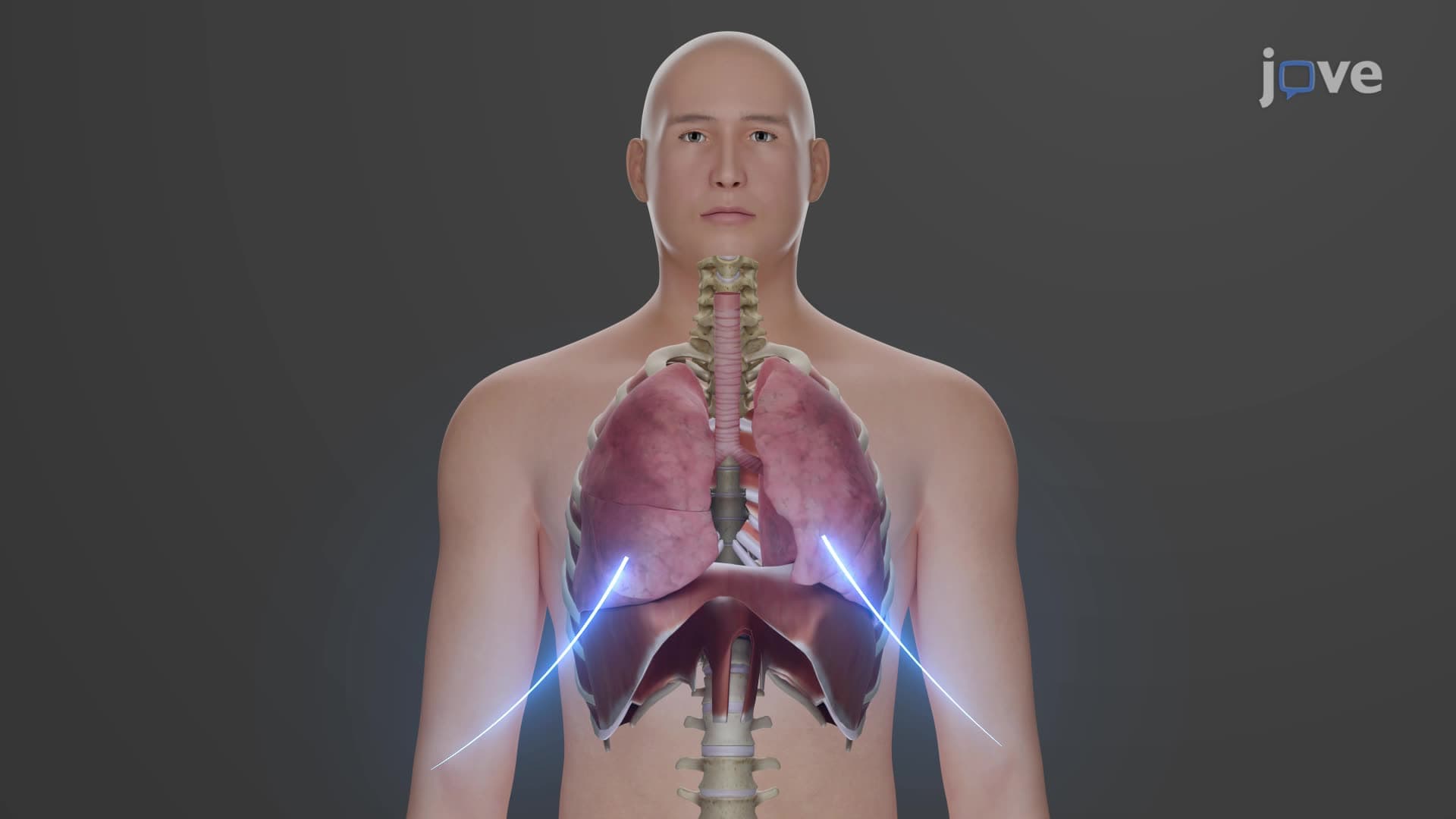
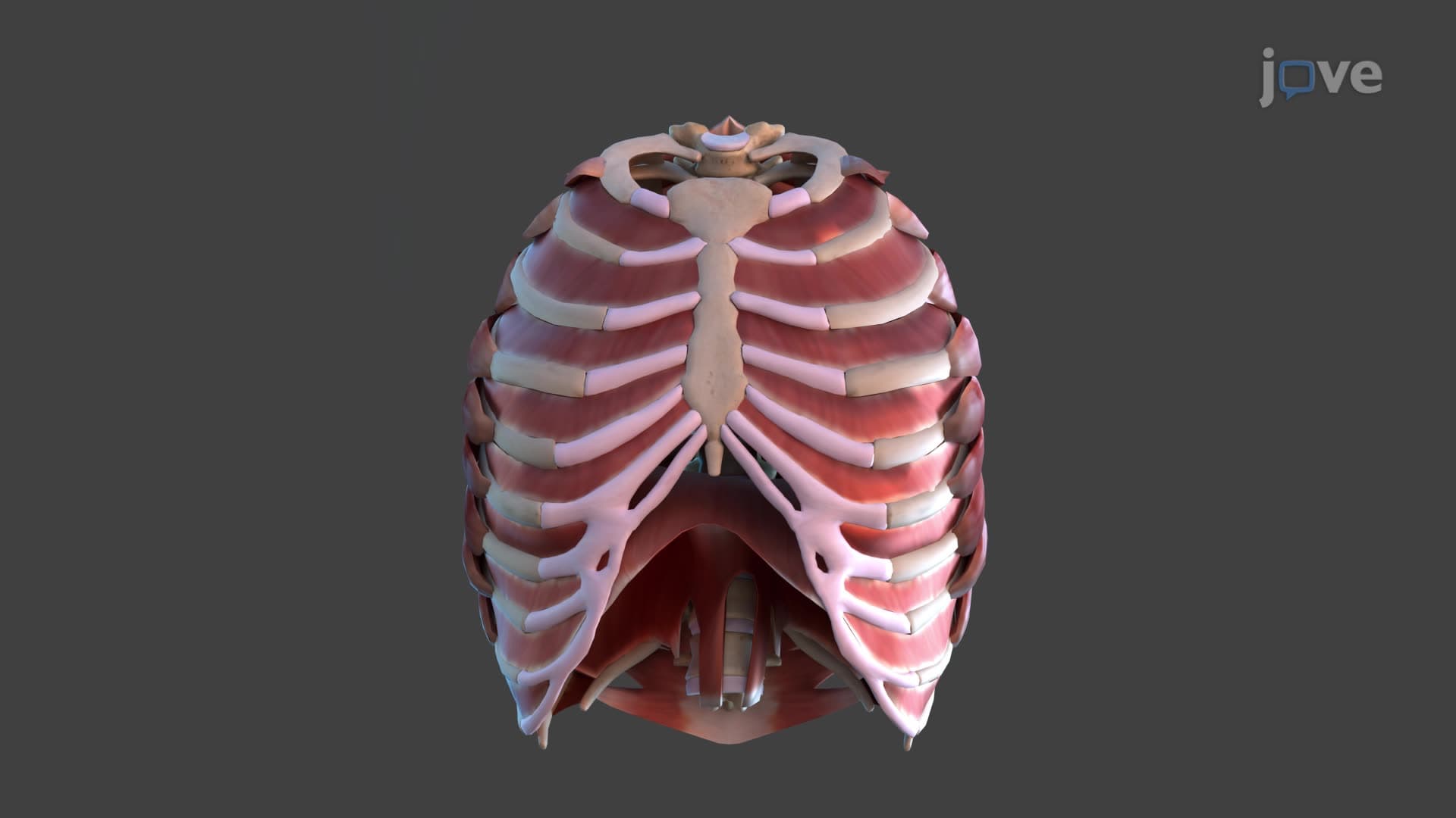
Pleural pressure breathing involves complex pressure differentials that drive respiratory function. During inspiration, the diaphragm and intercostal muscles expand the chest cavity, creating negative pressure within the pleural space. This pressure change, typically -5 to -8 mmHg during quiet breathing, pulls the visceral pleura outward, expanding the lungs.
Medical students studying for the MCAT encounter pleural pressure calculations regularly, as understanding these gradients explains how mechanical ventilators work in intensive care units nationwide. The pressure relationships also explain why certain lung diseases cause breathing difficulties.
Pleuritis pleural inflammation represents one of the most common pleural pathologies encountered in American healthcare settings. When bacterial pneumonia or viral infections affect the pleura, the normally smooth membrane surfaces become roughened and inflamed, causing the characteristic sharp, stabbing chest pain that worsens with deep breathing.
Healthcare professionals use this anatomical knowledge to diagnose conditions through physical examination techniques like percussion and auscultation, skills emphasized in nursing programs and medical schools across the United States. Understanding normal pleural anatomy helps clinicians identify abnormal findings like pleural effusions or adhesions.
Related Micro-courses