Loading subject page…

Micro-course: The Respiratory System
84,701 views
Learn key concepts and earn your certificate.



Trachea

00:00
01:22
Video Summary: What Is Trachea
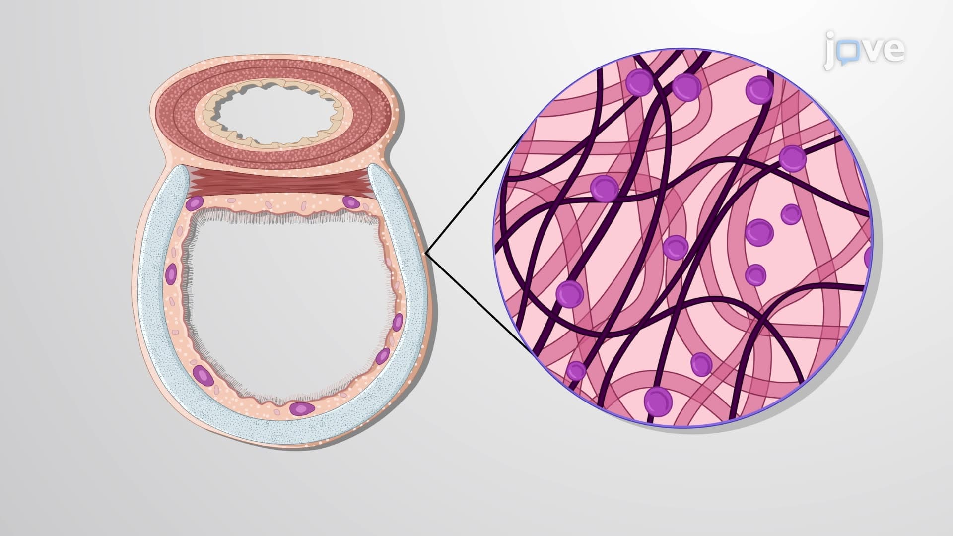
Ever wondered how your throat stays open when you swallow a big bite of food? The trachea anatomy biology reveals a remarkable engineering feat-incomplete cartilage rings that flex to accommodate your esophagus while keeping your airway rigid. In US emergency rooms, understanding tracheal structure is critical when paramedics perform life-saving procedures like emergency tracheotomies. The What is Trachea involves four distinct wall layers working together to filter air, produce mucus, and maintain structural integrity during every breath you take. Watch the full video on JoVE Coach to master this concept with expert-led visuals and step-by-step explanations.
- Understand What is Trachea and identify its precise anatomical location from larynx to bronchial bifurcation in the respiratory system
- Analyze the four-layer wall structure including mucosa, submucosa, hyaline cartilage, and adventitia and their specific protective functions
- Identify the 16-20 C-shaped cartilage rings and explain how their incomplete design prevents airway collapse while allowing esophageal expansion
- Recognize the ciliated pseudostratified columnar epithelium's role in trapping and removing airborne particles through mucociliary clearance
- Evaluate the carina's strategic position and its critical function in triggering protective cough reflexes when foreign objects enter the airway
- Apply knowledge of tracheal anatomy to understand emergency medical procedures like tracheotomy, intubation, and the Heimlich maneuver
- Compare tracheal flexibility mechanisms with rigid support structures and analyze how this balance maintains optimal respiratory function
- Explore clinical implications of tracheal obstruction and connect anatomical features to real-world medical interventions
Understanding What is Trachea: The Body's Essential Airway
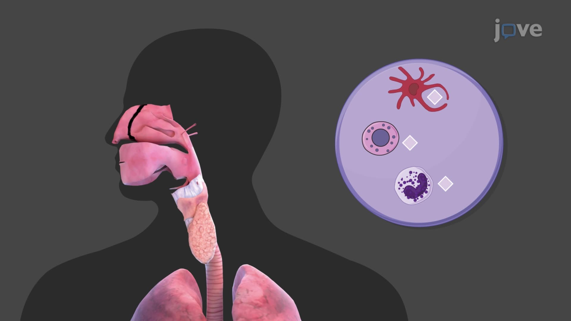
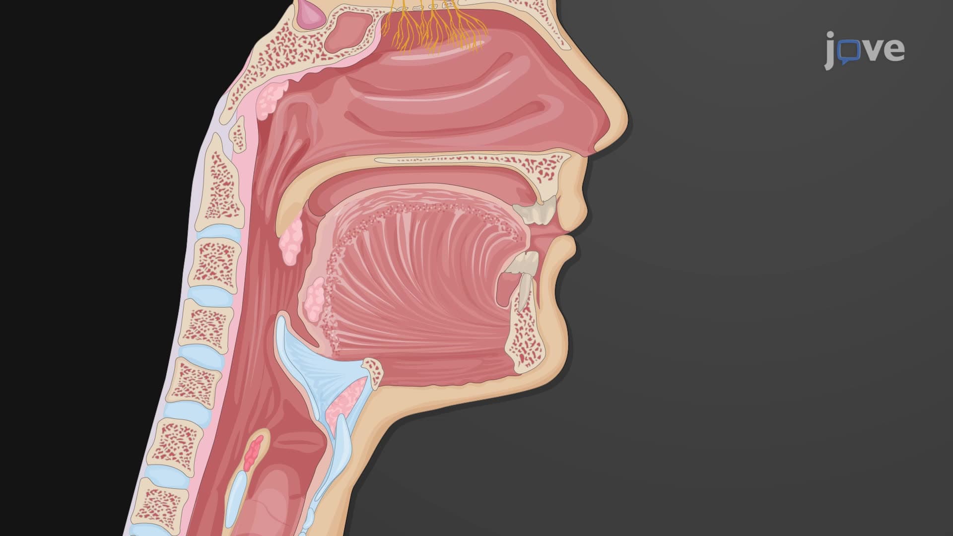
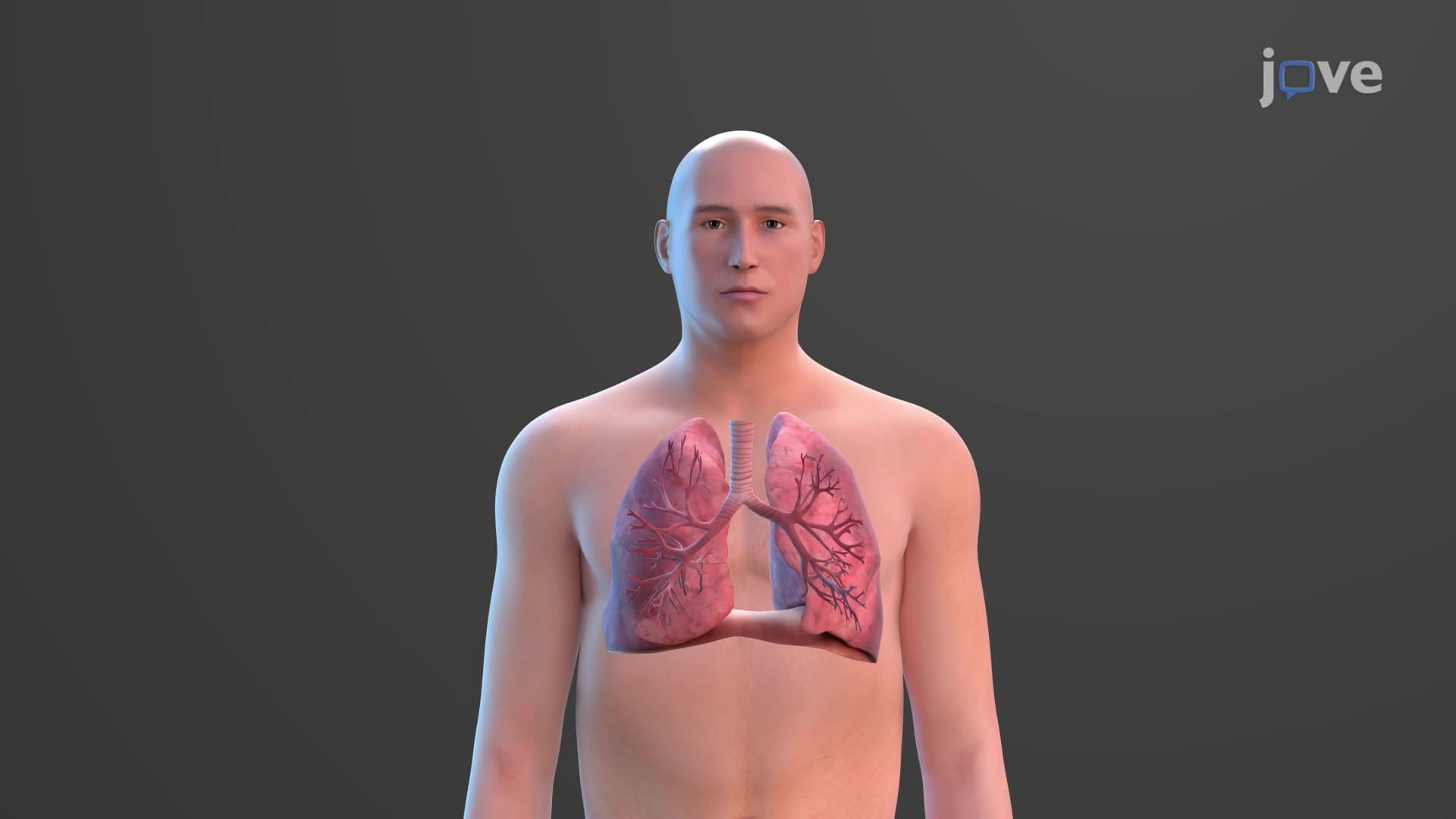
The trachea represents one of the most ingeniously designed structures in human anatomy. This 4-5 inch tube serves as your body's primary air highway, connecting your larynx (voice box) to your lungs through the bronchi. What makes the trachea anatomy biology so fascinating is its dual requirement: it must remain rigid enough to stay open during powerful inhalation, yet flexible enough to accommodate the expanding esophagus during swallowing.
Tracheal Wall Architecture: Four-Layer Protection System
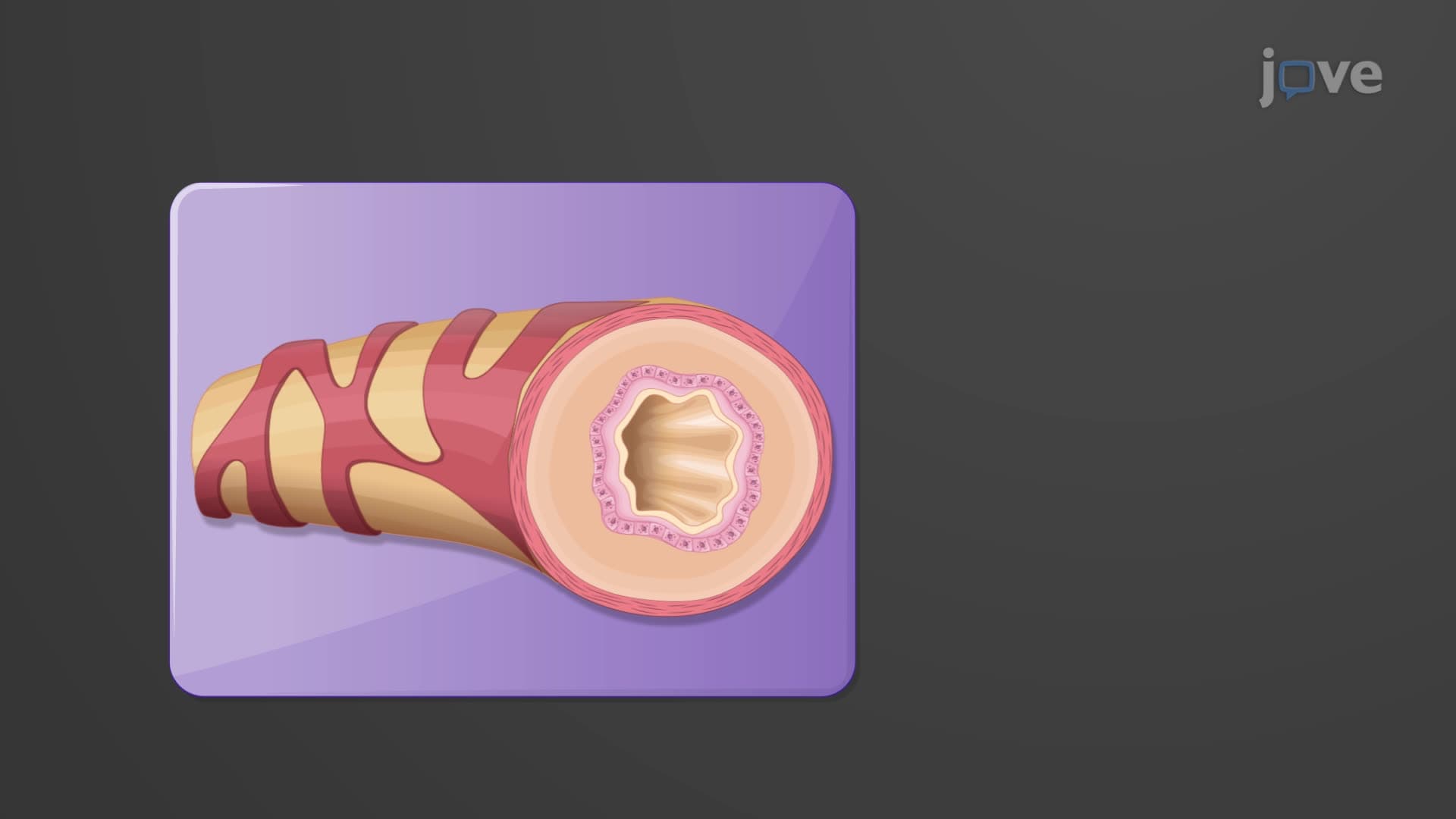
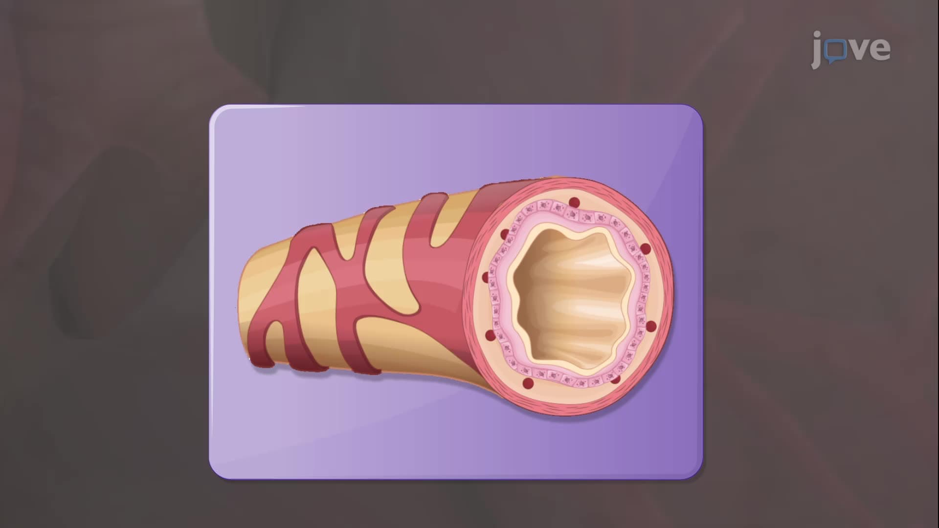
The windpipe trachea explained through its four-layer construction reveals sophisticated biological engineering. The innermost mucosa layer features ciliated pseudostratified columnar epithelium-tiny hair-like projections that beat approximately 1,000 times per minute, creating an "escalator effect" that moves trapped particles upward and out of your respiratory system. This mechanism is so efficient that healthy lungs clear most inhaled particles within 24 hours.
The submucosa contains seromucous glands that produce the sticky mucus essential for particle trapping. In the third layer, 16-20 C-shaped hyaline cartilage rings provide semi-rigid support. These incomplete rings are crucial-their open posterior section allows your esophagus to expand when swallowing large food boluses, preventing choking. The outermost adventitia anchors the trachea to surrounding structures while maintaining flexibility.
Clinical Significance and Emergency Applications
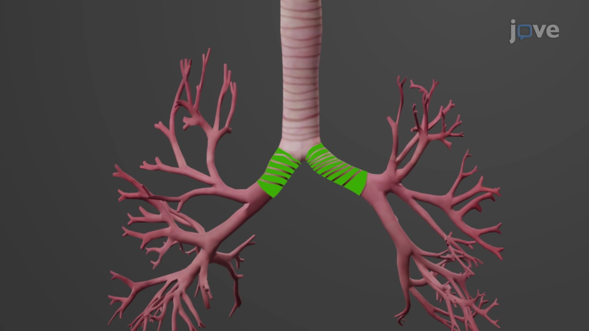
Understanding what is the trachea in respiratory anatomy becomes critical in medical emergencies. The carina-where the trachea bifurcates into main bronchi-contains specialized receptors that trigger violent coughing when foreign objects approach. This location is also where emergency physicians perform bronchoscopy procedures to remove aspirated objects.
For students preparing for the MCAT or AP Biology exams, remember that tracheal cartilage differs from other body cartilages. Its C-shape design appears frequently in multiple-choice questions testing anatomical structure-function relationships. In clinical settings, the tracheal cartilage C-ring structure guides proper tracheostomy placement-typically performed between the second and fourth tracheal rings to avoid major blood vessels.
Real-World Applications in Healthcare
US paramedics rely on detailed knowledge of tracheal anatomy length when performing emergency intubations. The standard adult trachea measures approximately 10-12 centimeters, with the carina trachea bifurcation located at the level of the fifth thoracic vertebra. This anatomical landmark helps healthcare providers position endotracheal tubes correctly-too deep risks entering a main bronchus and ventilating only one lung.
Frequently Asked Questions
The trachea is a 4-5 inch flexible tube that serves as your main airway, extending from the larynx in your neck down to the carina where it splits into two main bronchi. It's positioned in front of your esophagus and behind your sternum, making it easily accessible for emergency procedures when necessary.
MCAT questions frequently test the structure-function relationship of C-shaped cartilage rings, asking why they're incomplete posteriorly. AP Biology exams often include diagrams where you must identify the ciliated epithelium and explain its role in respiratory defense mechanisms.
These rings prevent airway collapse during inspiration while allowing esophageal expansion during swallowing-a perfect example of form following function. USMLE and medical school exams test this concept because understanding it explains both normal physiology and emergency airway management.
EMTs and paramedics use tracheal anatomy knowledge daily-the cartilage rings guide proper tracheostomy placement, while understanding the carina's location helps with correct endotracheal tube positioning. This knowledge directly saves lives in emergency situations.
Not at all! The trachea's structure is actually quite intuitive once you understand its job-think of it as a flexible vacuum cleaner hose with built-in filters and structural support. Starting with its basic function makes the anatomical details much easier to grasp.
Use the mnemonic "My Students Have Arrived" for Mucosa, Submucosa, Hyaline cartilage, Adventitia (inside to outside). Connect each layer to its function-mucosa moves particles, submucosa makes mucus, cartilage provides support, adventitia attaches to neighbors.
Focus on the bronchial tree branching pattern and alveolar structure, as these build directly on tracheal concepts. Understanding how the trachea's design principles continue throughout the respiratory system creates a comprehensive foundation for advanced pulmonology topics.
These exams emphasize clinical applications-you might see scenarios about airway obstruction where you must identify which anatomical features make certain emergency procedures possible. They test practical knowledge rather than just memorization.
Conditions like severe throat cancer or prolonged mechanical ventilation may require bypassing the upper airway entirely. The trachea's superficial neck location and cartilage ring structure provide stable landmarks for surgeons to create this alternative breathing pathway safely.
Related Micro-courses