Loading subject page…


78,918 views

Video Summary: What are Photoreceptors and Visual Pathways
Ever wonder why you can't distinguish colors in moonlight, yet owls hunt perfectly in darkness? Photoreceptors visual pathways explain this fascinating phenomenon through specialized cells in your retina. Students at Johns Hopkins University studying vision science learn how rod and cone photoreceptors work differently-rods excel in dim conditions while cones enable color perception. Understanding what are photoreceptors and visual pathways reveals how light transforms into the vivid visual world you experience daily. Watch the full video on JoVE Coach to master this concept with expert-led visuals and step-by-step explanations.
Photoreceptors visual pathways represent one of biology's most sophisticated sensory systems, converting electromagnetic radiation into meaningful visual perception. This intricate process begins in your retina and culminates in conscious visual experience through your brain's visual cortex.
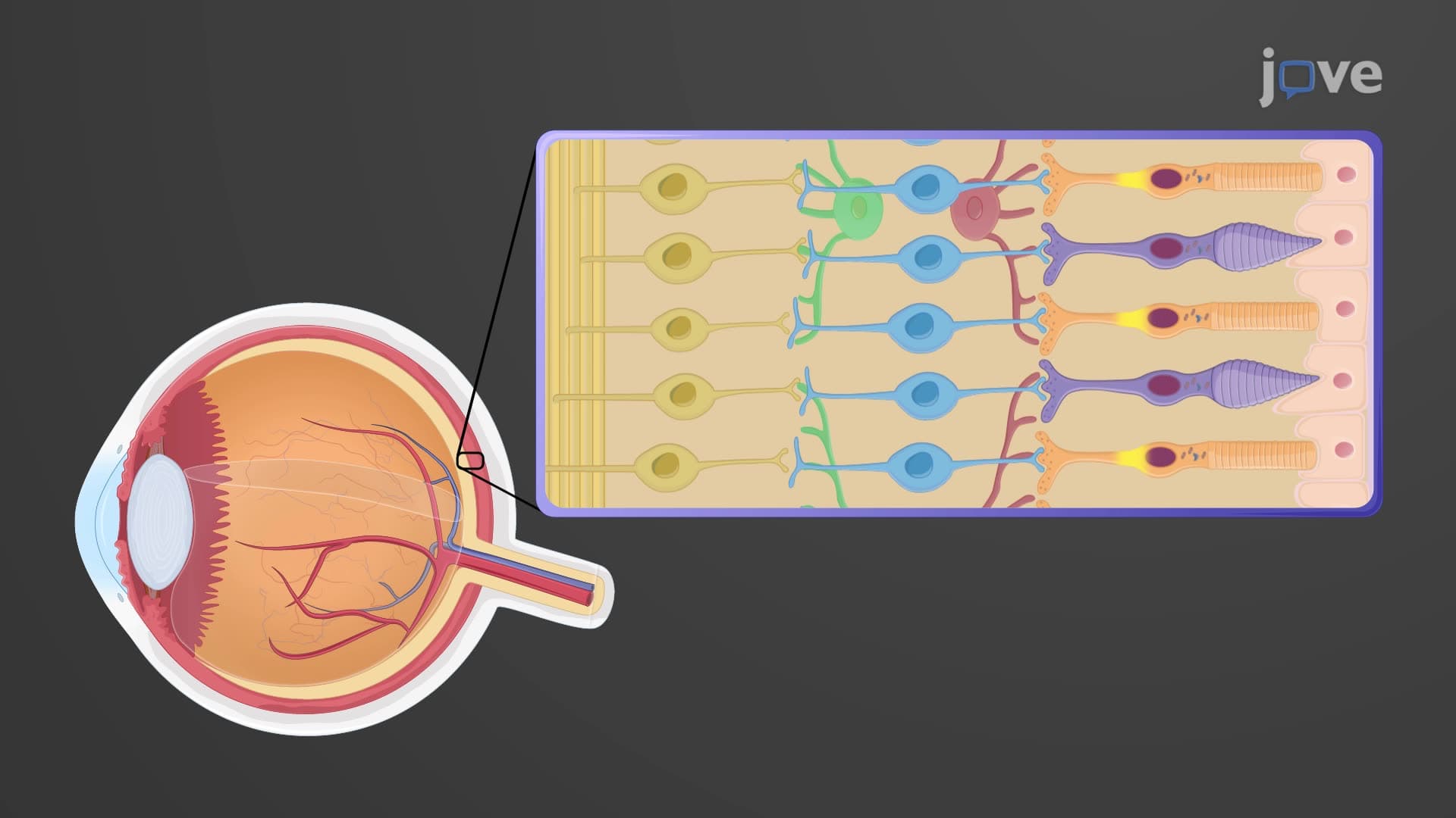
The eye photoreceptor visual pathway starts with two distinct cell types strategically positioned in your retina's neural layer. Rod photoreceptors, numbering approximately 120 million per eye, contain rhodopsin-a photopigment combining retinal (vitamin A derivative) and opsin protein. These cells excel in low-light conditions, explaining why security guards working night shifts rely heavily on rod-mediated vision.
Cone photoreceptors, totaling about 6 million per eye, exist in three varieties based on their opsin proteins: L-cones (red-sensitive), M-cones (green-sensitive), and S-cones (blue-sensitive). This trichromatic system enables humans to distinguish approximately 10 million color variations-crucial for professions like graphic design at companies like Adobe or medical imaging at Cleveland Clinic.
When light strikes photoreceptors, phototransduction photoreceptor mechanisms activate through a complex biochemical cascade. Photons cause retinal to change from 11-cis to all-trans configuration, triggering opsin conformational changes. This molecular event initiates hyperpolarization-unusual since most sensory cells depolarize when stimulated.
The visual processing pathway continues as photoreceptors synapse with bipolar cells, which then connect to ganglion cells. This three-neuron chain creates the foundation for all visual information processing, with horizontal and amacrine cells providing lateral inhibition and signal refinement.
Ganglion cell axons form the optic nerve chiasm tract, where approximately 50% of fibers cross at the optic chiasm-ensuring each brain hemisphere receives information from both eyes' contralateral visual fields. This anatomical arrangement proves critical for depth perception and binocular vision.
The visual cortex pathway terminates in the primary visual cortex (V1) in your occipital lobe, where orientation-selective neurons first described by Hubel and Wiesel (Nobel Prize winners) begin processing complex visual features. Students preparing for the MCAT frequently encounter questions about this pathway's clinical significance-damage at different levels produces characteristic visual field defects diagnostically important for neurologists at institutions like Mayo Clinic.
Understanding these pathways proves essential for AP Biology students, particularly when studying sensory systems, and provides foundational knowledge for pre-med students preparing for medical school coursework in neuroscience and ophthalmology.
Related Micro-courses