Loading subject page…

- Anatomy and Physiology
- The Special Senses
- Auditory Pathway
Micro-course: The Special Senses
78,917 views
Learn key concepts and earn your certificate.





Auditory Pathway


00:00
01:15
Video Summary: What Is Auditory Pathway
Ever wondered how your brain instantly recognizes your favorite song from the first note? The auditory pathway brain anatomy transforms sound waves into meaningful neural signals through a complex journey from ear to brain. When a patient at Johns Hopkins loses hearing after a stroke affecting their temporal lobe, doctors trace the damage through this intricate pathway to understand exactly where processing failed. What is Auditory Pathway represents one of neuroscience's most elegant examples of sensory processing. Watch the full video on JoVE Coach to master this concept with expert-led visuals and step-by-step explanations.
- Understand the complete What is Auditory Pathway from sound wave detection to conscious perception in the brain
- Identify the key anatomical structures including cochlear nuclei, inferior colliculus, medial geniculate nucleus, and auditory cortex
- Analyze how mechanical sound vibrations convert to electrical neural signals through hair cell transduction
- Compare the roles of different brainstem nuclei in processing and relaying auditory information
- Evaluate how pathway damage at different levels affects hearing and sound perception
- Apply knowledge of tonotopic organization to understand frequency-specific processing
- Recognize clinical implications when auditory pathway components are damaged by stroke or disease
- Explore connections between auditory processing and language comprehension in the brain
Understanding What is Auditory Pathway: From Sound to Perception
The auditory pathway brain anatomy represents a sophisticated neural highway that transforms mechanical sound vibrations into conscious auditory experiences. Unlike simple sensory systems, this hearing pathway neuroscience involves multiple processing stations that analyze, filter, and interpret acoustic information before it reaches conscious awareness.
Peripheral Processing: The Cochlear Foundation
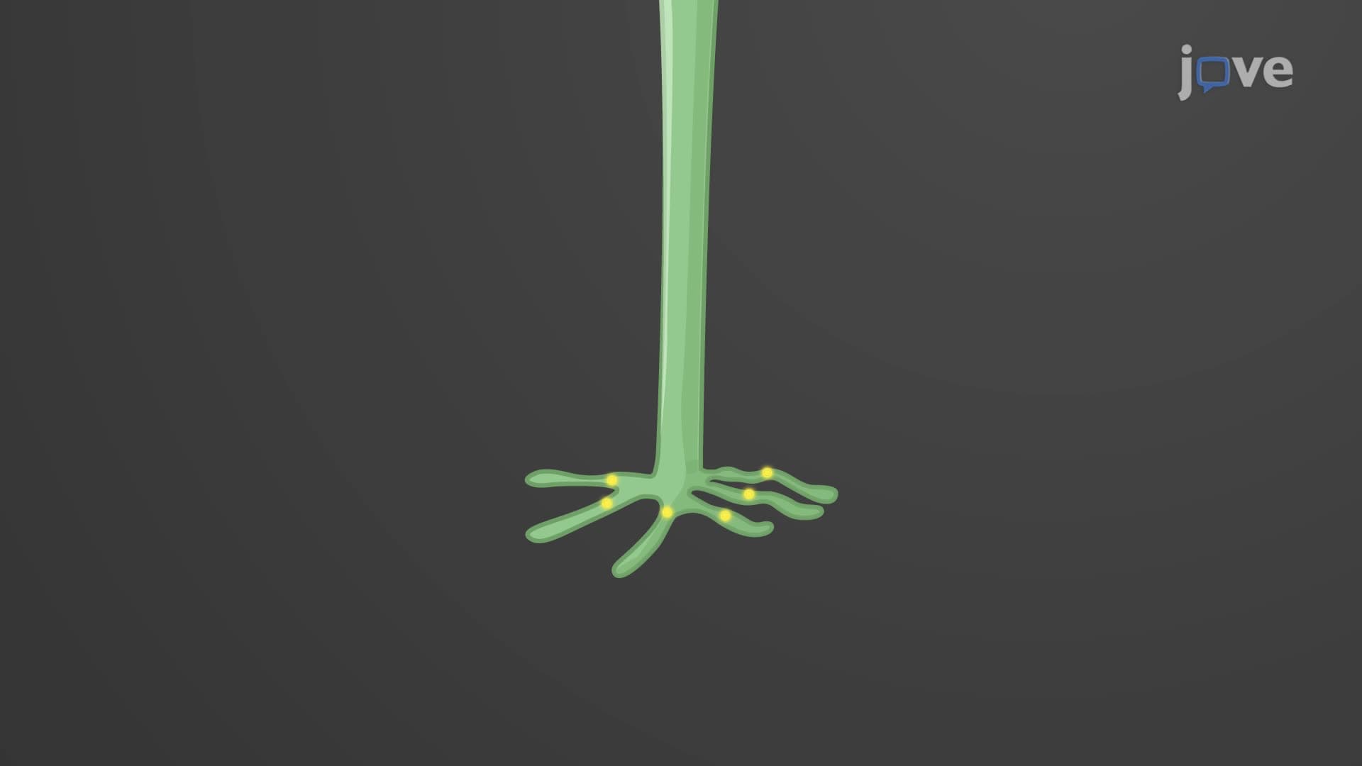
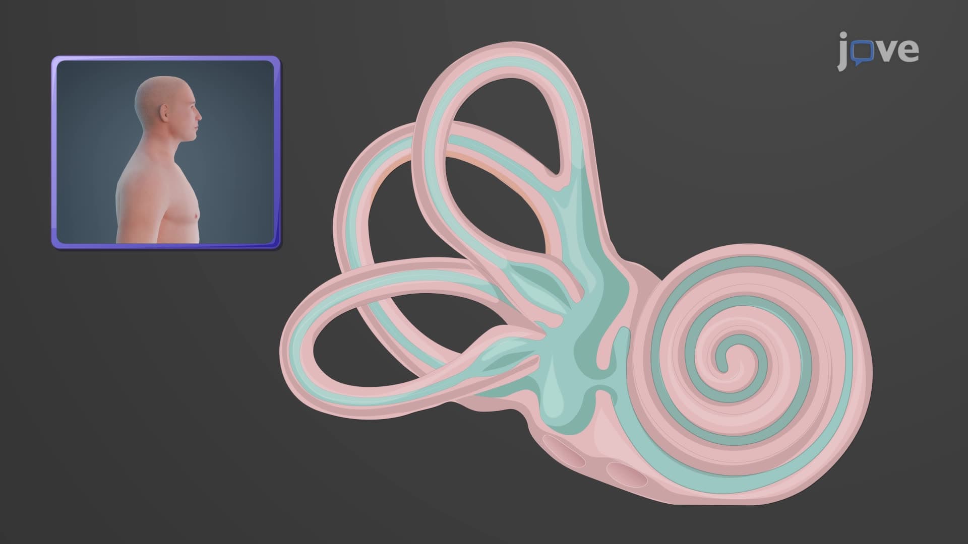
The journey begins in the cochlea, where specialized inner hair cells convert mechanical energy into electrical signals. These sensory receptors exhibit remarkable precision-they can detect movements smaller than the width of an atom. When hair cells depolarize, they release glutamate neurotransmitter onto spiral ganglion neurons, initiating the cochlear nerve pathway. This first-order neuron carries tonotopically organized information, meaning different frequencies activate specific nerve fibers in a systematic arrangement.
Central Auditory Processing: Brainstem Integration
The cochlear nerve synapses at the cochlear nuclei in the medulla, where what is the auditory pathway in neuroscience becomes increasingly complex. Here, second-order neurons perform initial sound processing including timing analysis and intensity coding. Information then travels to the superior olivary complex, which computes binaural cues essential for sound localization-critical for understanding why we can pinpoint a fire truck's direction even with our eyes closed.
The inferior colliculus auditory processing center serves as the primary integration hub in the midbrain. This structure receives converging inputs from both ears and performs sophisticated calculations about sound timing, intensity, and frequency content. Students studying for the MCAT should understand that damage here can cause profound difficulties in sound localization and auditory scene analysis.
Thalamic Relay and Cortical Processing
The medial geniculate thalamus acts as the final relay station before cortical processing. Like other thalamic nuclei, it doesn't merely pass information but actively gates and modulates auditory signals based on attention and arousal states. The auditory cortex pathway in the temporal lobe represents the culmination of this sound processing pathway, where conscious perception occurs and complex sounds like speech are decoded.
In AP Biology and college neuroscience courses, students learn that the primary auditory cortex maintains tonotopic organization, with different regions specialized for different frequency ranges. Secondary auditory areas integrate this information with memory and language systems, explaining why hearing loss can significantly impact cognitive function in elderly patients.
Clinical Relevance and Exam Applications
Understanding this pathway proves essential for healthcare professionals. On the USMLE and NCLEX exams, questions often focus on localizing lesions based on hearing deficits. For example, unilateral sensorineural hearing loss might indicate cochlear nerve damage, while bilateral high-frequency loss could suggest age-related changes in hair cells.
Frequently Asked Questions
The auditory pathway is the neural network that processes sound from the ear to the brain, involving the cochlear nerve, brainstem nuclei, thalamus, and auditory cortex. Understanding this pathway is crucial because it explains how mechanical sound waves become meaningful perceptions, and helps diagnose hearing disorders by identifying where processing breaks down.
MCAT questions often test pathway organization, especially cochlear transduction mechanisms and central processing locations. AP Biology focuses more on sensory receptor function and basic neural pathways. Both exams may ask about frequency discrimination, sound localization, and how pathway damage affects perception.
USMLE frequently tests lesion localization using hearing deficits as clues. Key concepts include distinguishing conductive versus sensorineural hearing loss, understanding Weber and Rinne test results, and recognizing how brainstem strokes affect auditory processing. Questions often involve acoustic neuroma effects on the eighth cranial nerve.
Audiologists at Mayo Clinic use pathway understanding to interpret hearing tests and recommend appropriate interventions. For example, if a patient shows normal outer hair cell function but poor speech discrimination, the problem likely involves central processing rather than peripheral hearing, guiding treatment decisions toward auditory processing therapy rather than hearing aids.
No advanced math is required, though basic wave properties help with frequency concepts. The focus should be on biological mechanisms rather than acoustic physics. Understanding membrane potentials and synaptic transmission from general biology provides sufficient background for mastering auditory pathway concepts.
Create a flowchart tracing the pathway from cochlea to cortex, including key structures and their functions. Practice clinical scenarios that test pathway localization, and use mnemonics for remembering brainstem nuclei order. Focus on understanding functional relationships rather than memorizing isolated facts.
Auditory pathways share organizational principles with other sensory systems, so learning one helps understand others. Many students find starting with visual pathways easier due to more familiar anatomy, but auditory pathways offer excellent examples of frequency coding and binaural processing that appear throughout neuroscience.
The auditory cortex connects extensively with language areas in the left hemisphere, particularly Wernicke's area for speech comprehension. Speech-language pathologists use this knowledge to understand why some patients with stroke can hear sounds but cannot understand speech, indicating damage to auditory-language connections rather than basic hearing pathways.
Related Micro-courses