Loading subject page…


665,904 views

Video Summary: What Is Skeletal Muscle Anatomy
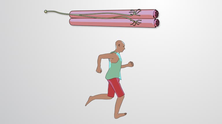
Ever wondered how your bicep contracts when you flex your arm at the gym? Skeletal muscle anatomy reveals the intricate layers within muscles like the biceps brachii, from the protective epimysium coating to the microscopic sarcomeres that power every movement. Understanding what is skeletal muscle anatomy helps explain how Olympic sprinters generate explosive power through coordinated muscle fiber contractions. Watch the full video on JoVE Coach to master this concept with expert-led visuals and step-by-step explanations.
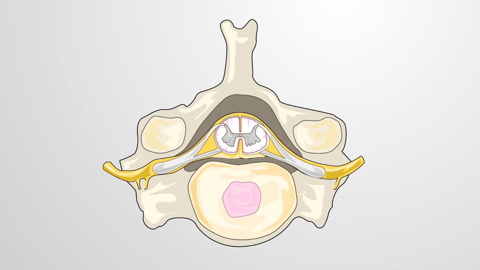
Skeletal muscle anatomy represents one of biology's most elegant examples of structure-function relationships. Unlike cardiac or smooth muscle, skeletal muscles operate under conscious control through the somatic nervous system, enabling everything from delicate piano playing to powerlifting competitions. This voluntary control system makes skeletal muscle anatomy particularly relevant for students studying human physiology and kinesiology.
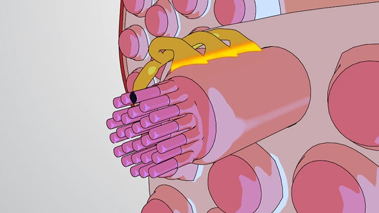
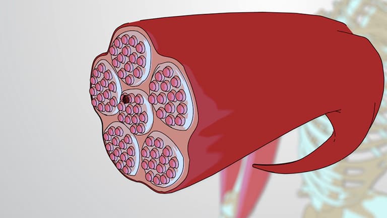
The hierarchical organization of skeletal muscles begins with the epimysium, a tough connective tissue sheath that encases the entire muscle and distinguishes it from surrounding organs. Think of major muscles like the quadriceps femoris in your thigh-the epimysium creates clear boundaries between this massive muscle group and adjacent structures. Within this outer covering, muscles subdivide into fascicles, bundles of muscle cells wrapped in perimysium. This organization appears clearly in cuts of meat, where you can observe the grain patterns that represent fascicle arrangements.
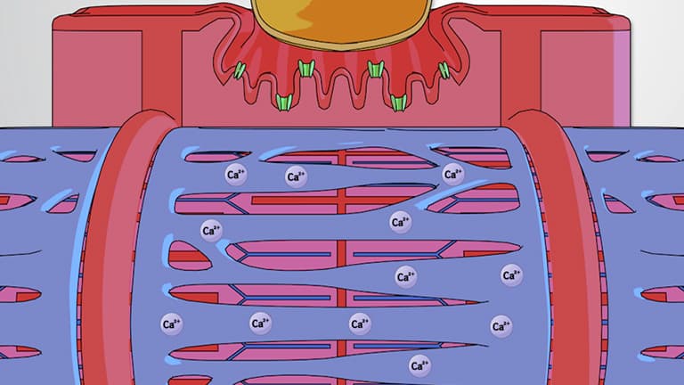
Individual muscle cells, properly called muscle fibers, possess unique characteristics that set them apart from typical cells. Each fiber is surrounded by the sarcolemma, a specialized plasma membrane capable of conducting electrical signals throughout the cell. These multinucleated cells can extend the entire length of a muscle, sometimes reaching several inches in muscles like the sartorius.
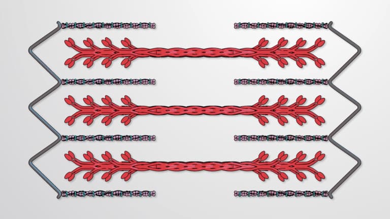
The ultimate functional unit of skeletal muscle anatomy is the sarcomere, contained within thread-like myofibrils that fill each muscle fiber. Sarcomeres contain precisely arranged actin (thin) and myosin (thick) filaments that slide past each other during contraction. This sliding filament mechanism, discovered through electron microscopy studies at institutions like Harvard Medical School, explains how muscles generate force and movement.
This intricate anatomy frequently appears on AP Biology exams, MCAT questions, and college physiology courses. Students often encounter skeletal muscle anatomy when studying exercise physiology, rehabilitation science, or preparing for health profession programs. Understanding these structural relationships proves essential for comprehending muscle disorders, athletic performance, and therapeutic interventions used in American healthcare settings.
Related Micro-courses