Loading subject page…


665,902 views

Video Summary: What Is the Spinal Cord
Ever wondered how your brain instantly knows when you stub your toe, or how it commands your hand to pull away from a hot stove? The spinal cord serves as your body's primary information superhighway, connecting your brain to every part of your body. When NFL players suffer spinal injuries, medical teams immediately assess spinal cord function because this critical structure controls both movement and sensation. What is the spinal cord, and how does this remarkable bundle of nervous tissue coordinate millions of signals every second? Watch the full video on JoVE Coach to master this concept with expert-led visuals and step-by-step explanations.
The spinal cord represents one of the most sophisticated communication networks in the human body. This cylindrical bundle of nervous tissue extends from the brainstem through the foramen magnum, traveling down the vertebral column until it terminates around the L1-L2 lumbar level. Unlike a simple cable, the spinal cord actively processes information while serving as the critical pathway between the brain and peripheral nervous system.
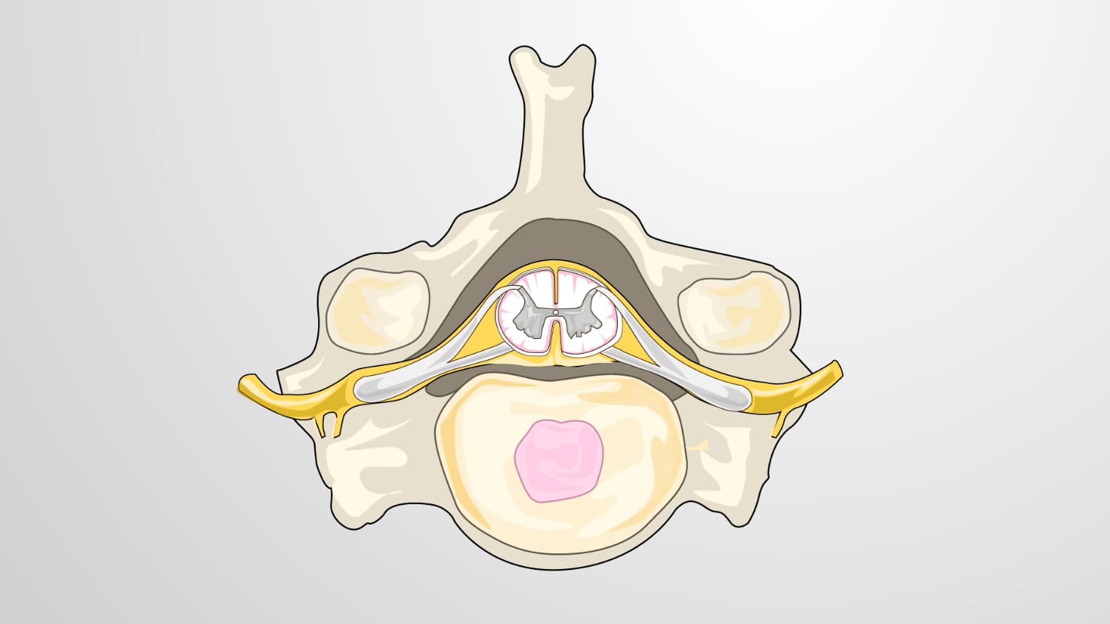
In cross-section, the spinal cord reveals a distinctive butterfly-shaped core of gray matter surrounded by white matter. The gray matter contains cell bodies of neurons, including the crucial motor neurons located in the ventral (anterior) horn. These motor neurons directly control skeletal muscle contraction, making them essential for all voluntary movement. The surrounding white matter consists of myelinated axons organized into ascending and descending tracts-think of these as express lanes carrying specific types of information up to the brain or down from it.
The spinal cord divides into functional segments corresponding to vertebral levels, each giving rise to paired spinal nerves. The cervical region (C1-C8) controls neck, shoulder, arm, and hand function-critical knowledge for MCAT students studying neuroanatomy. Each spinal nerve splits into ventral roots (motor control) and dorsal roots (sensory input), creating the functional organization that allows precise neurological testing. When physicians at Johns Hopkins or Mayo Clinic assess patients with suspected spinal injuries, they use dermatome maps to pinpoint exactly which spinal segments might be affected.
Below L2, the spinal cord gives way to the cauda equina-a bundle of nerve roots resembling a horse's tail. This anatomical arrangement is crucial for medical procedures like lumbar punctures, commonly performed at L3-L4 to avoid damaging the actual spinal cord. Understanding this anatomy proves essential for pre-med students preparing for the MCAT and medical students studying for USMLE Step 1 examinations.
Related Micro-courses