Loading subject page…


222,112 views
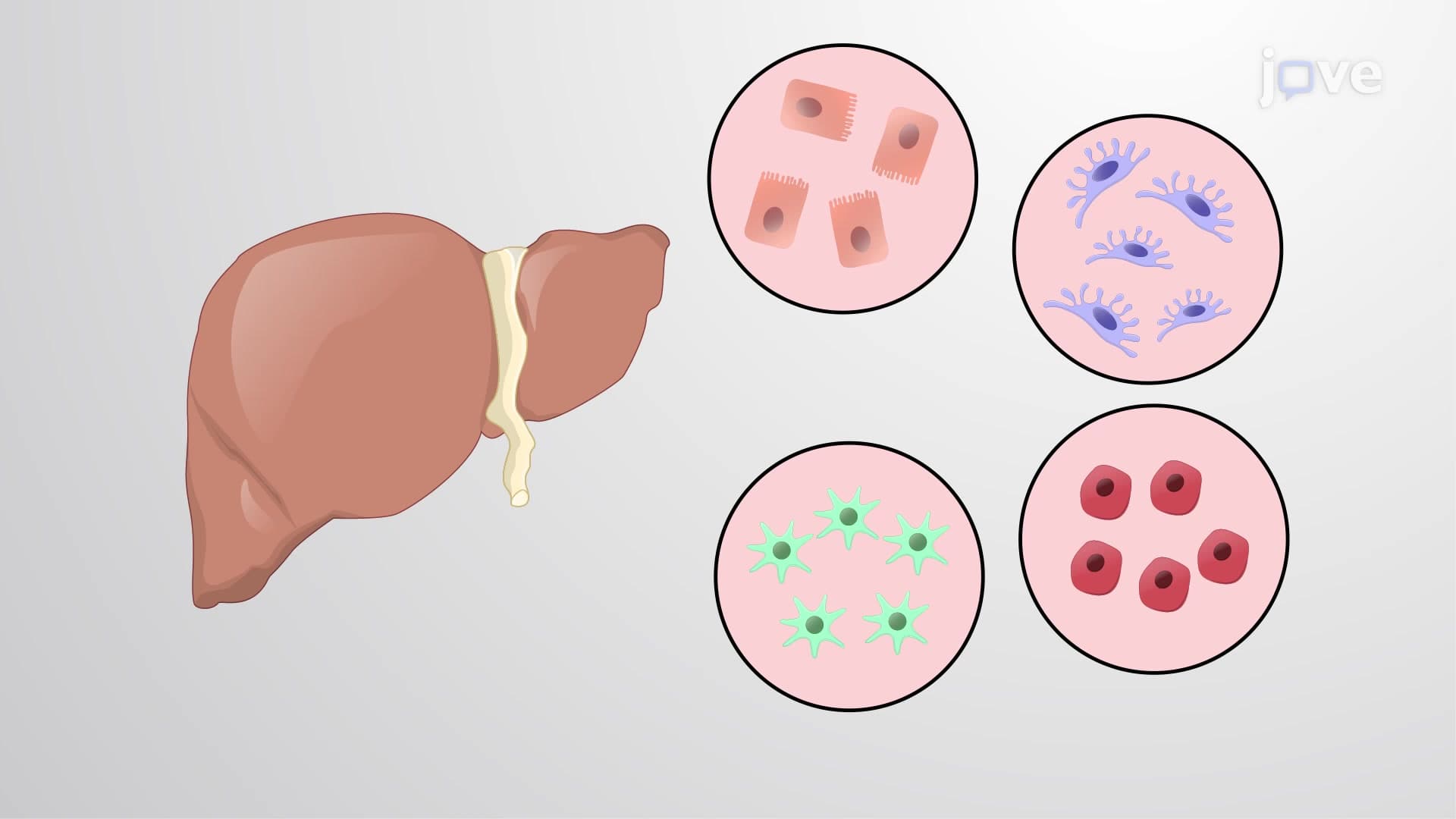

Video Summary: Overview of Cell Separation and Isolation Explained
Did you know that scientists can extract a single type of cell from millions in a blood sample? This overview of cell separation and isolation reveals how researchers use sophisticated techniques like density gradient centrifugation and fluorescence-activated cell sorting (FACS) to purify specific cell populations. For example, medical labs across the US routinely isolate T-cells from patient blood samples to diagnose immune disorders. Watch the full video on JoVE Coach to master this concept with expert-led visuals and step-by-step explanations.
Overview of cell separation and isolation encompasses the essential laboratory techniques that allow scientists to extract and purify specific cell types from complex biological samples. This process forms the backbone of countless research applications, from cancer research at the National Institutes of Health to stem cell therapy development at leading US medical centers like Mayo Clinic and Cleveland Clinic.
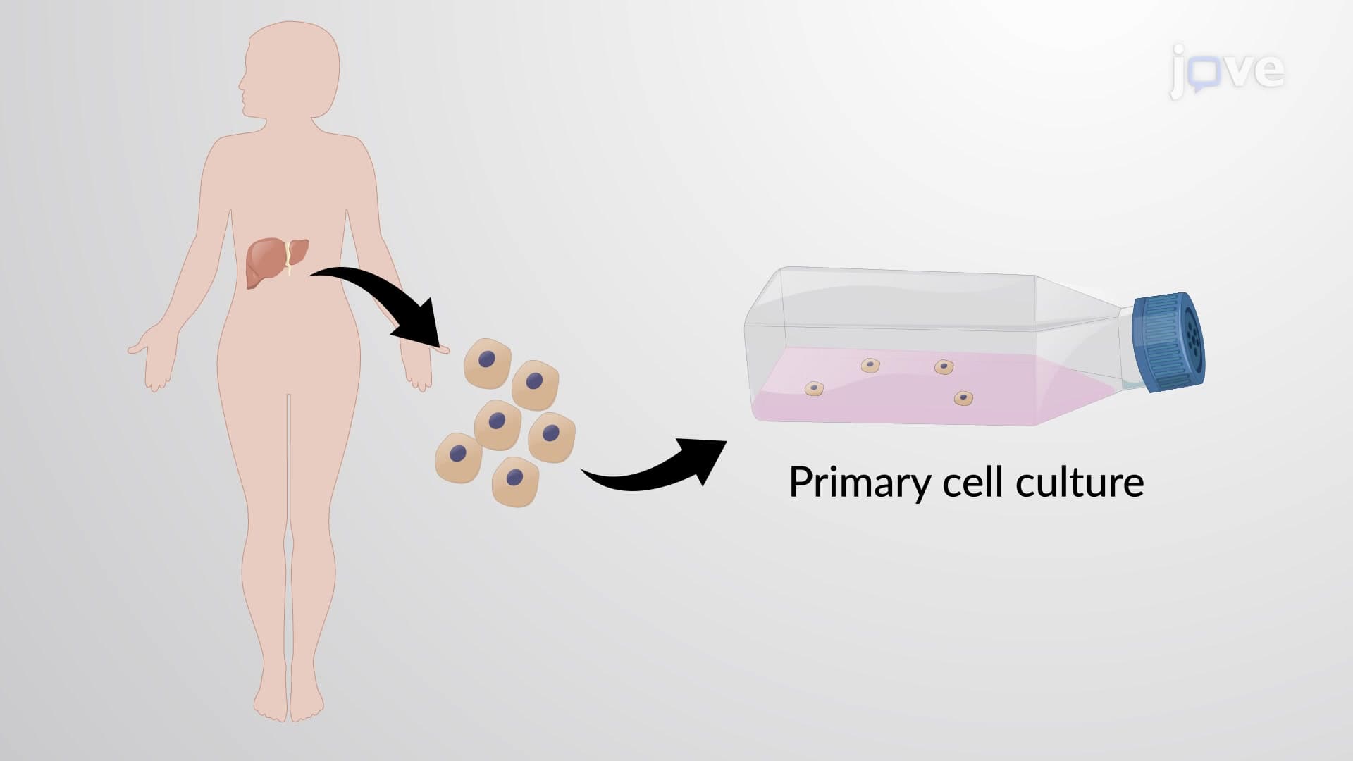
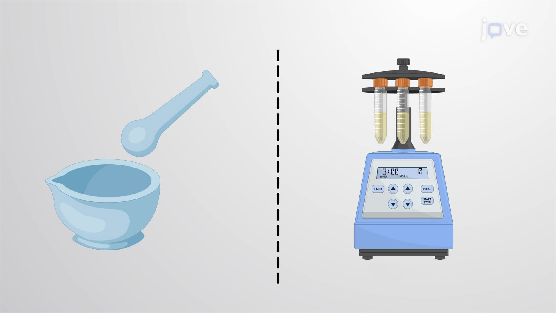
The journey begins with sample preparation, where solid tissues undergo mechanical disruption followed by enzymatic treatment. Proteolytic enzymes like collagenase and trypsin break down the extracellular matrix that holds cells together, creating a single-cell suspension ready for separation.
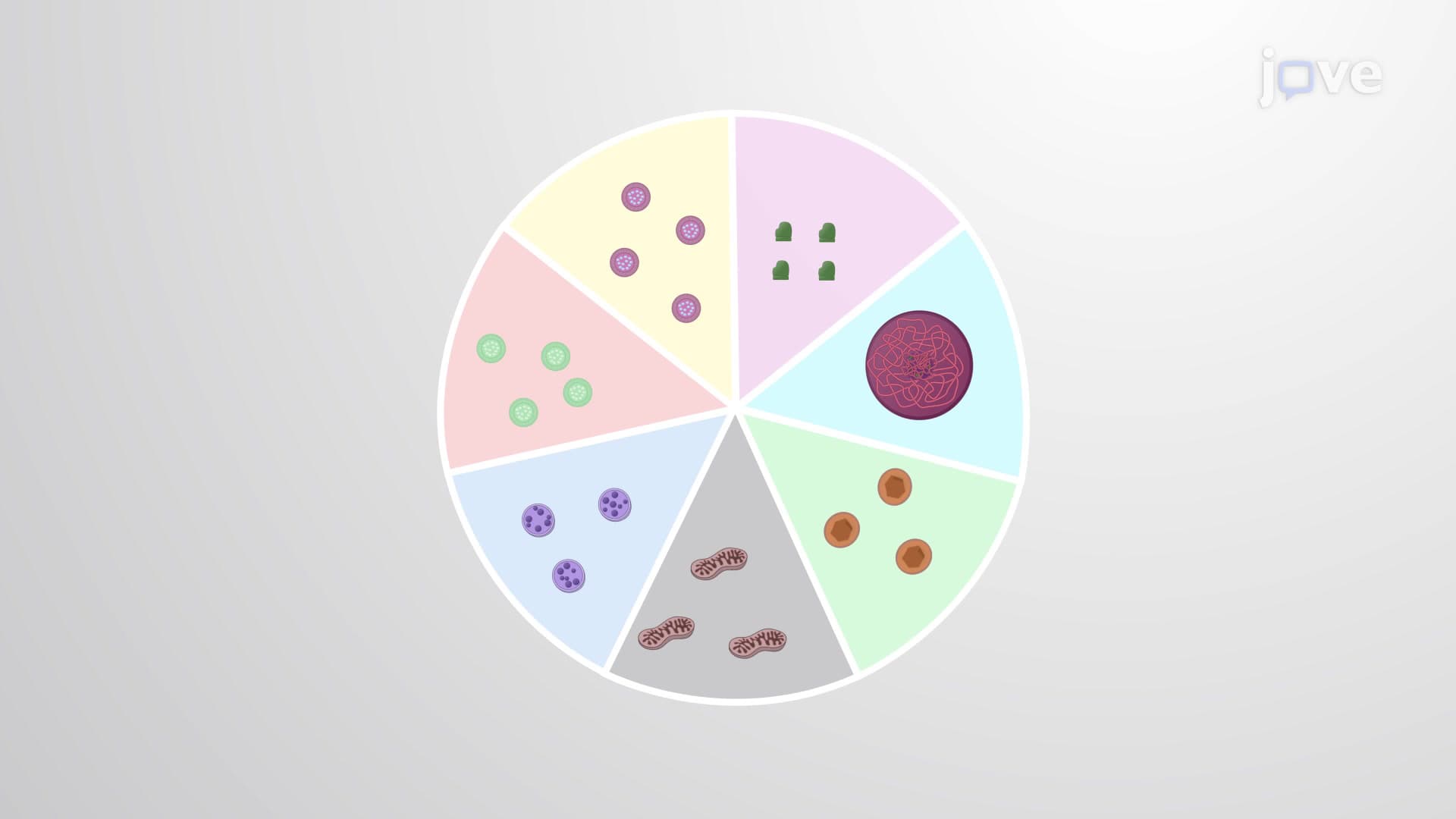
Density gradient centrifugation represents one of the most widely used approaches in cell separation. This technique exploits the natural density differences between cell types, using media like Ficoll-Paque or Percoll to create gradient layers. When spun in a centrifuge, cells migrate to their isodensity point-heavier cells settle deeper while lighter cells remain near the surface.
This method proves particularly valuable in clinical settings. US hospitals routinely use Ficoll gradients to separate peripheral blood mononuclear cells (PBMCs) from whole blood, enabling doctors to analyze immune cell populations in patients with autoimmune diseases or monitor transplant rejection.
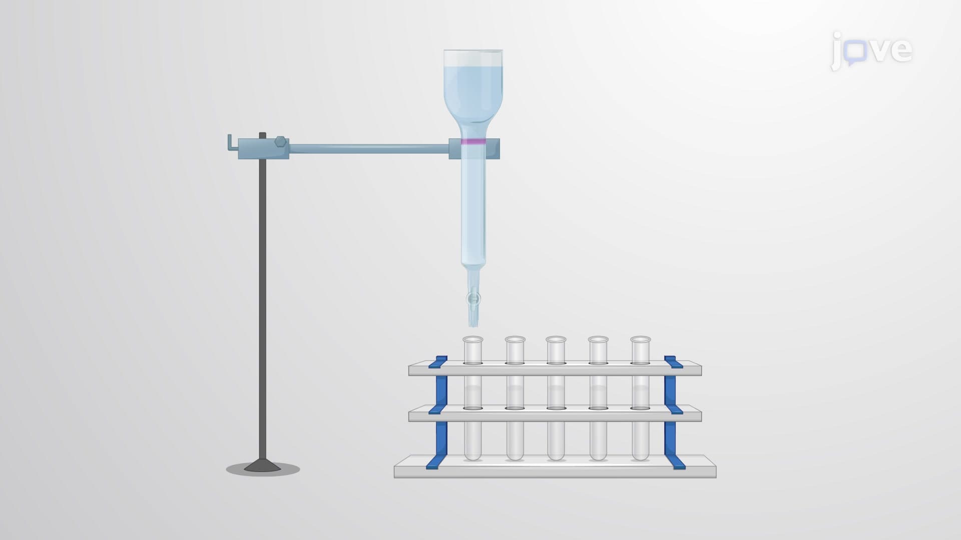
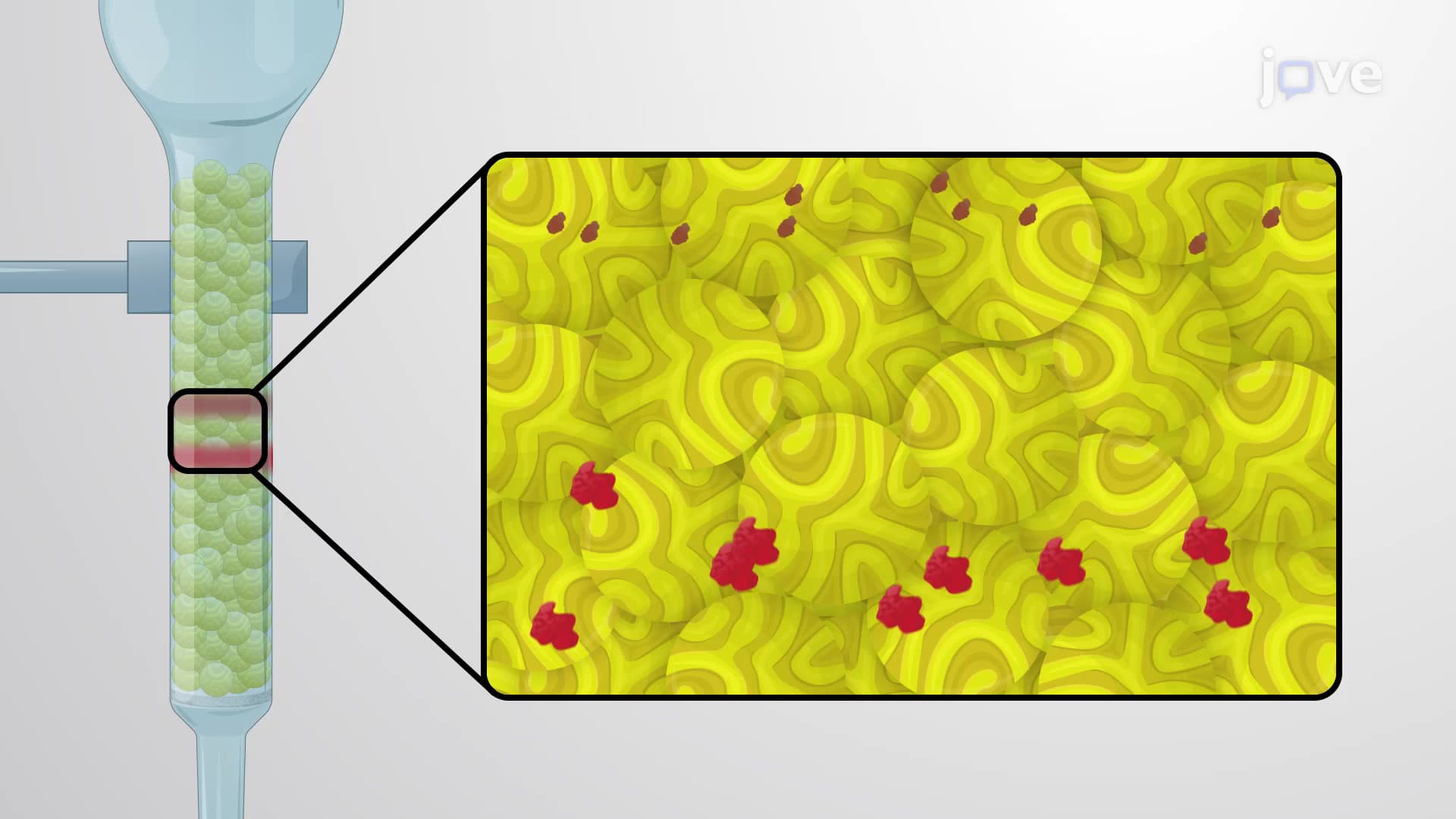
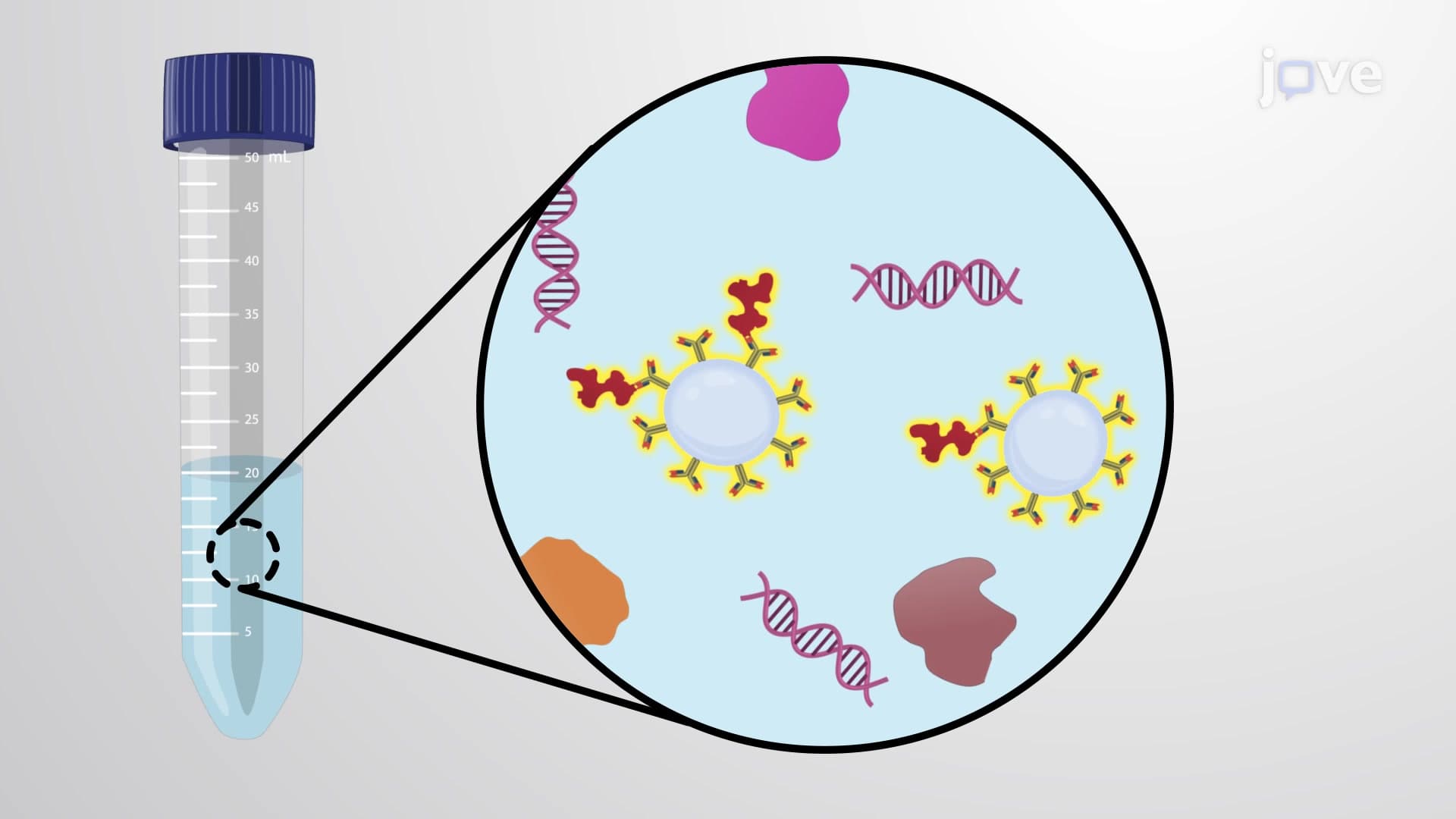
Modern cell separation has evolved beyond density differences to target specific molecular signatures. Cell affinity chromatography employs immobilized antibodies or other ligands to capture cells expressing particular surface markers. The target cells bind to the column matrix while unwanted cells wash through, resulting in highly purified populations.
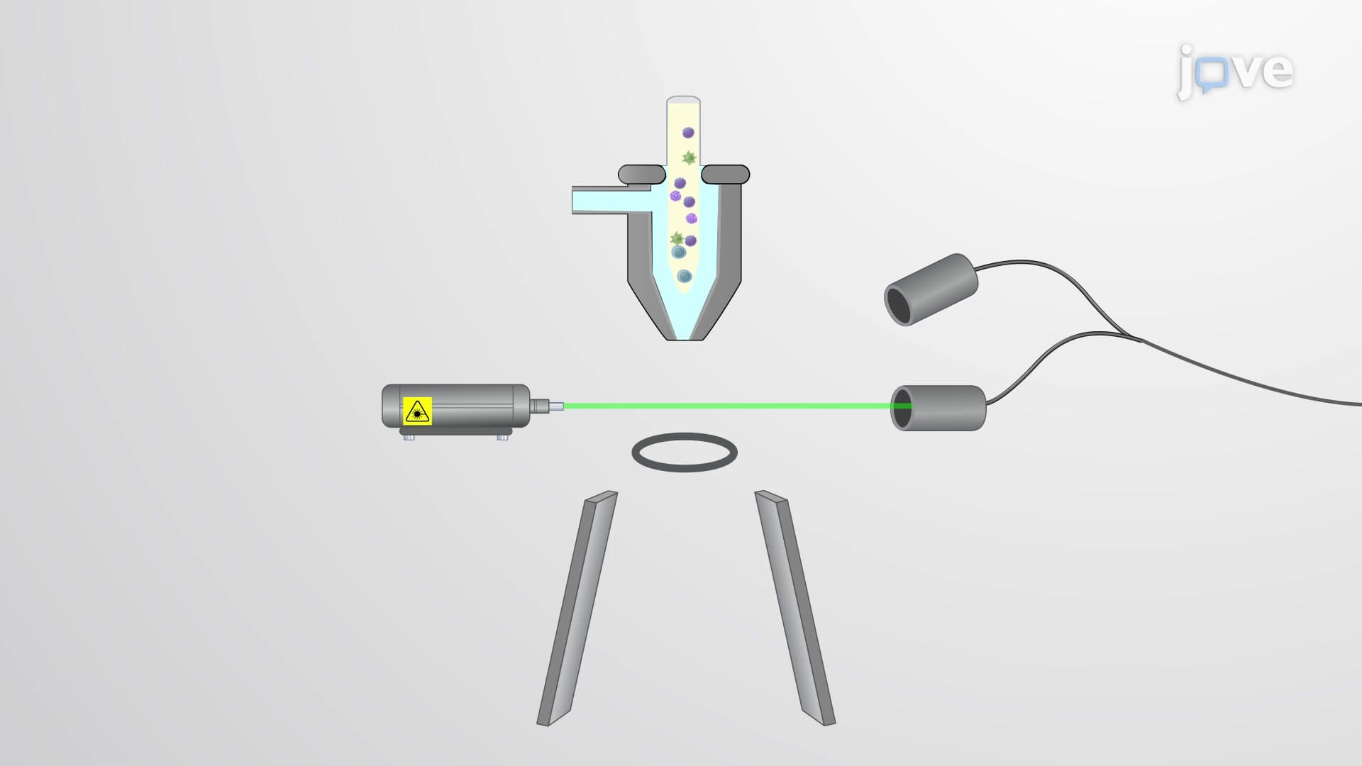
Fluorescence-activated cell sorting (FACS) takes this concept further by using fluorescently-labeled antibodies to identify and physically separate individual cells. Flow cytometers analyze thousands of cells per second, directing each cell to collection tubes based on its fluorescence profile. This technology enables researchers at institutions like Stanford University and Harvard Medical School to isolate rare cell populations with unprecedented precision.
These techniques directly impact human health through applications in personalized medicine and regenerative therapy. For students preparing for the MCAT or AP Biology exams, understanding cell separation principles provides crucial context for topics ranging from immunology to cancer biology. College-level cell biology courses frequently include laboratory exercises using these methods, making hands-on familiarity essential for STEM majors.
Related Micro-courses