Loading subject page…

- Cell Biology
- Analyzing Cells and Proteins
- Flow Cytometry
Micro-course: Analyzing Cells and Proteins
222,112 views
Learn key concepts and earn your certificate.


Flow Cytometry

00:00
01:23
Video Summary: What Is Flow Cytometry
Did you know that a single drop of blood contains over 5 million white blood cells, each with unique characteristics that doctors can analyze to diagnose diseases like leukemia? Flow cytometry is the powerful laboratory technique that makes this cellular analysis possible by measuring individual cell properties as they flow through laser beams. At major US medical centers like Johns Hopkins and Mayo Clinic, flow cytometry helps diagnose blood cancers and monitor immune system health in thousands of patients daily. Watch the full video on JoVE Coach to master this concept with expert-led visuals and step-by-step explanations.
- Understand what is flow cytometry and how this analytical technique measures individual cell characteristics in heterogeneous populations
- Identify the key components of flow cytometry systems, including lasers, detectors, and fluidics that enable single-cell analysis
- Analyze forward scatter and side scatter data to differentiate cell types based on size and internal complexity
- Compare different cell populations, such as distinguishing granulocytes from lymphocytes using scatter plot analysis
- Evaluate how fluorescent-activated cell sorting (FACS) uses antibody markers to identify and separate specific cell types
- Apply flow cytometry principles to understand clinical applications in immunology and hematology diagnostics
- Recognize the role of sheath fluid and hydrodynamic focusing in creating single-cell streams for accurate measurement
- Explore how electromagnetic deflection systems sort cells based on fluorescent labeling and electrical charging
What is Flow Cytometry: Fundamentals and Principles
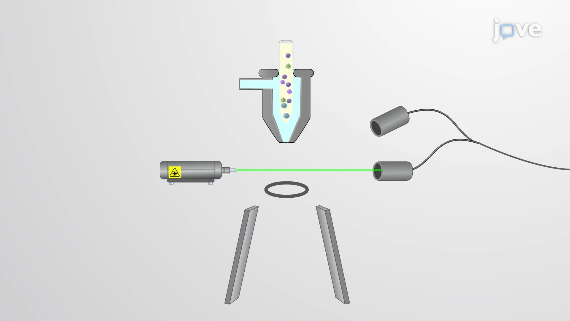
Flow cytometry represents one of the most versatile analytical techniques in modern biology and medicine, enabling researchers and clinicians to analyze thousands of individual cells per second. This technology combines fluidics, optics, and electronics to measure multiple characteristics of cells simultaneously as they pass through laser beams in single file.
The fundamental principle relies on hydrodynamic focusing, where a sample containing cells is injected into a stream of sheath fluid. This creates a narrow, single-cell stream that ensures each cell passes individually through the interrogation point where laser light interacts with cellular components. This precise positioning is crucial for accurate measurements and reproducible results.
Light Scattering: Forward and Side Scatter Analysis
When laser light encounters a cell, it scatters in multiple directions, providing valuable information about cellular characteristics. Forward scatter (FSC) occurs when light bends slightly as it passes around the cell, correlating with cell size. Larger cells like monocytes produce more forward scatter than smaller lymphocytes.
Side scatter (SSC) results from light refracting through internal cellular structures, indicating cellular complexity or granularity. Neutrophils, packed with granules containing antimicrobial compounds, exhibit high side scatter, while smooth lymphocytes show lower values. This dual-parameter analysis allows researchers to distinguish cell populations on scatter plots, forming the foundation for cellular identification.
Fluorescence Detection and FACS Technology
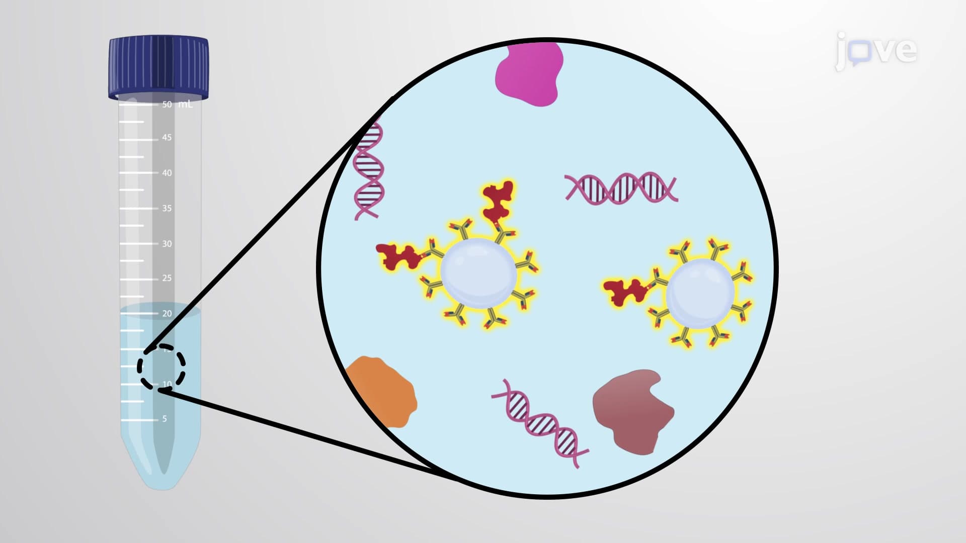
Fluorescent-activated cell sorting (FACS) extends basic flow cytometry by incorporating fluorescent markers. Antibodies conjugated to fluorescent dyes bind specifically to cellular markers like CD4 on helper T cells or CD19 on B cells. When excited by appropriate laser wavelengths, these fluorophores emit light at characteristic wavelengths detected by photomultiplier tubes.
The sorting capability comes from an electrostatic charging system that applies specific charges to droplets containing cells of interest. Charged plates then deflect these droplets into collection tubes, enabling physical separation of cell populations. This technology proves essential in research applications and clinical procedures like isolating stem cells for transplantation.
Clinical Applications and Educational Relevance
Flow cytometry plays critical roles in US healthcare, from diagnosing leukemias at institutions like MD Anderson Cancer Center to monitoring HIV progression through CD4+ T cell counts. Students preparing for MCAT exams encounter flow cytometry in immunology and cell biology sections, while AP Biology courses include this technique in laboratory investigation curricula.
Understanding flow cytometry concepts helps students grasp broader principles of cell biology, immunology, and analytical chemistry that appear across standardized tests and college coursework in life sciences programs.
Frequently Asked Questions
Flow cytometry is a laboratory technique that analyzes individual cells by passing them through laser beams to measure their size, complexity, and fluorescent properties. It's like having a microscopic assembly line that can examine thousands of cells per second, providing detailed information about cell populations in blood, tissue, or research samples.
The MCAT frequently includes flow cytometry questions in biological sciences sections, focusing on data interpretation from scatter plots and fluorescence histograms. AP Biology exams test understanding of the technique's principles and applications in immunology labs. Students should practice interpreting flow cytometry graphs showing different cell populations and understanding experimental design using fluorescent markers.
Flow cytometry is the broader analytical technique, while FACS (fluorescent-activated cell sorting) is a specialized type of flow cytometry that can physically separate cells. Regular flow cytometers only analyze and count cells, but FACS instruments add sorting capability by charging and deflecting cells into collection tubes. Think of FACS as flow cytometry plus a separation system.
US medical centers use flow cytometry extensively for diagnosing blood cancers, monitoring immune system disorders, and tracking HIV progression through CD4+ cell counts. For example, Children's Hospital of Philadelphia uses flow cytometry to diagnose pediatric leukemias, while transplant centers nationwide employ it to match organ donors and recipients by analyzing immune cell markers.
No advanced chemistry background is required to grasp fundamental flow cytometry concepts. The technique builds on basic biology principles like antibody-antigen binding and light scattering that high school students learn. Understanding requires familiarity with cell biology and basic physics concepts about light behavior, making it accessible to students in introductory college biology courses.
Focus on understanding the relationship between physical cell properties and measurement parameters - larger cells produce more forward scatter, granular cells show higher side scatter. Practice interpreting scatter plots and histograms, and memorize common cell surface markers like CD4, CD8, and CD19. Create concept maps linking cell types to their characteristic scatter and fluorescence patterns.
Flow cytometry complements microscopy, PCR, and immunoassays in comprehensive cellular analysis. Students should explore fluorescence microscopy to understand fluorophore principles, learn about monoclonal antibody production for marker specificity, and study cell culture techniques for sample preparation. These interconnected methods form the foundation of modern cell biology research and clinical diagnostics.
Flow cytometry expertise opens doors to clinical laboratory science, immunology research, pharmaceutical development, and biotechnology careers. Clinical laboratory scientists at major US hospital systems operate flow cytometers daily, while research scientists at companies like Genentech and Amgen use advanced flow cytometry for drug development and quality control in biomanufacturing processes.
Related Micro-courses
17 Concepts
11 Concepts

17 Concepts