Loading subject page…


222,112 views

Video Summary: What Is Immunoprecipitation
Did you know that researchers at Stanford University can fish out a single protein from thousands of others in a cell, much like finding a specific person in a crowded stadium? Immunoprecipitation makes this molecular detective work possible by using antibodies as highly specific "fishing hooks" to capture target proteins from complex biological samples. This technique has revolutionized cancer research and drug development across American laboratories. What is immunoprecipitation becomes clear when you see how antibodies attached to beads selectively bind and isolate proteins through a simple centrifugation process. Watch the full video on JoVE Coach to master this concept with expert-led visuals and step-by-step explanations.
Immunoprecipitation represents one of biochemistry's most elegant solutions to a complex problem: how do you isolate a single protein from the thousands present in a living cell? This technique harnesses the exquisite specificity of antibodies-the same molecules your immune system uses to recognize invaders-to selectively capture target proteins from biological samples.
The process relies on the lock-and-key relationship between antibodies and their target proteins (antigens). When researchers at institutions like Johns Hopkins or UCLA need to study a specific protein involved in cancer or neurological diseases, they use immunoprecipitation to pull that protein out of a cellular soup containing thousands of other molecules.
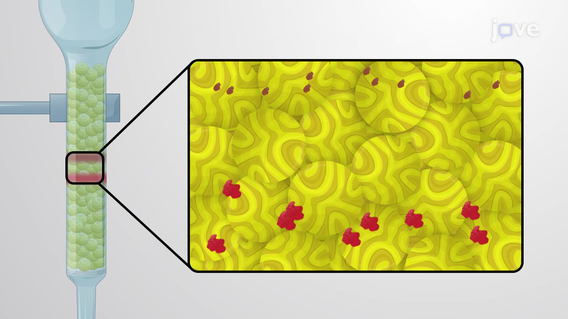
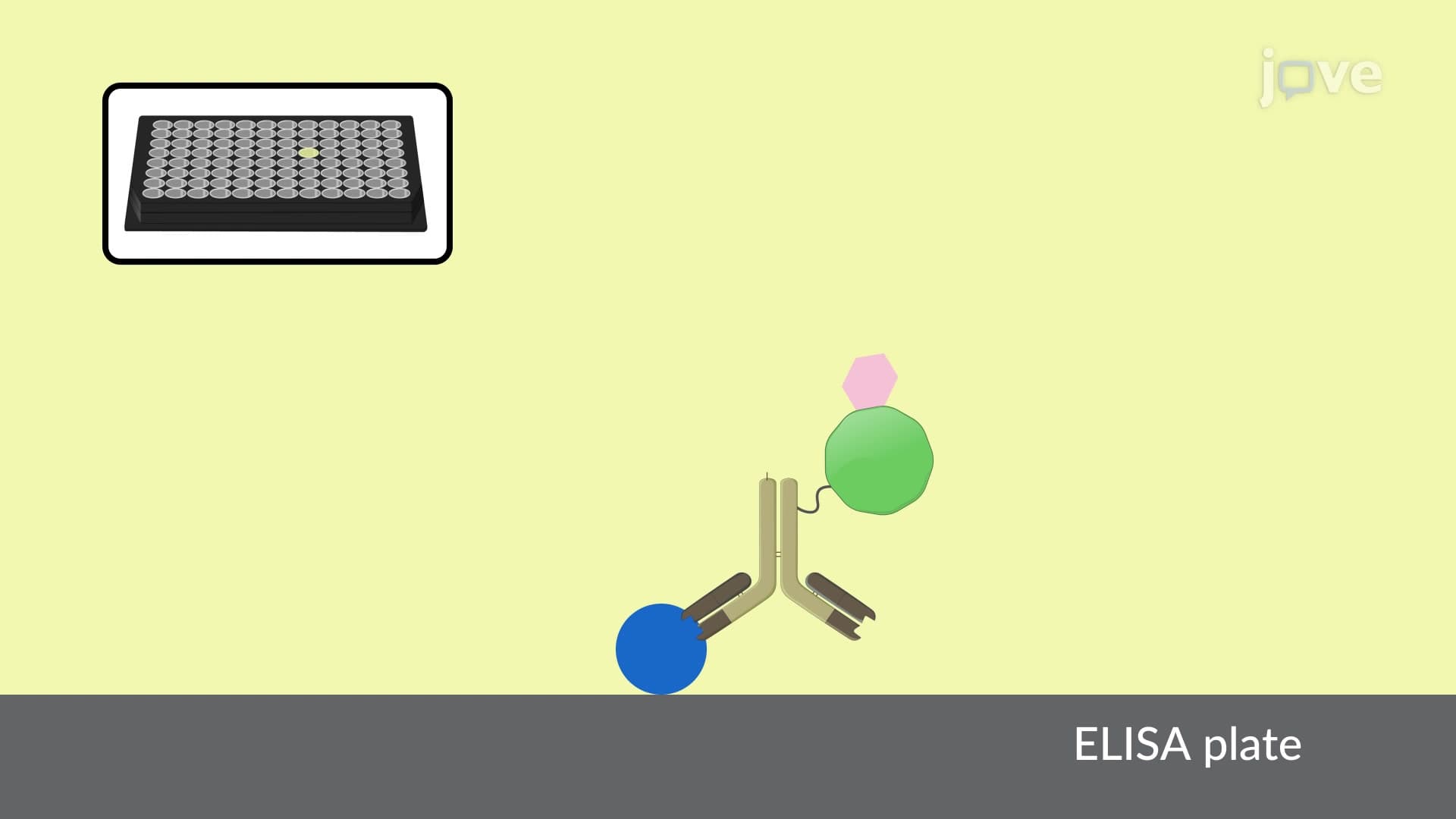
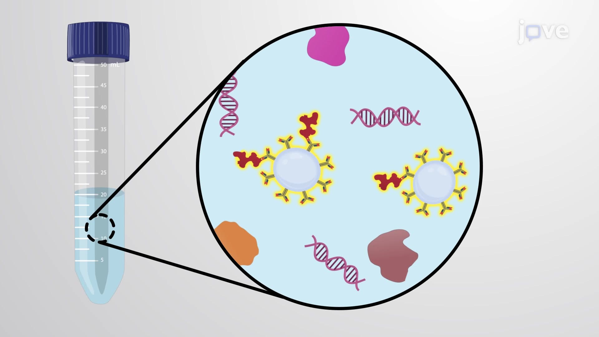
The technique employs a clever two-part system. First, antibodies specific to the target protein are attached to solid supports-either magnetic beads or agarose gel beads. This attachment often occurs through helper proteins called protein A or protein G, which act as molecular adapters that bind strongly to antibodies while remaining attached to the bead surface.
These protein A and G molecules, originally discovered in bacteria, have become indispensable tools in American research laboratories. They provide a standardized way to orient antibodies correctly on bead surfaces, ensuring maximum binding efficiency. When the antibody-coated beads encounter a protein mixture, they selectively bind only to their target protein, forming an antibody-protein complex.
The beauty of immunoprecipitation lies in its simplicity after the initial binding step. Low-speed centrifugation-typically around 1,000-3,000 rpm-causes the bead-antibody-protein complexes to settle into a pellet at the bottom of the tube. This gentle spinning preserves protein structure while effectively separating bound proteins from everything else in the sample.
The final step involves strategic chemistry: researchers use buffers with altered pH (usually acidic) or high salt concentrations to disrupt the antibody-protein interaction. This controlled disruption releases the target protein into solution while leaving the antibodies still attached to their beads. A second round of gentle centrifugation separates the beads from the now-purified protein solution.
Immunoprecipitation has become essential for studying protein interactions in diseases like Alzheimer's, where researchers at institutions such as the Mayo Clinic use it to isolate and analyze proteins involved in brain cell death. The technique also plays a crucial role in developing new medications, as pharmaceutical companies use it to understand how potential drugs interact with their protein targets.
Students preparing for advanced placement biology exams or MCAT sections on molecular biology should understand that immunoprecipitation often appears in questions about protein purification techniques and antibody applications. The concept connects directly to broader themes in cell biology, including protein structure-function relationships and the specificity of biological recognition systems.
Related Micro-courses