Loading subject page…

Micro-course: Analyzing Cells and Proteins
222,112 views
Learn key concepts and earn your certificate.





SDS-PAGE


00:00
01:27
Video Summary: What Is Sds Page
Ever wondered how scientists identify specific proteins in crime scene DNA analysis or diagnose genetic diseases? SDS PAGE is the gold-standard laboratory technique that separates proteins by size, making it essential for everything from medical diagnostics to pharmaceutical research. This molecular weight-based method uses specialized chemicals to unfold proteins and run them through a gel, similar to how a sieve sorts different sized particles. Major US biotech companies like Genentech rely on what is SDS PAGE methodology for drug development and quality control. Watch the full video on JoVE Coach to master this concept with expert-led visuals and step-by-step explanations.
- Understand what is SDS PAGE and how this electrophoresis technique separates proteins based on molecular weight
- Identify the key components including SDS detergent, β-mercaptoethanol reducing agent, and polyacrylamide gel matrix
- Analyze how the two-gel system works, comparing the stacking gel's large pores to the resolving gel's smaller pores
- Recognize the role of SDS in denaturing proteins and providing uniform negative charge distribution
- Apply knowledge of protein migration patterns to interpret gel results and determine molecular weights
- Evaluate how different acrylamide concentrations affect protein separation resolution
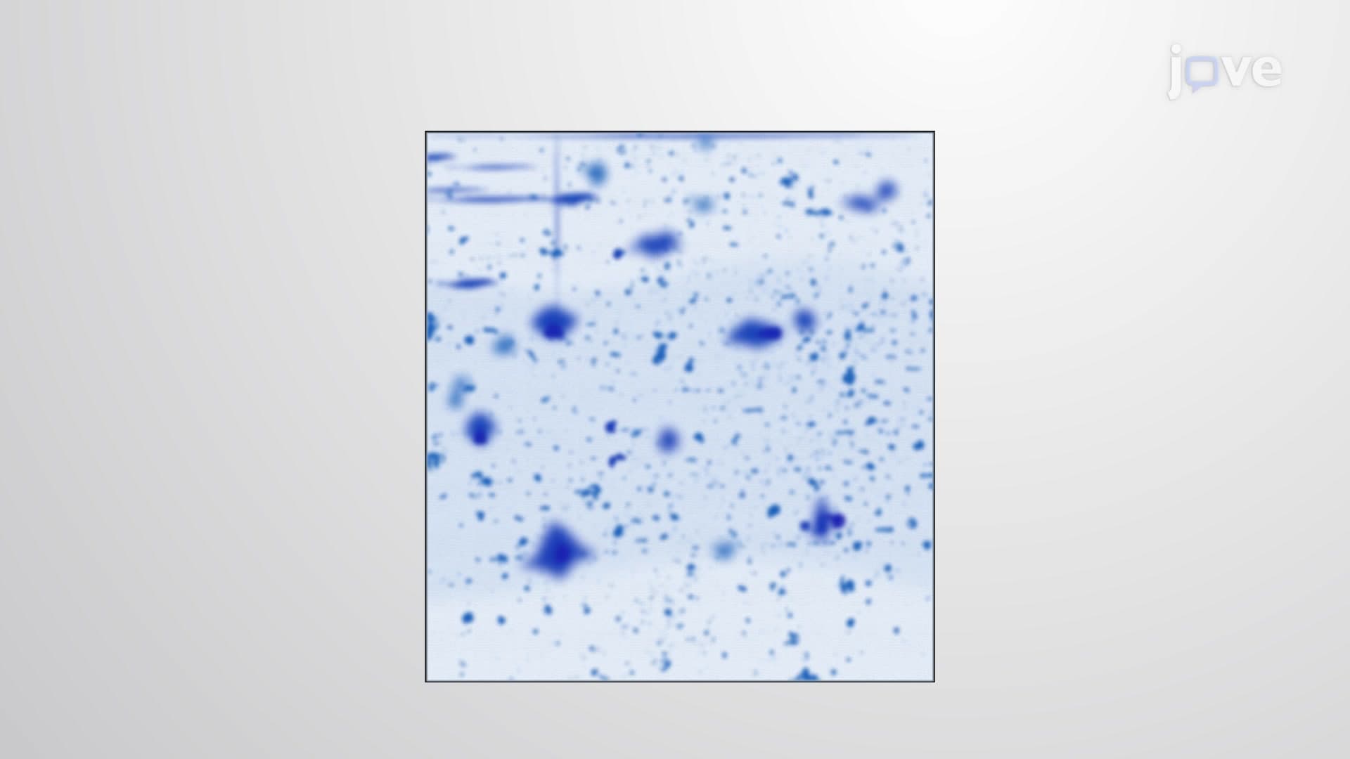
- Explore visualization methods like Coomassie blue staining for detecting separated protein bands
- Compare SDS PAGE results with molecular weight markers to quantify unknown proteins
Understanding What is SDS PAGE: The Foundation of Protein Analysis
SDS PAGE (Sodium Dodecyl Sulfate-Polyacrylamide Gel Electrophoresis) represents one of the most fundamental techniques in biochemistry and molecular biology laboratories across the United States. This powerful method enables scientists to separate complex protein mixtures based solely on molecular weight, making it indispensable for research at institutions like the National Institutes of Health and major pharmaceutical companies including Pfizer and Moderna.
The Chemistry Behind SDS PAGE: Breaking Down Protein Structure
The SDS PAGE definition centers on controlled protein denaturation and size-based separation. The process begins with sample preparation using two critical chemicals. β-mercaptoethanol acts as a reducing agent, systematically breaking disulfide bonds that maintain protein tertiary structure. Meanwhile, SDS (sodium dodecyl sulfate) serves a dual purpose: this anionic detergent binds to hydrophobic protein regions, completely unfolding the molecules while coating them with uniform negative charges at a consistent ratio of approximately one SDS molecule per two amino acids.
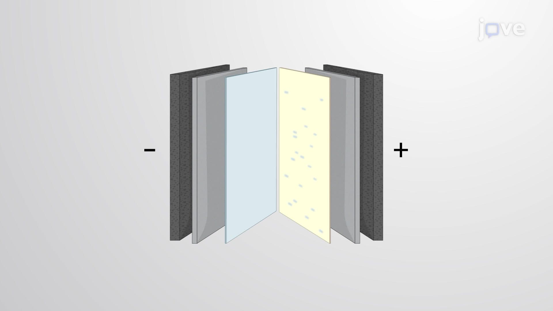
This chemical treatment transforms all proteins into linear, negatively charged rods with identical charge-to-mass ratios. Consequently, when subjected to an electric field, proteins migrate solely based on their size-larger proteins encounter more resistance and move slower, while smaller proteins navigate more easily through the gel matrix.
The Two-Gel System: Stacking and Resolving for Optimal Separation
What is SDS PAGE in detail involves understanding the ingenious two-gel design. The upper stacking gel contains a low acrylamide concentration (typically 4-5%), creating large pores that allow proteins to compress into tight, uniform bands-imagine multiple lanes of traffic merging into a single file before entering a tunnel. This "stacking" effect ensures all proteins enter the resolving gel simultaneously, maximizing separation quality.
The lower resolving gel features higher acrylamide concentrations (8-15%), forming smaller pores that act as a molecular sieve. Students preparing for AP Biology or college biochemistry courses should understand this concept as analogous to sorting marbles through progressively smaller screens-larger proteins become trapped in the gel matrix while smaller proteins migrate further toward the positive electrode.
Real-World Applications: From Clinical Diagnostics to Biotechnology
US medical laboratories routinely employ SDS PAGE for diagnosing genetic disorders, monitoring treatment responses, and quality control in pharmaceutical manufacturing. For example, clinical labs use this technique to analyze hemoglobin variants in sickle cell disease diagnosis, while biotech companies verify protein purity in therapeutic drug production. Students encountering this concept in MCAT preparation or college coursework should recognize its significance extends far beyond academic exercises-it's a cornerstone technology driving American healthcare innovation and drug discovery.
Frequently Asked Questions
SDS PAGE is a laboratory technique that separates proteins by size using an electric field and specialized gel. It's crucial because it allows scientists to identify, analyze, and purify specific proteins from complex biological samples. This method is fundamental to medical research, drug development, and clinical diagnostics across US healthcare systems.
MCAT questions often test understanding of protein denaturation, gel electrophoresis principles, and data interpretation from gel images. College exams typically include mechanism questions, troubleshooting scenarios, and calculations involving migration distances. Students should practice identifying protein bands and correlating molecular weights with migration patterns.
The stacking gel has lower acrylamide concentration (4-5%) creating larger pores, while the resolving gel has higher concentration (8-15%) with smaller pores. The stacking gel compresses proteins into tight bands for uniform entry, while the resolving gel separates them by size. This two-step process dramatically improves separation quality and band sharpness.
US clinical laboratories use SDS PAGE to diagnose hemoglobinopathies, analyze cerebrospinal fluid proteins for neurological disorders, and monitor kidney function through urine protein analysis. Hospital labs also employ it for identifying abnormal proteins in blood samples and verifying therapeutic protein integrity in drug manufacturing.
Not at all! While the chemistry involves multiple steps, the core concept resembles everyday sorting processes-like separating different sized objects through screens. High school students with basic chemistry knowledge can master SDS PAGE principles by focusing on the size-separation concept rather than complex molecular interactions.
Practice interpreting gel images and calculating molecular weights using standard curves. Focus on understanding why proteins migrate differently and how chemical treatments affect protein structure. Create flowcharts showing the step-by-step process from sample preparation through visualization, and work through practice problems involving data analysis.
Mastering SDS PAGE provides the foundation for understanding protein purification, Western blotting, and mass spectrometry techniques. It introduces key concepts like protein denaturation, electrophoretic mobility, and molecular weight determination that appear throughout advanced biochemistry coursework. This knowledge directly applies to research methods courses and laboratory work.
Students should explore Native PAGE (non-denaturing conditions), isoelectric focusing (separation by charge), and two-dimensional gel electrophoresis (separation by both charge and size). Western blotting builds directly on SDS PAGE principles by adding antibody detection steps for specific protein identification.
Related Micro-courses

17 Concepts
11 Concepts

17 Concepts