Loading subject page…

- Cell Biology
- Analyzing Cells and Proteins
- Subcellular Fractionation
Micro-course: Analyzing Cells and Proteins
222,112 views
Learn key concepts and earn your certificate.


Subcellular Fractionation

00:00
01:32
Video Summary: What Is Subcellular Fractionation
Ever wondered how researchers at Johns Hopkins isolate specific cell parts to study diseases like Alzheimer's? Subcellular fractionation is the laboratory technique that separates different organelles from broken-open cells, allowing scientists to study mitochondria, nuclei, and other cellular components individually. This process is essential for understanding how cellular dysfunction contributes to conditions ranging from cancer to metabolic disorders. Watch the full video on JoVE Coach to master this concept with expert-led visuals and step-by-step explanations.
- Understand what subcellular fractionation is and why it's fundamental to cellular biology research
- Identify the two primary methods used in subcellular fractionation: differential centrifugation and density gradient centrifugation
- Analyze how centrifugal force speeds (measured in x g) determine which organelles separate at each step
- Compare the effectiveness of differential versus density gradient centrifugation for separating different organelle types
- Evaluate the limitations of size-based separation when organelles have similar physical properties
- Apply knowledge of organelle characteristics to predict separation outcomes in fractionation protocols
- Recognize real-world applications in medical research, drug development, and disease diagnosis
- Explore how fractionation techniques contribute to our understanding of cellular metabolism and disease pathogenesis
Understanding Subcellular Fractionation Fundamentals
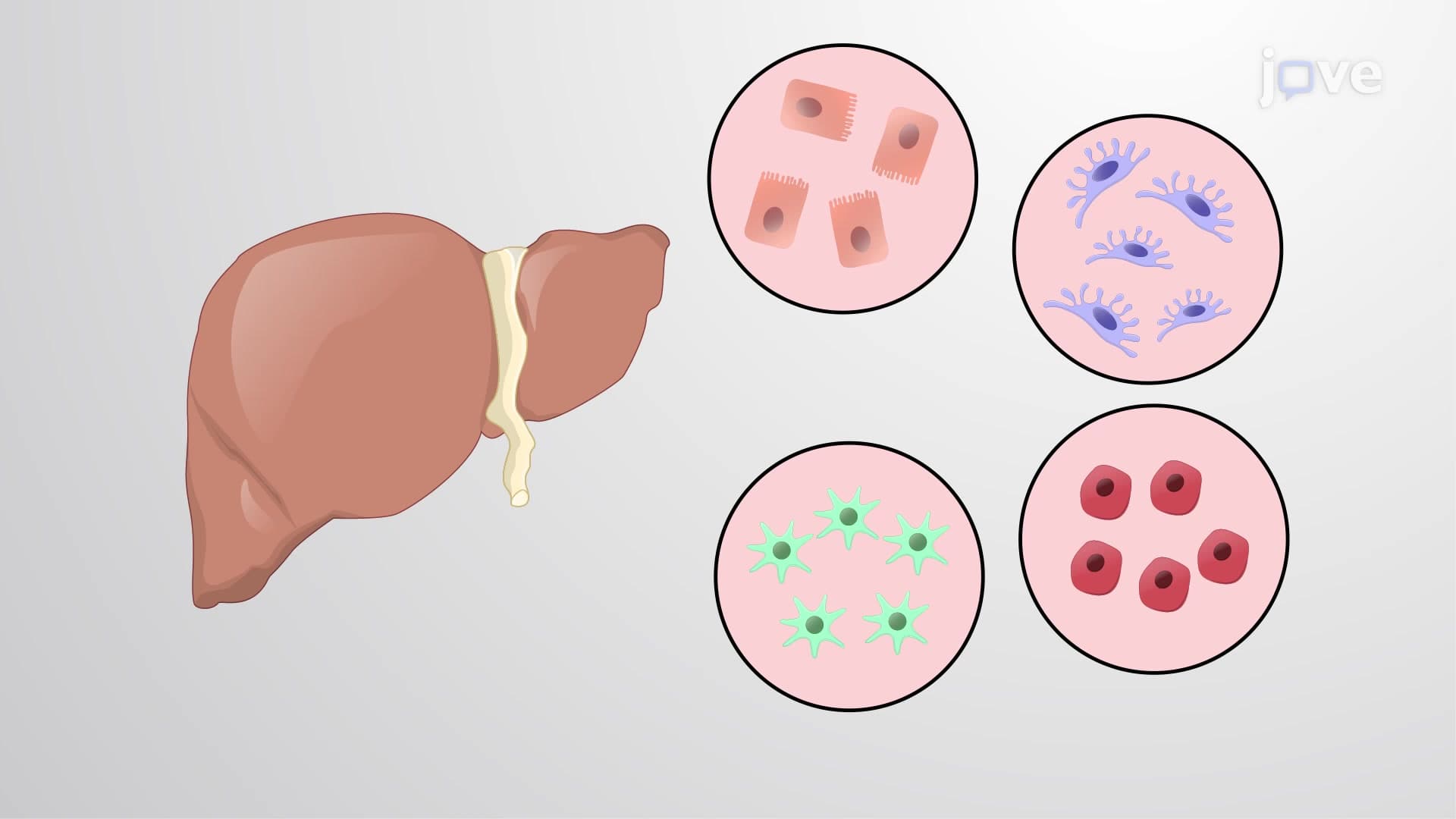
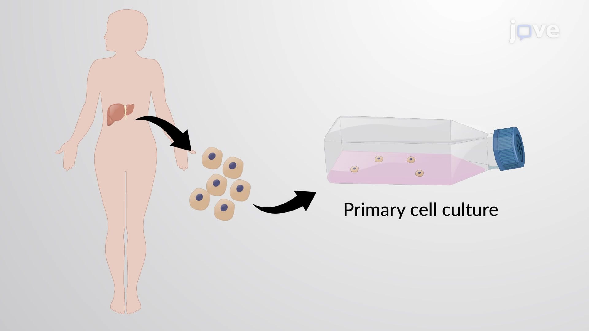
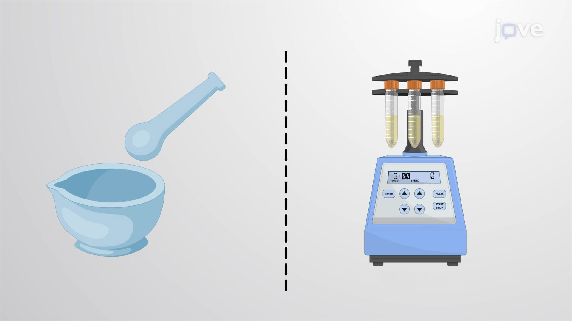
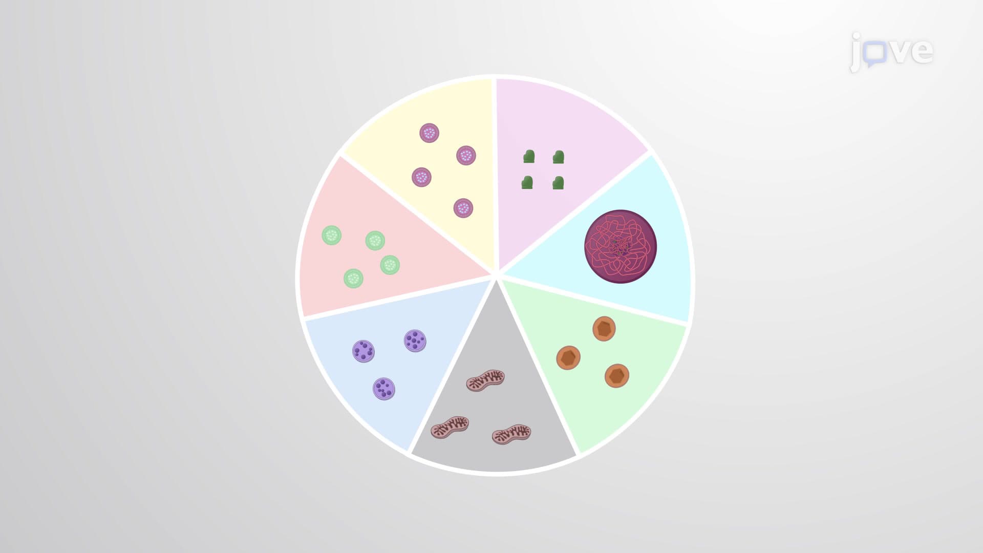
Subcellular fractionation serves as a cornerstone technique in cell biology, enabling researchers to isolate and study individual cellular components in unprecedented detail. This process begins with creating a cell lysate-essentially breaking open cells to release their internal contents while preserving organelle structure and function. The technique has revolutionized our understanding of cellular processes and remains essential in both academic research and pharmaceutical development.
Differential Centrifugation: The Sequential Approach
Differential centrifugation operates on the principle that larger, denser organelles will sediment faster than smaller ones when subjected to centrifugal force. The process follows a systematic approach: initial low-speed centrifugation (400-600 x g) removes nuclei and large cellular debris, creating the first fraction. The remaining supernatant undergoes medium-speed centrifugation (10,000-20,000 x g) to pellet mitochondria, lysosomes, and peroxisomes. Finally, ultracentrifugation at speeds exceeding 80,000 x g separates microsomes, membrane fragments, and ribosomes.
This method proves particularly valuable in clinical laboratories studying metabolic disorders. For instance, researchers at the Cleveland Clinic use differential centrifugation to isolate mitochondria from patient muscle biopsies, helping diagnose mitochondrial diseases that affect energy production in cells.
Density Gradient Centrifugation: Precision Separation
When organelles share similar sizes-like mitochondria and peroxisomes-differential centrifugation reaches its limitations. Density gradient centrifugation overcomes this challenge by using chemical gradients, typically sucrose or glycerol solutions of varying concentrations. During centrifugation, organelles migrate to positions where their density matches that of the surrounding gradient medium, creating distinct bands that can be individually collected.
This technique has proven invaluable in studying cellular responses to diseases. Researchers at the National Institutes of Health employ density gradient centrifugation to separate different types of membrane vesicles, advancing our understanding of how cells communicate and respond to therapeutic interventions.
Applications in Modern Biology and Medicine
Subcellular fractionation techniques appear frequently in standardized exams, particularly the MCAT and AP Biology assessments. Students should understand not only the mechanical processes but also the biological rationale behind organelle separation. College-level biochemistry courses often include laboratory exercises using these techniques, providing hands-on experience with centrifugation protocols and fraction analysis.
In pharmaceutical research, subcellular fractionation enables drug developers to study how medications affect specific cellular compartments, contributing to more targeted and effective treatments for conditions ranging from cancer to neurodegenerative diseases.
Frequently Asked Questions
Subcellular fractionation is a laboratory technique that separates different organelles and cellular components from broken-open cells, allowing scientists to study each part individually. It's crucial for understanding how specific organelles function, how they're affected by diseases, and how potential treatments might work. This technique has been instrumental in discovering cellular processes and developing medical treatments.
The MCAT frequently tests understanding of centrifugation principles, organelle properties, and experimental design involving fractionation. AP Biology exams may include questions about organelle separation based on size and density, or ask students to interpret experimental results from fractionation studies. Students should understand both the techniques and their applications in research.
Differential centrifugation separates organelles based primarily on size and mass using sequential speed increases, while density gradient centrifugation uses chemical gradients to separate based on density differences. For exams, remember that differential centrifugation is faster but less precise, while density gradient centrifugation can separate organelles with similar sizes but different densities.
Major medical centers like Mayo Clinic and Johns Hopkins use subcellular fractionation to study disease mechanisms and test new treatments. Researchers isolate mitochondria to study metabolic disorders, separate nuclear fractions to examine gene expression changes, and isolate membrane components to understand how drugs interact with cells. This technique is essential for developing personalized medicine approaches.
Basic high school chemistry and biology provide sufficient background to understand subcellular fractionation concepts. You'll need to know about cell organelles, their relative sizes, and basic physics concepts like centrifugal force. The mathematical calculations are typically straightforward, focusing more on understanding principles than complex equations.
Create a step-by-step flowchart showing the fractionation process, including centrifugation speeds and which organelles separate at each step. Practice interpreting experimental results and explaining why certain organelles appear in specific fractions. Focus on connecting organelle properties (size, density, function) to separation outcomes.
Mitochondria and peroxisomes present the greatest separation challenge because they're similar in size and density. This is why density gradient centrifugation was developed-to provide the precision needed when differential centrifugation isn't sufficient. Lysosomes and peroxisomes can also be challenging to separate completely using differential centrifugation alone.
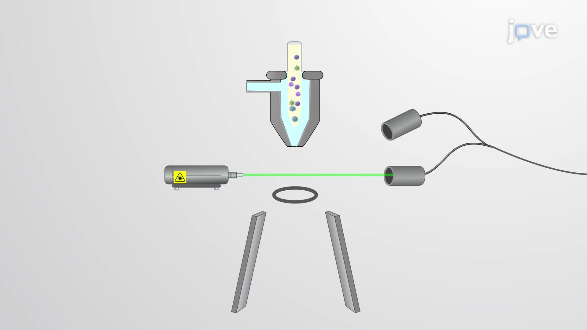
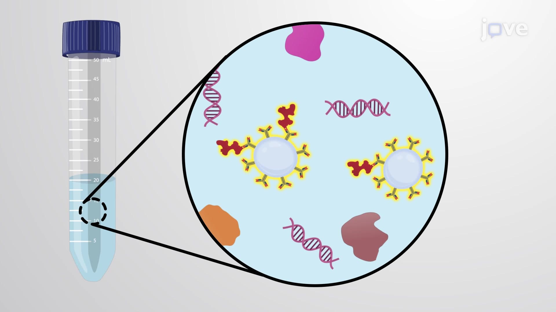
Consider studying immunoaffinity purification, which uses antibodies to isolate specific proteins or organelles, and flow cytometry for analyzing cellular components. Advanced courses might cover proteomics applications, where fractionation helps identify protein locations within cells, and metabolomics studies that examine metabolic pathways in isolated organelles.
Related Micro-courses
17 Concepts
11 Concepts

17 Concepts