Loading subject page…


222,112 views


Video Summary: What Is Western Blotting
Ever wondered how researchers at the CDC identify specific proteins in disease samples? Western blotting is the gold-standard analytical technique that detects target proteins from complex mixtures, enabling scientists to diagnose conditions like HIV or study cancer biomarkers. This powerful method combines protein separation with antibody-based detection to pinpoint exact proteins of interest. What is Western Blotting becomes clear through its step-by-step process of separation, transfer, and visualization. Watch the full video on JoVE Coach to master this concept with expert-led visuals and step-by-step explanations.
Western blotting stands as one of the most widely used techniques in molecular biology laboratories across the United States. From university research labs at Harvard Medical School to pharmaceutical companies like Pfizer, this method serves as the backbone for protein identification and quantification. The technique earned its name as part of a series of "blotting" methods, following Southern blotting (for DNA) and Northern blotting (for RNA).
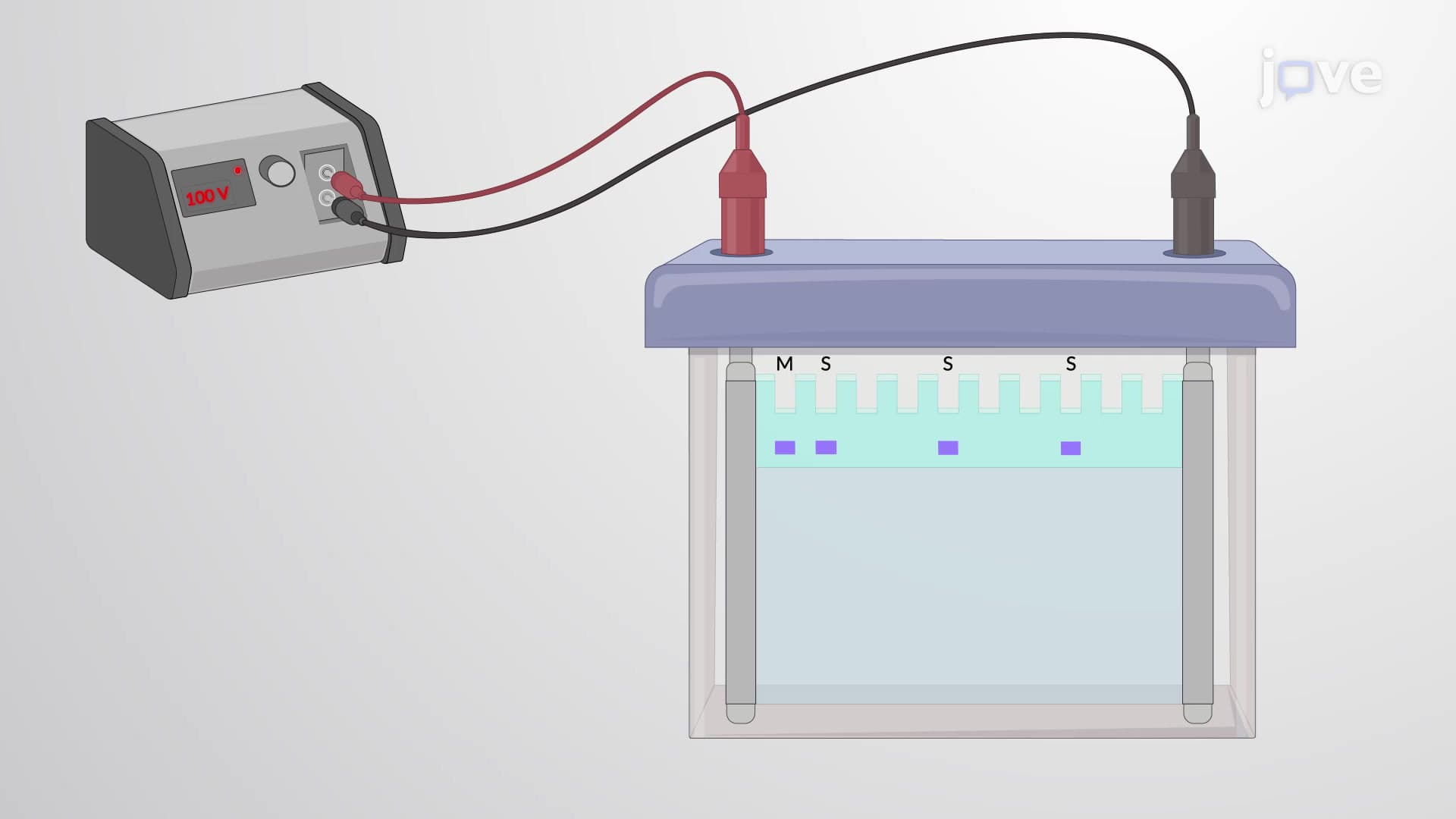
The western blotting process begins with SDS-PAGE (Sodium Dodecyl Sulfate-Polyacrylamide Gel Electrophoresis), where proteins migrate through a gel matrix based on their molecular weight. The SDS detergent denatures proteins and coats them with negative charges, ensuring separation occurs purely by size rather than charge differences. This principle appears frequently on AP Biology exams and college biochemistry courses.
Following separation, proteins transfer from the gel to a nitrocellulose or PVDF membrane through electroblotting. This transfer preserves the spatial arrangement established during electrophoresis, creating a protein "map" on the membrane. The negatively charged proteins migrate toward the positive electrode, effectively printing their positions onto the membrane surface.
The detection phase distinguishes western blotting from simple protein separation. Primary antibodies, designed to recognize specific protein targets, bind to their corresponding antigens on the membrane. These antibodies often come from companies like Cell Signaling Technology or Abcam, which maintain rigorous validation standards.
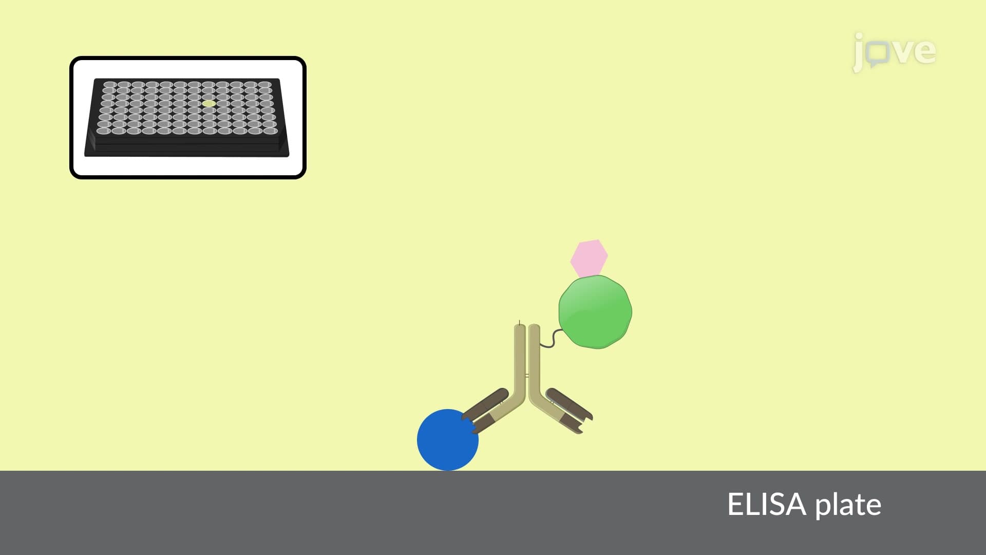
Secondary antibodies, conjugated to detection systems like horseradish peroxidase (HRP) or fluorescent tags, amplify the signal. When exposed to appropriate substrates, HRP produces chemiluminescent signals detectable by imaging systems. This amplification strategy allows researchers to detect even minute quantities of target proteins.
Western blotting plays crucial roles in clinical diagnostics and drug development. The FDA relies on western blotting for HIV confirmation testing, while cancer researchers use it to study tumor suppressor proteins like p53. Pharmaceutical companies employ this technique throughout drug development, from initial target validation to monitoring treatment effects. Students preparing for the MCAT will encounter western blotting concepts in both biochemistry and molecular biology sections.
Related Micro-courses