Loading subject page…

- Cell Biology
- Analyzing Cells and Proteins
- X-ray Diffraction of Biological Samples
Micro-course: Analyzing Cells and Proteins
222,112 views
Learn key concepts and earn your certificate.



X-ray Diffraction of Biological Samples

00:00
01:10
Video Summary: X Ray Diffraction of Biological Samples Explained
Did you know that the famous "Photo 51" X-ray diffraction image taken by Rosalind Franklin at King's College London helped reveal DNA's double helix structure? X ray diffraction of biological samples is a powerful non-destructive technique that reveals the atomic-level architecture of proteins, nucleic acids, and other biomolecules. Today, researchers at institutions like Harvard Medical School use this method to determine protein structures for drug development. Watch the full video on JoVE Coach to master this concept with expert-led visuals and step-by-step explanations.
- Understand how X ray diffraction of biological samples works by analyzing the interference patterns created when X-rays interact with crystalline structures
- Identify the key components of an XRD analyzer including the X-ray source, sample holder, and electronic detector system
- Analyze diffraction patterns to determine three-dimensional molecular structures of proteins and nucleic acids
- Compare single-crystal XRD techniques used in protein crystallography with powder XRD methods for pharmaceutical analysis
- Evaluate the historical significance of X-ray diffraction in discovering DNA's double helix structure
- Apply knowledge of diffraction patterns to interpret concentric ring formations in polycrystalline samples
- Recognize how phase and purity analysis is conducted by comparing experimental data with reference databases
- Explore real-world applications in drug discovery and structural biology research
Understanding X Ray Diffraction of Biological Samples
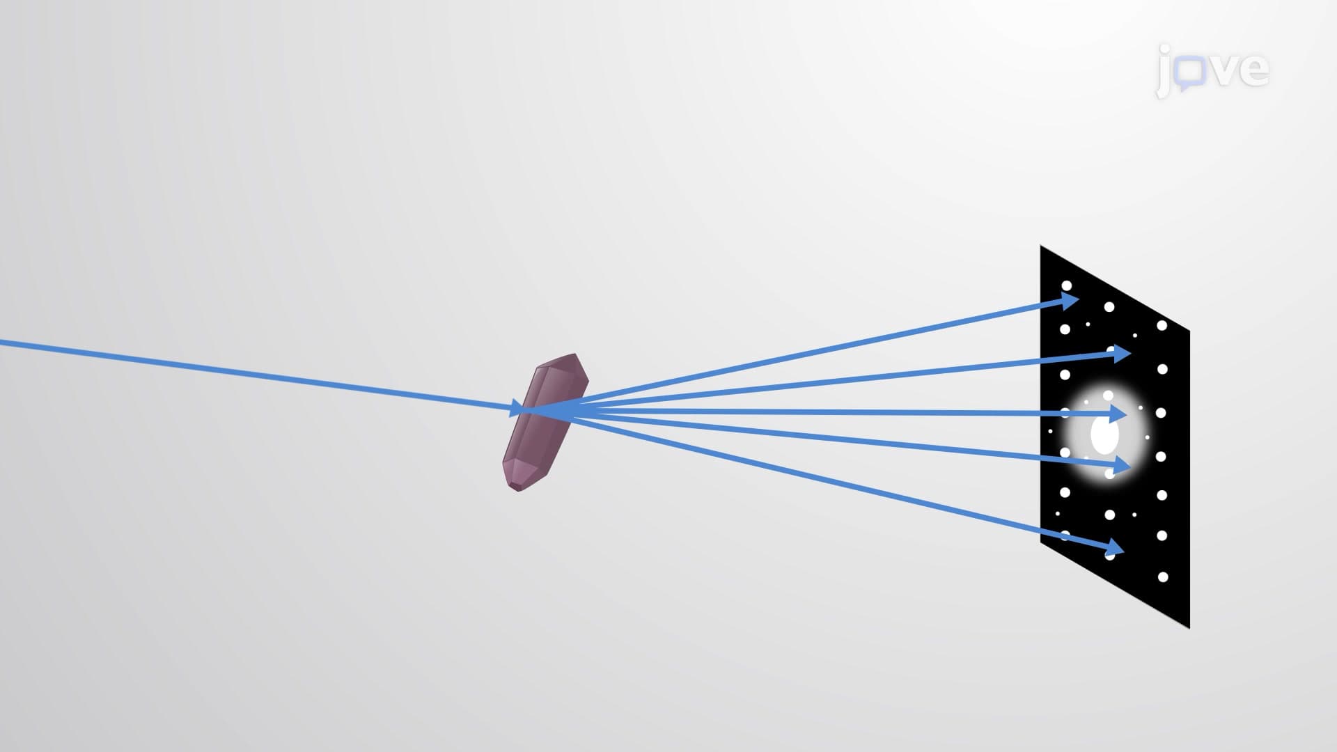
X ray diffraction of biological samples represents one of the most revolutionary analytical techniques in modern science, fundamentally transforming our understanding of molecular biology. When X-rays encounter a crystallized biological sample, they scatter in predictable patterns based on the precise arrangement of atoms within the crystal lattice. This scattering creates an interference pattern that serves as a molecular "fingerprint," revealing structural details invisible to other analytical methods.
The Science Behind Biological X-Ray Diffraction
The technique relies on Bragg's Law, which describes how X-rays reflect off parallel planes of atoms within a crystal. When the path difference between reflected rays equals a whole number of wavelengths, constructive interference occurs, producing bright spots on the detector. For biological samples, this process becomes particularly complex because proteins and nucleic acids contain thousands of atoms arranged in intricate three-dimensional patterns.
At Stanford University's SLAC National Accelerator Laboratory, researchers use synchrotron radiation to generate extremely bright X-ray beams for studying protein structures. The intensity and precision of these beams allow scientists to examine even the smallest biological crystals, revealing details about enzyme active sites and drug binding locations that are crucial for pharmaceutical development.
Applications in Modern Biological Research
Single-crystal XRD has become indispensable in protein crystallography, enabling researchers to determine the exact positions of thousands of atoms within a single protein molecule. This information directly impacts drug design-for example, scientists at Pfizer used XRD data to understand how their COVID-19 antiviral drug Paxlovid binds to viral proteins.
Powder XRD serves different but equally important purposes in biological research. When studying pharmaceutical formulations, researchers can verify the crystalline form of active ingredients, ensuring consistent drug performance. The technique also helps identify polymorphs-different crystalline forms of the same compound that may have vastly different biological activities.
Relevance for Students and Exam Preparation
Understanding x ray diffraction of biological samples tutorial concepts is essential for students preparing for advanced biology and chemistry courses. The MCAT frequently includes questions about protein structure determination methods, while AP Biology exams may test knowledge of how scientific techniques contribute to biological discoveries. College biochemistry courses extensively cover XRD applications in structural biology, making this knowledge fundamental for pre-med and biochemistry majors.
Frequently Asked Questions
X ray diffraction of biological samples is a technique that uses X-ray beams to determine the three-dimensional structure of biological molecules like proteins and DNA. It's crucial because it reveals atomic-level details that help scientists understand how biological molecules function and interact. This knowledge directly contributes to drug development, disease research, and our fundamental understanding of life processes.
The MCAT often includes passages about protein structure determination methods, requiring you to interpret diffraction patterns or understand how XRD contributed to major biological discoveries. You might encounter questions about Bragg's Law applications or how structural biology techniques support drug design. Focus on understanding the relationship between crystal structure and diffraction patterns rather than memorizing complex mathematical formulations.
Absolutely-AP Biology exams may test your knowledge of how scientific methods contributed to major discoveries like DNA structure determination. College biochemistry courses extensively cover XRD in protein structure units. Expect questions about interpreting diffraction data, understanding resolution limits, and connecting structural information to biological function.
Major US pharmaceutical companies like Merck, Johnson & Johnson, and Bristol Myers Squibb use XRD throughout drug development. They determine target protein structures to design better drugs, analyze crystal forms of medications to ensure consistent performance, and verify the purity of drug formulations. For example, determining the structure of viral proteins helped develop COVID-19 treatments.
While the underlying physics involves complex mathematics, you can understand the biological applications without advanced calculus or quantum mechanics. Focus on conceptual understanding-how X-rays interact with crystals, why diffraction patterns form, and how scientists interpret these patterns. Most biology and pre-med courses emphasize applications rather than mathematical derivations.
Create visual aids showing the relationship between molecular structure and diffraction patterns, and practice interpreting real diffraction images from famous discoveries like DNA structure determination. Connect XRD to other structural biology techniques you're learning, and focus on understanding how structural information impacts biological function rather than memorizing technical details.
Explore other structural biology techniques like NMR spectroscopy and cryo-electron microscopy, which complement XRD in determining molecular structures. Study protein folding mechanisms and how structural changes affect biological activity. Understanding these connections will deepen your appreciation for how structure determines function in biological systems.
The basic concepts are very accessible to high school students, especially when presented through historical discoveries and real-world applications. Start with understanding how X-rays interact with matter, then build up to interpreting diffraction patterns. The key is focusing on the biological significance rather than getting overwhelmed by the physics and mathematics behind the technique.
Related Micro-courses

17 Concepts
11 Concepts

17 Concepts