Loading video…


 10 Concepts
 3.6K views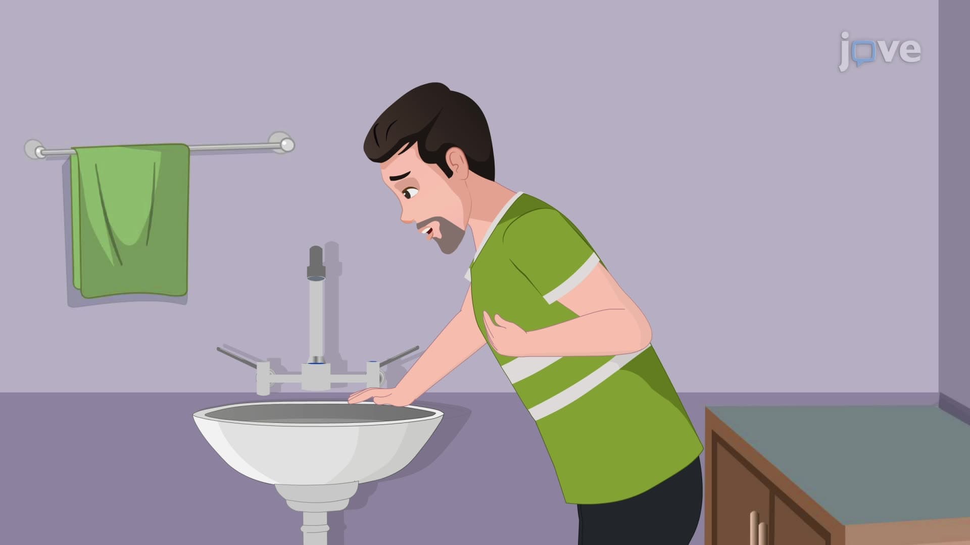

Video Summary: Esophageal Perforation Ii Clinical Manifestations Explained
Did you know that a small tear in your esophagus can trigger life-threatening complications within hours? Esophageal perforation ii clinical manifestations vary dramatically based on where the injury occurs-from neck pain in cervical perforations to severe chest pain in thoracic cases. Consider a patient at Johns Hopkins Emergency Department presenting with sudden chest pain after forceful vomiting, requiring immediate ICU intervention. Esophageal Perforation II Clinical Manifestations Explained demonstrates how location determines symptoms and treatment urgency. Watch the full video on JoVE Coach to master this concept with expert-led visuals and step-by-step explanations.
Esophageal perforations represent one of the most urgent gastrointestinal emergencies encountered in US hospitals, with clinical presentations that vary significantly based on anatomical location. The esophagus spans three distinct regions-cervical, thoracic, and abdominal-each producing unique symptom patterns that guide diagnosis and treatment decisions.
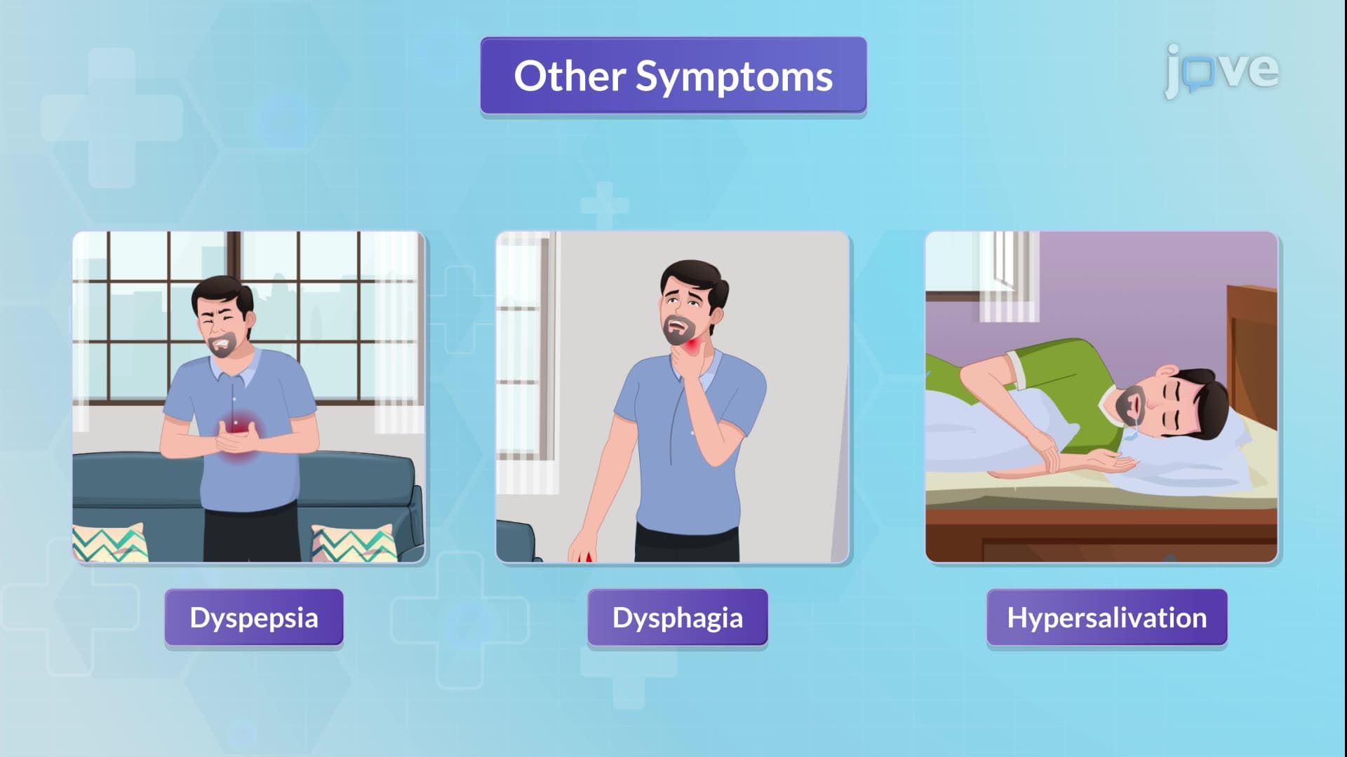
Cervical esophageal perforations typically manifest with neck pain, difficulty swallowing (dysphagia), painful swallowing (odynophagia), and voice changes (dysphonia). These symptoms reflect the proximity to vocal cords and cervical structures. Patients may also develop subcutaneous emphysema, creating a crackling sensation under the skin.
Thoracic perforations present with severe retrosternal chest pain, often mimicking heart attack symptoms. This pain frequently follows episodes of forceful vomiting or retching, a classic scenario seen in Boerhaave syndrome cases at major US trauma centers like Massachusetts General Hospital. The chest pain may radiate to the back and worsen with breathing or movement.
Abdominal esophageal perforations cause epigastric pain that characteristically radiates to the shoulder, reflecting diaphragmatic irritation. This referred pain pattern helps clinicians distinguish esophageal injuries from other abdominal emergencies.
Hemodynamic instability, respiratory distress, or signs of sepsis indicate life-threatening complications requiring immediate intensive care. Emergency departments across the US follow standardized protocols including nothing-by-mouth orders, intravenous fluid resuscitation, broad-spectrum antibiotics, and proton pump inhibitors to reduce gastric acid production.
When perforations extend through all esophageal layers (transmural), patients risk developing acute peritonitis-a surgical emergency. Early recognition prevents progression to septic shock, which carries mortality rates exceeding 30% in US clinical studies.
Treatment selection depends on perforation size, location, and patient stability. Stable patients may benefit from minimally invasive endoscopic interventions like stent placement or clip closure, techniques increasingly used at leading US medical centers. Unstable patients require surgical intervention, potentially including esophageal diversion or resection procedures.
This knowledge proves essential for students preparing for healthcare careers, appearing frequently on MCAT practice exams and undergraduate physiology assessments. Understanding these clinical manifestations builds foundational knowledge for advanced medical coursework and clinical rotations.
Related Micro-courses