Loading subject page…


3,573 views

Video Summary: Esophageal Strictures Ii Clinical Features Explained
Did you know that difficulty swallowing affects nearly 15 million Americans annually, often signaling serious esophageal conditions? Esophageal strictures II clinical features present a progressive pattern where patients initially struggle with solid foods before liquids become problematic. Consider a college athlete who notices increasing difficulty eating pizza or sandwiches at the campus dining hall-this classic progression from solids to liquids exemplifies how Esophageal Strictures II Clinical Features Explained manifests in real patients. Healthcare providers use endoscopy, barium swallow tests, and targeted biopsies to diagnose these narrowed esophageal segments. Watch the full video on JoVE Coach to master this concept with expert-led visuals and step-by-step explanations.
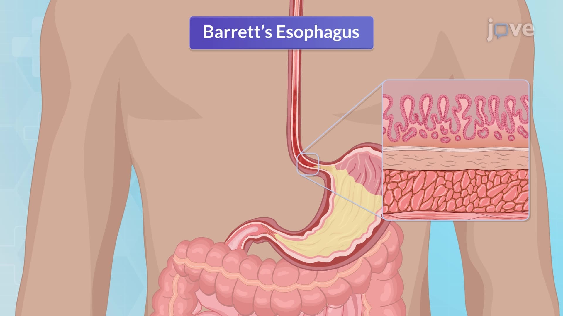
Esophageal strictures represent abnormal narrowing of the esophageal lumen, creating significant challenges for normal swallowing function. The clinical presentation follows a predictable pattern that healthcare providers recognize across major US medical centers, from Johns Hopkins to Mayo Clinic. This systematic approach to clinical features helps distinguish esophageal strictures from other swallowing disorders commonly encountered in emergency departments and gastroenterology clinics.
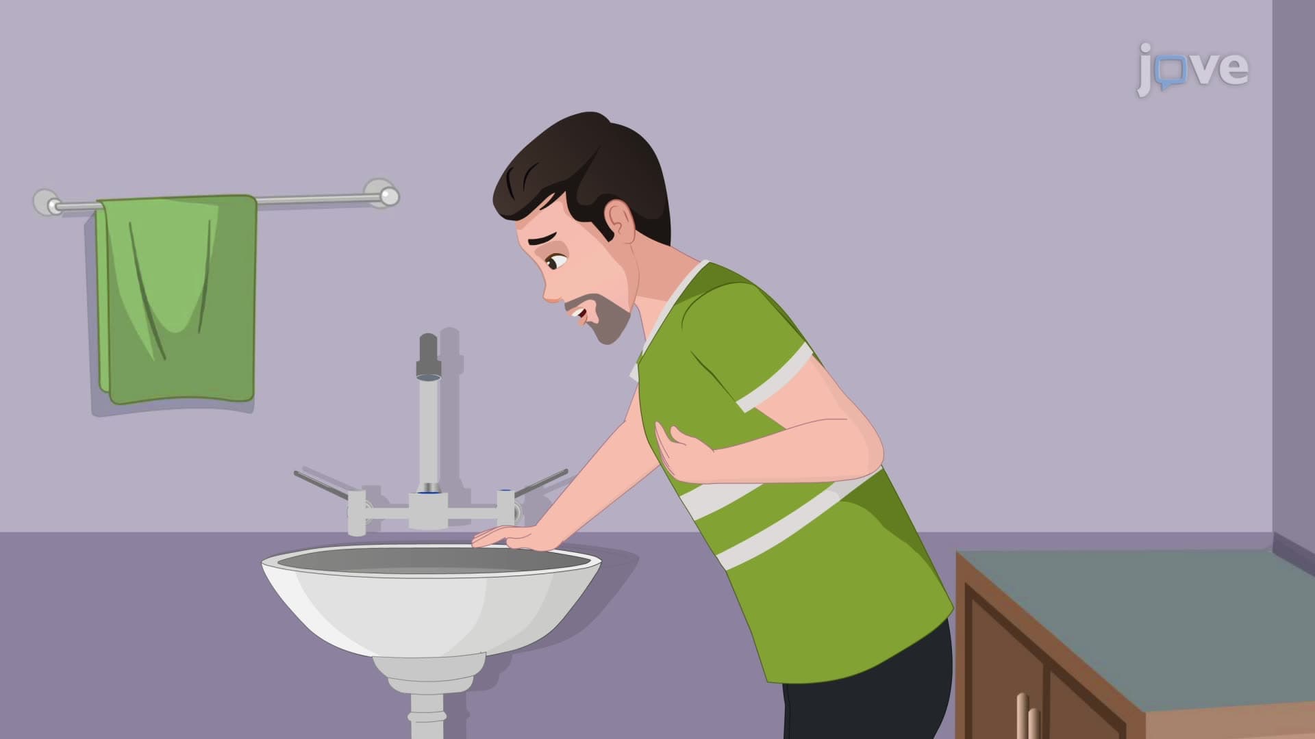
The most characteristic symptom involves progressive difficulty swallowing, beginning with solid foods and eventually affecting liquids. Patients typically describe initial problems with foods like steak or bread rolls, progressing to softer items like pasta or rice. Eventually, even liquids become difficult to swallow. This progression occurs because solids require a wider esophageal diameter than liquids-as strictures worsen, the available space becomes increasingly inadequate. Students preparing for the MCAT or USMLE should recognize this progression pattern as pathognomonic for esophageal strictures.
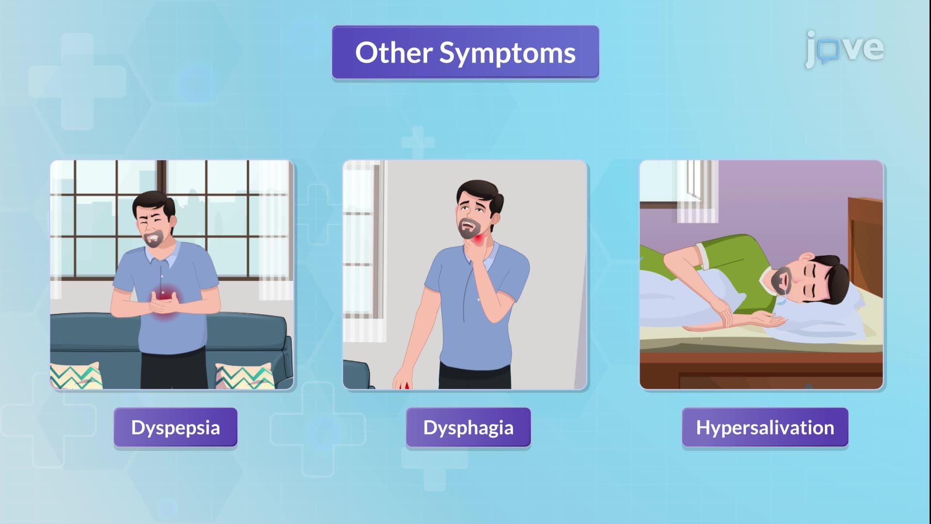
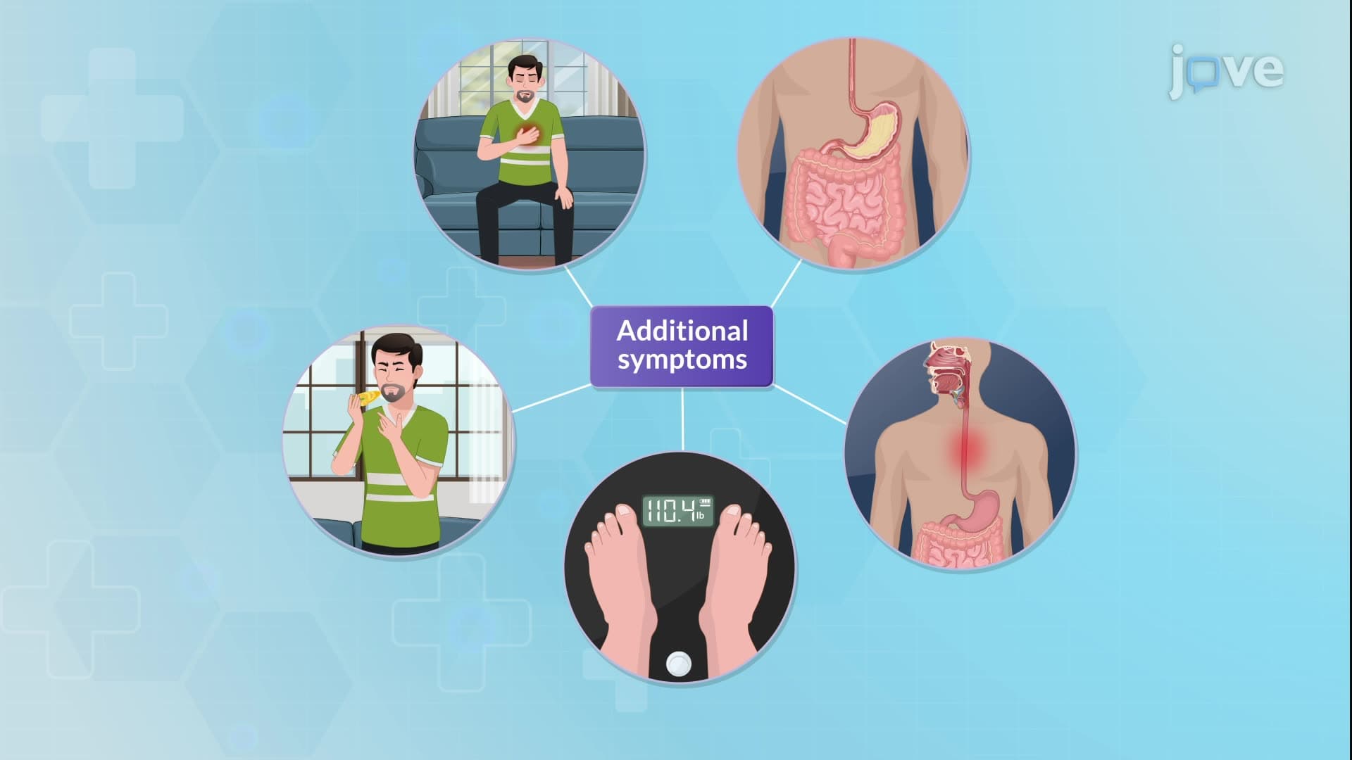
Beyond dysphagia, patients experience chest pain, particularly retrosternal discomfort during swallowing attempts. Regurgitation occurs when food accumulates above the strictured area, unable to pass into the stomach. Heartburn reflects underlying gastroesophageal reflux disease (GERD), often the primary cause of benign strictures. Unintended weight loss results from reduced oral intake, while coughing or choking during meals indicates aspiration risk-a serious complication requiring immediate medical attention.
US medical centers employ systematic diagnostic protocols beginning with comprehensive history and physical examination. Upper endoscopy serves as the gold standard, allowing direct visualization of esophageal narrowing while enabling tissue sampling when malignancy is suspected. Barium swallow studies provide complementary information, particularly useful for detecting subtle strictures or ruling out conditions like hiatal hernias. These imaging studies appear frequently on radiology boards and internal medicine shelf exams, making them essential knowledge for medical students.
Management centers on esophageal dilation using balloon dilators or bougie systems inserted through endoscopes. This procedure, performed in outpatient endoscopy centers nationwide, provides immediate symptom relief in most cases. Malignant strictures may require metallic stent placement to maintain luminal patency, while severe cases unresponsive to dilation may necessitate esophagectomy-major surgery typically reserved for specialized centers like MD Anderson or Memorial Sloan Kettering.
Related Micro-courses