Loading video…


 8 Concepts
 5.7K views


Video Summary: Peptic Ulcer Disease Iii Clinical Explained
Did you know that over 4 million Americans develop peptic ulcers annually, yet many dismiss their symptoms as simple heartburn? Peptic ulcer disease III clinical manifestations vary dramatically based on ulcer location-gastric ulcers cause post-meal burning while duodenal ulcers trigger delayed cramping pain. Emergency departments across major US hospitals like Johns Hopkins regularly treat complications including perforation and bleeding. Peptic Ulcer Disease III Clinical Explained covers diagnostic approaches from endoscopy to H. pylori testing. Watch the full video on JoVE Coach to master this concept with expert-led visuals and step-by-step explanations.
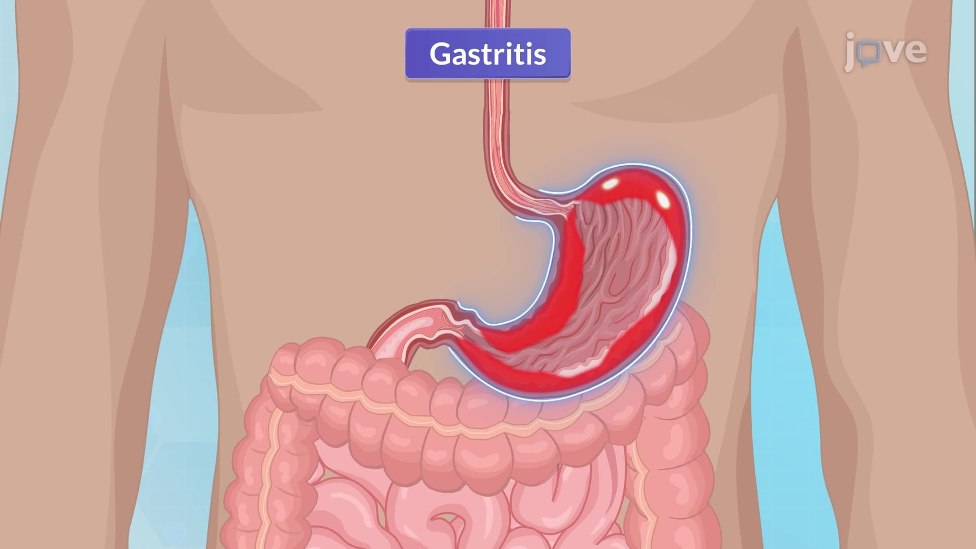
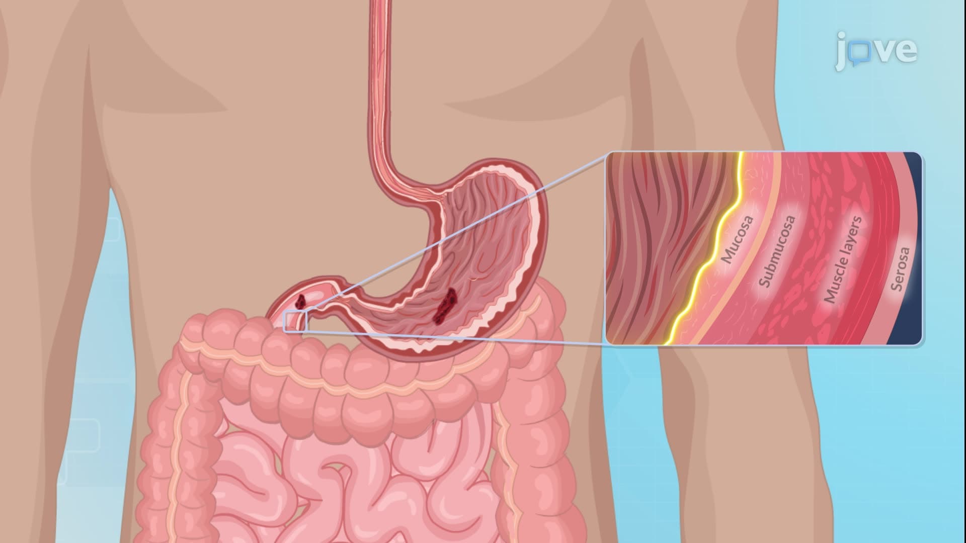
Peptic ulcer disease III clinical presentations follow predictable patterns that help clinicians pinpoint ulcer locations. Gastric ulcers typically cause burning or gaseous epigastric discomfort immediately following meals, as food directly contacts the ulcerated gastric mucosa. This immediate response contrasts sharply with duodenal ulcers, which produce cramping mid-epigastric pain 2-3 hours post-meal when gastric acid reaches the duodenum during the digestive process.
Beyond basic pain patterns, peptic ulcer disease basics encompass numerous gastrointestinal symptoms. Patients frequently experience sour eructation (acid reflux), bloating, and early satiety. Pyrosis (heartburn) occurs as gastric contents reflux into the esophagus. Bowel habits may change, with some patients developing constipation while others experience diarrhea due to altered gastric motility and acid secretion.
Critical complications require immediate recognition. Hematemesis (vomiting blood) and melena (black, tarry stools) indicate active bleeding, common in approximately 15% of peptic ulcer cases treated at major US medical centers like Mayo Clinic and Cleveland Clinic. Gastric outlet obstruction develops when pyloric muscle spasms or mechanical scarring impede gastric emptying, causing projectile vomiting and severe dehydration.
Understanding peptic ulcer disease III clinical diagnosis involves multiple complementary approaches. Upper endoscopy remains the gold standard, allowing direct visualization and tissue sampling for H. pylori testing. Barium contrast studies, though less invasive, effectively identify structural abnormalities and gastric outlet obstruction patterns.
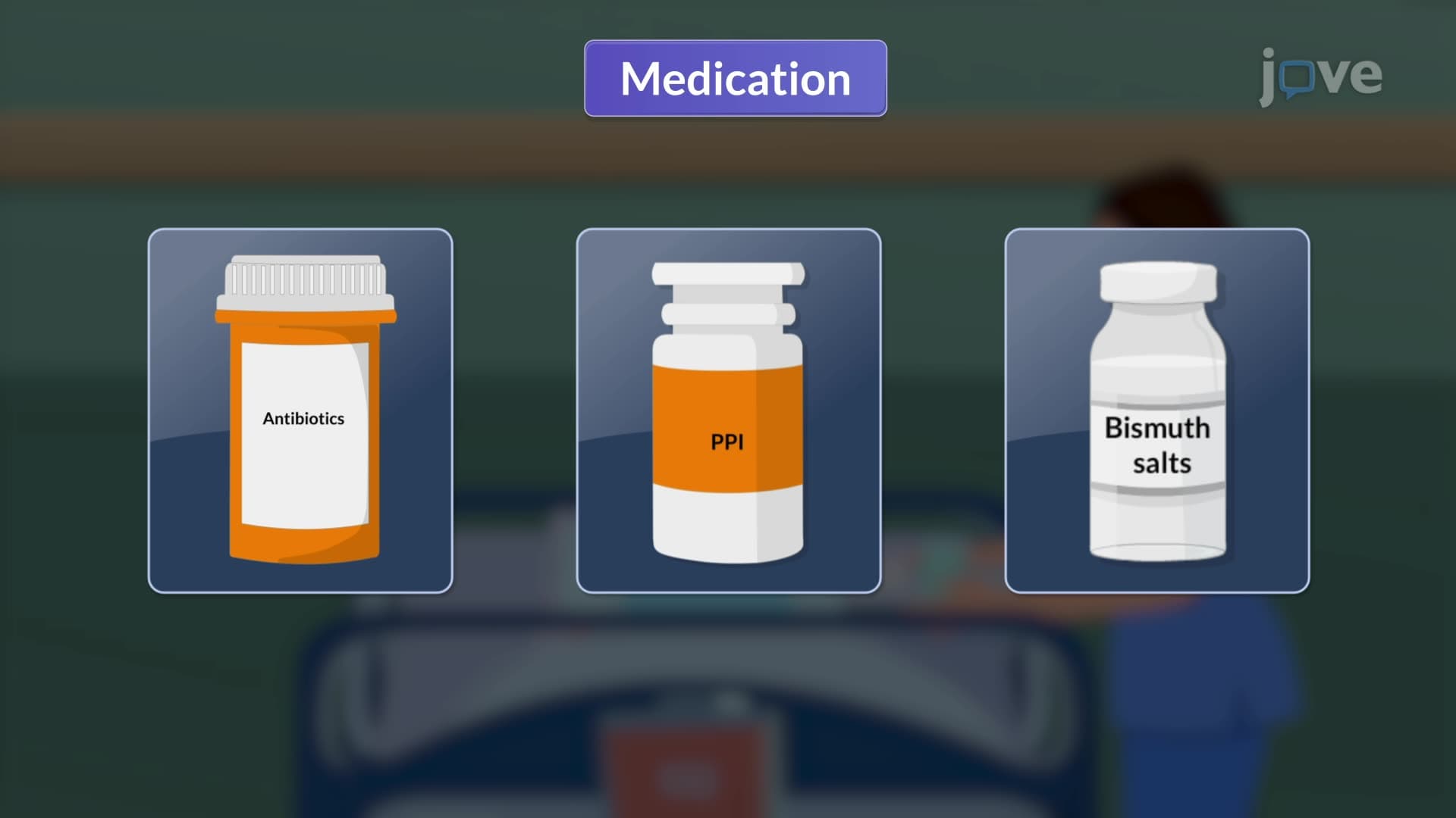
H. pylori detection employs several methods crucial for MCAT and USMLE preparation. Rapid urease testing of antral mucosal biopsies provides immediate results during endoscopy. Non-invasive options include serology for H. pylori antibodies, stool antigen tests, and urea breath tests using carbon-13 or carbon-14 isotopes.
The most dangerous complication-perforation-presents as sudden, severe upper abdominal pain radiating to the shoulder, indicating peritoneal irritation. This surgical emergency affects roughly 5% of ulcer patients and requires immediate intervention at trauma centers nationwide.
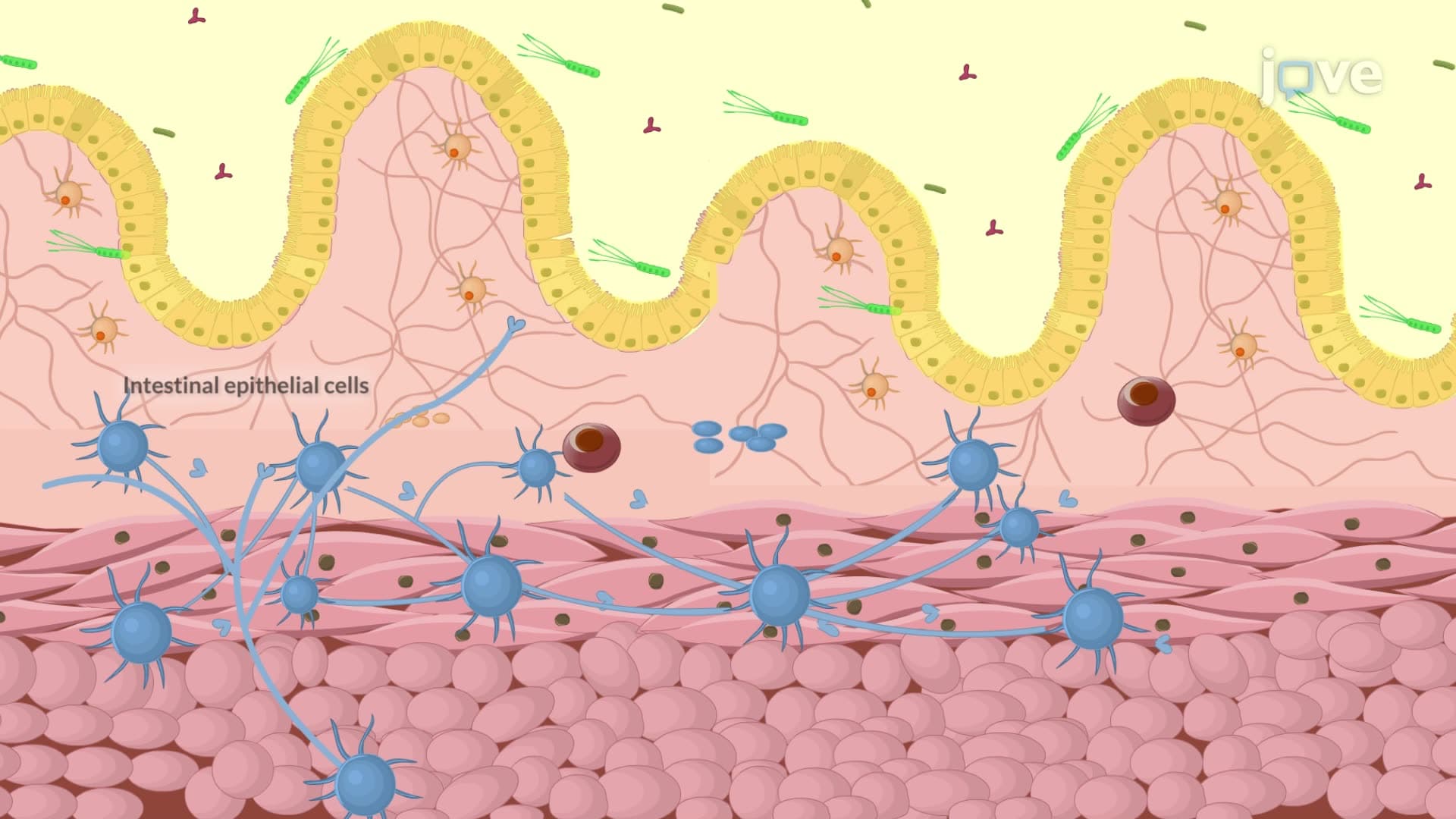
For students preparing for AP Biology or college pathophysiology courses, understanding these clinical patterns reinforces concepts about digestive physiology, acid-base balance, and microbial pathogenesis. Medical students studying for USMLE Step 1 must master the correlation between H. pylori pathophysiology and clinical presentation patterns that distinguish peptic ulcer disease from other gastrointestinal disorders.
Related Micro-courses