Loading video…


 8 Concepts
 5.7K views

Video Summary: Peptic Ulcer Disease V Surgical Explained
Did you know that over 4 million Americans develop peptic ulcers annually, with some requiring life-saving surgical intervention? Peptic ulcer disease v surgical procedures include vagotomy and gastrectomy techniques that can dramatically alter digestive anatomy. At Cleveland Clinic, surgeons perform these complex reconstructions when medications fail to heal severe ulcers. Understanding Peptic Ulcer Disease V Surgical Explained concepts prepares you for advanced healthcare studies. Watch the full video on JoVE Coach to master this concept with expert-led visuals and step-by-step explanations.
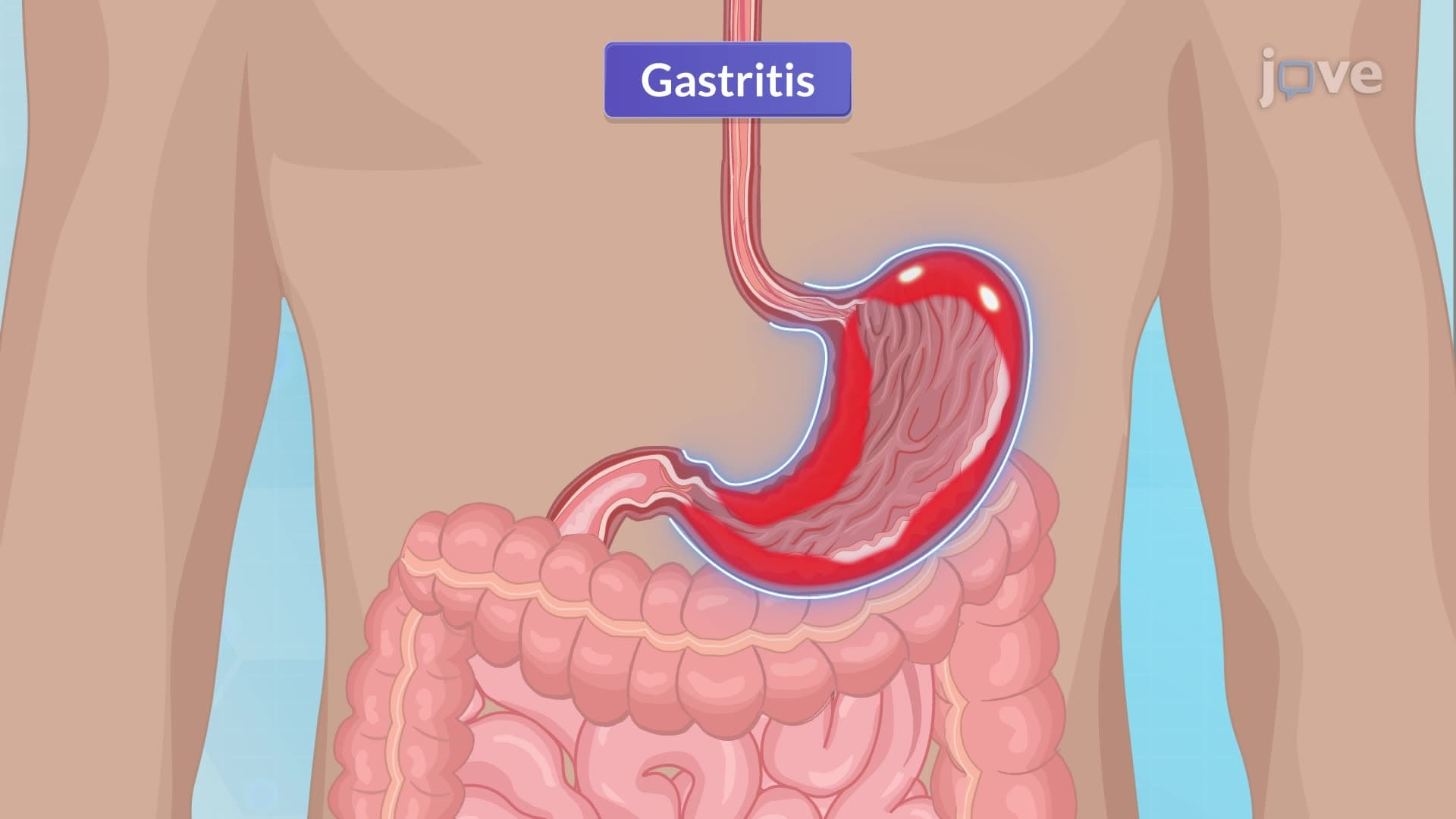
Surgical treatment for peptic ulcer disease represents a critical intervention when conservative medical management fails. These procedures fundamentally alter gastric physiology to reduce acid production and improve digestive function. Understanding these surgical approaches is essential for students pursuing healthcare careers, particularly those preparing for the MCAT, NCLEX, or advanced anatomy courses.
Vagotomy procedures target the vagus nerve's role in gastric acid stimulation. Surgeons can perform truncal vagotomy (cutting the main vagal trunks), selective vagotomy (preserving hepatic and celiac branches), or highly selective vagotomy (affecting only parietal cell innervation). Each approach offers different benefits and complications. For instance, truncal vagotomy requires concurrent pyloroplasty because it disrupts normal pyloric function, while highly selective vagotomy preserves antral motility.
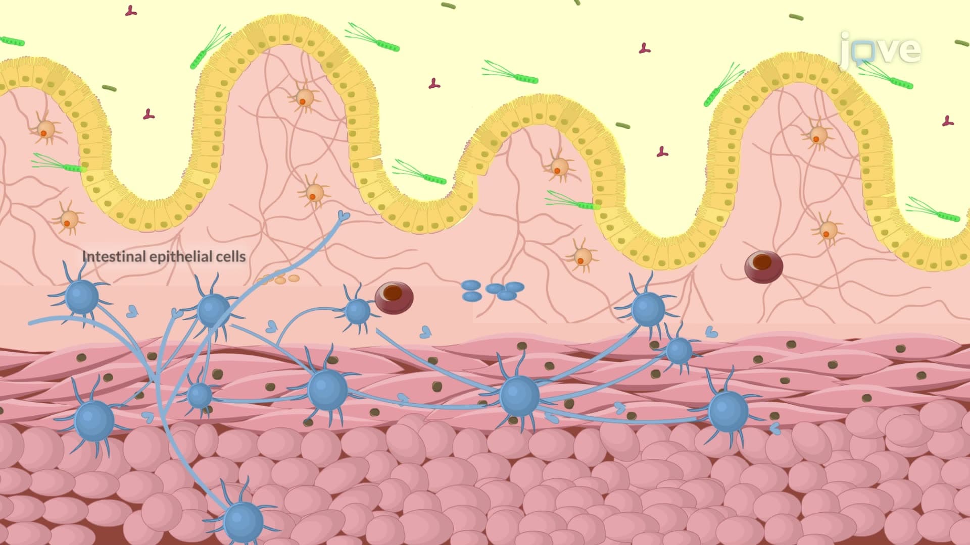
The physiological rationale involves interrupting parasympathetic stimulation of gastric acid secretion. Students studying for AP Biology or college-level physiology courses should understand how acetylcholine normally stimulates parietal cells through muscarinic receptors, and how vagotomy blocks this pathway.
Partial gastrectomy procedures remove diseased stomach portions while maintaining digestive continuity. Antrectomy involves removing the distal third of the stomach, eliminating the primary source of gastrin production. Subtotal gastrectomy extends this resection to include the middle third, reducing overall gastric capacity significantly.
Reconstruction techniques determine postoperative function. Billroth I reconstruction connects the remaining stomach directly to the duodenum, maintaining more physiological food flow. However, Billroth II reconstruction creates a gastrojejunal anastomosis, bypassing the duodenum entirely. Students preparing for medical school should understand how these anatomical changes affect nutrient absorption, particularly fat-soluble vitamins and vitamin B12.
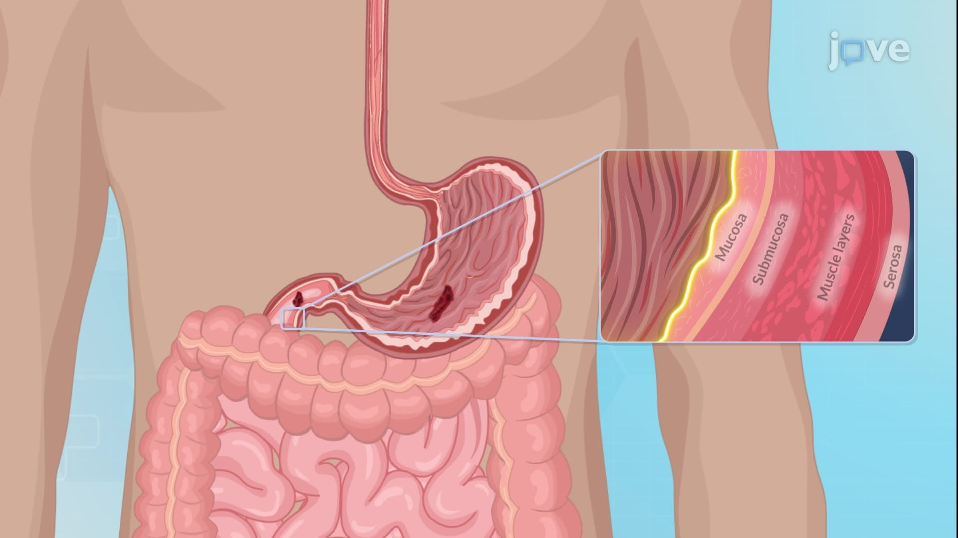
Modern surgical indications include perforation, obstruction, bleeding, and malignancy suspicion. At major medical centers like Johns Hopkins, these procedures require intensive postoperative monitoring. Nursing management involves maintaining NPO status until bowel function returns, managing nasogastric decompression, and monitoring for complications.
Critical complications include anastomotic leaks (presenting with fever, abdominal pain, and elevated white blood cell count), hemorrhage (manifesting as hematemesis or melena), and dumping syndrome (causing postprandial hypotension and diarrhea). Students studying for the NCLEX or HESI A2 should recognize these signs and appropriate interventions.
Related Micro-courses