Loading subject page…


9,805 views


Video Summary: Imaging Studies I Ct and Mri Explained
Did you know that a single CT scan at Johns Hopkins Hospital can capture over 1,000 cross-sectional images in just 30 seconds? Imaging studies I CT and MRI represent two revolutionary diagnostic technologies that allow doctors to see inside the human body without making a single incision. CT scans use rotating X-ray beams to create detailed bone and organ images, while MRI employs powerful magnetic fields to produce superior soft tissue visualization. Watch the full video on JoVE Coach to master this concept with expert-led visuals and step-by-step explanations.
Modern diagnostic imaging transformed medicine by allowing healthcare providers to visualize internal body structures without invasive procedures. Two cornerstone technologies-Computed Tomography (CT) and Magnetic Resonance Imaging (MRI)-each offer unique advantages for different clinical scenarios, making them essential tools in hospitals from Mayo Clinic to UCLA Medical Center.
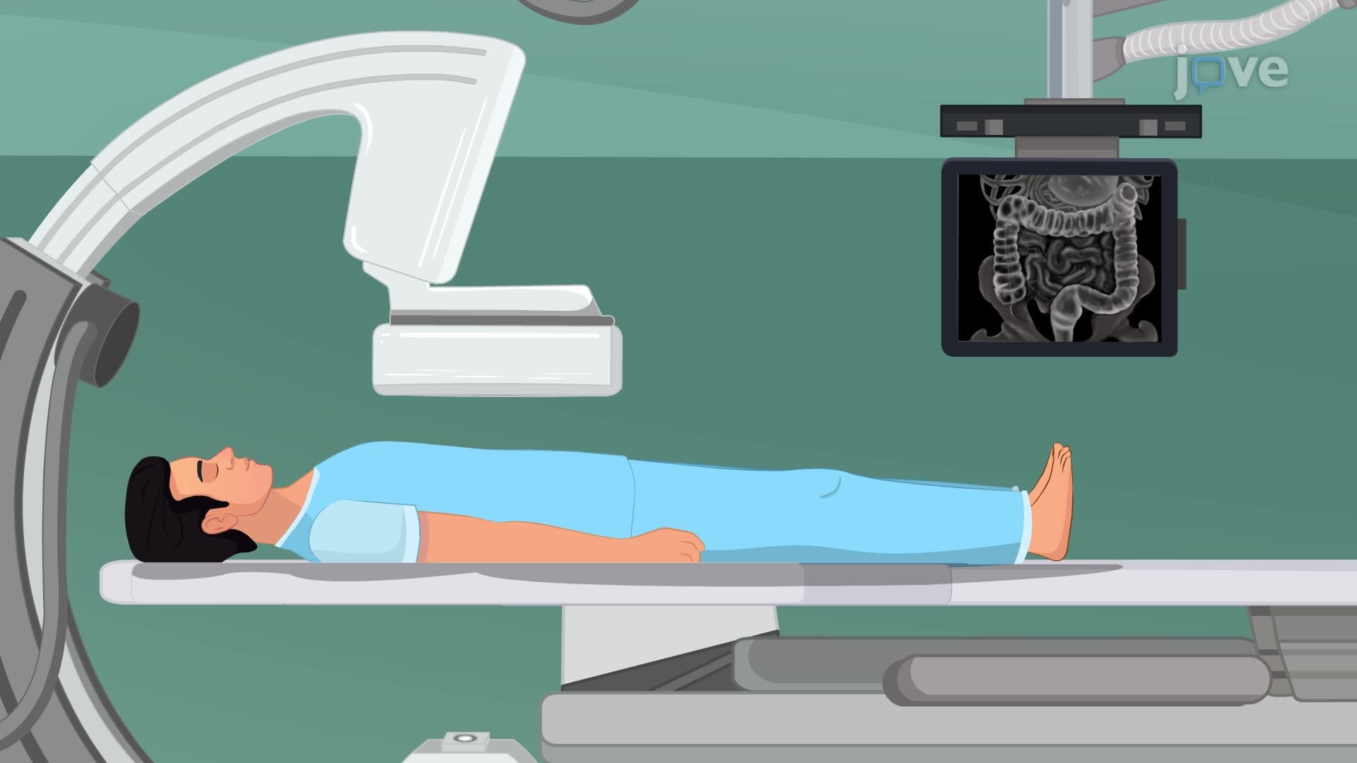
CT scans utilize sophisticated X-ray technology to generate cross-sectional images with remarkable detail. The patient lies on a motorized table that moves through a donut-shaped gantry containing an X-ray tube and detectors. As the X-ray source rotates 360 degrees around the patient, it emits radiation from multiple angles while detectors capture the transmitted X-rays. Advanced computer algorithms process this data to reconstruct detailed cross-sectional images, similar to looking at individual slices of bread in a loaf.
CT imaging excels in emergency medicine scenarios, particularly for trauma patients at centers like Cleveland Clinic's emergency department. The technology rapidly identifies bone fractures, internal bleeding, and organ damage. CT scans also prove invaluable for detecting lung diseases, abdominal conditions, and certain cancers. The entire scanning process typically takes 10-30 minutes, making it ideal for urgent diagnostic needs.
MRI technology operates on completely different principles, using powerful magnetic fields (typically 1.5 to 3 Tesla) and radiofrequency pulses instead of ionizing radiation. When patients enter the MRI scanner's magnetic field, hydrogen atoms in their body tissues align with the magnetic field. Radiofrequency pulses then disrupt this alignment, and as atoms return to their original state, they emit signals that specialized receivers detect and computers process into detailed images.
MRI's greatest strength lies in soft tissue differentiation. Neurologists at institutions like Stanford Medicine rely on MRI for detailed brain imaging, while orthopedic surgeons use it to evaluate joint structures, cartilage, and ligaments that CT cannot adequately visualize. MRI scans take longer than CT (30-90 minutes) but provide unparalleled detail for non-urgent diagnostic questions.
Understanding when to select CT versus MRI appears frequently on medical exams, including MCAT passages and AP Biology assessments. Students should remember that CT provides rapid imaging for emergencies and excellent bone detail, while MRI offers superior soft tissue contrast without radiation exposure. Both technologies represent fundamental concepts in medical imaging that prepare students for advanced healthcare careers and appear in college anatomy, physiology, and medical technology coursework.
Related Micro-courses