Loading subject page…


9,805 views

Video Summary: Imaging Studies Iii Gastrointestinal Motility Explained
Did you know that doctors can track your breakfast moving through your digestive system using radioactive tracers? Imaging studies III gastrointestinal motility techniques allow physicians at major medical centers like Mayo Clinic to diagnose conditions such as gastroparesis by watching how food travels through your stomach and intestines. These sophisticated diagnostic methods combine nuclear medicine with CT imaging to evaluate digestive function non-invasively. Watch the full video on JoVE Coach to master this concept with expert-led visuals and step-by-step explanations.
Imaging studies III gastrointestinal motility represents a sophisticated branch of nuclear medicine that evaluates how effectively the digestive system moves food and waste through the body. These diagnostic techniques have revolutionized gastroenterology by providing quantitative measurements of digestive function without requiring invasive procedures. For students preparing for the MCAT or advanced placement biology exams, understanding these concepts demonstrates the practical application of physics principles in medical diagnostics.
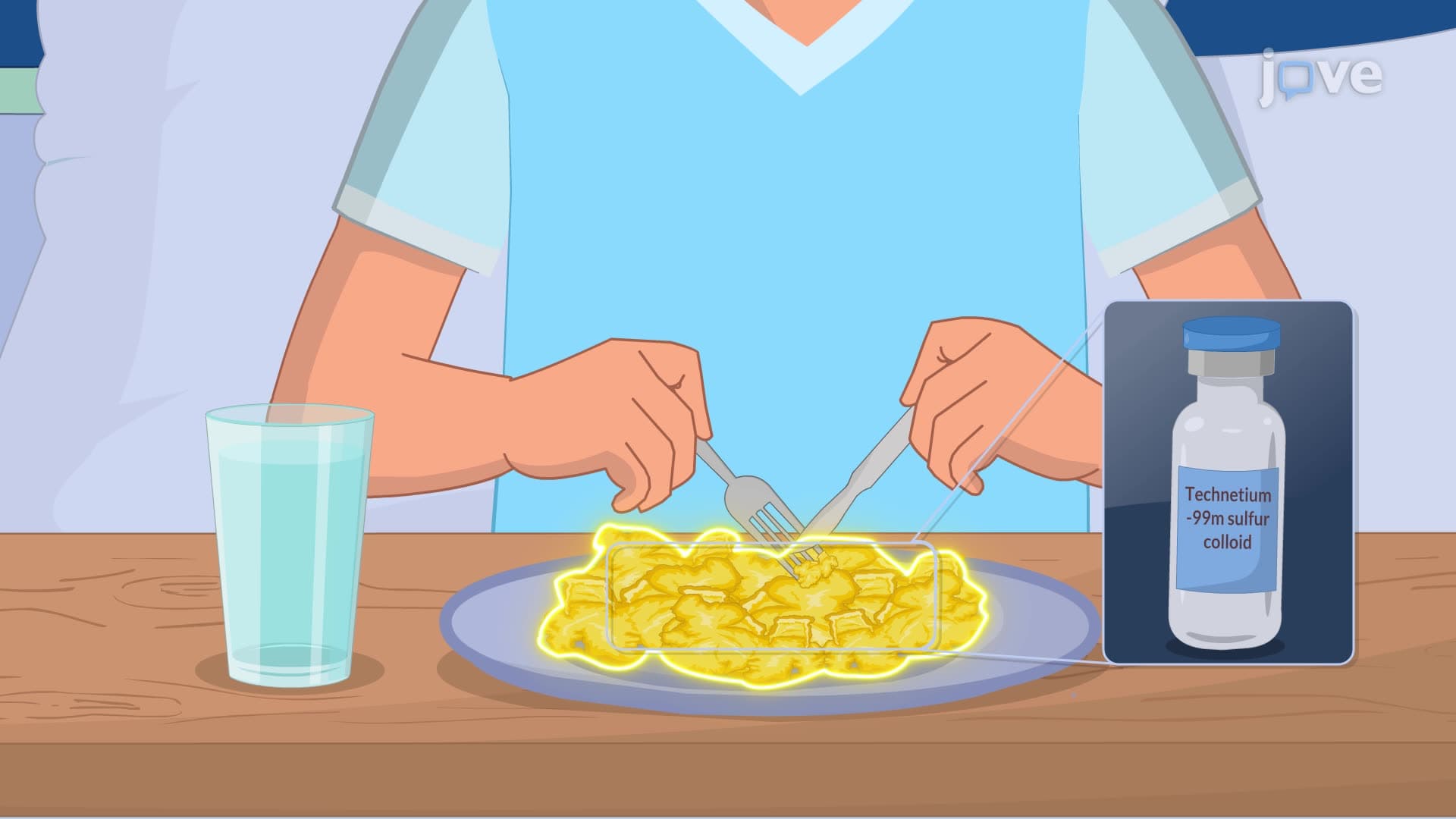
Gastric emptying studies exemplify the intersection of nuclear physics and clinical medicine. Patients consume a standardized meal containing technetium-99m sulfur colloid, a radioactive tracer with a half-life of 6.02 hours. The gamma rays emitted by this isotope are detected by scintillation cameras positioned over the patient's abdomen. Johns Hopkins Hospital and Cleveland Clinic routinely perform these studies to diagnose gastroparesis, a condition affecting approximately 4% of the US population, particularly diabetic patients.
The procedure typically involves imaging at 0, 1, 2, and 4 hours post-ingestion. Normal gastric emptying shows 90% of the meal leaving the stomach within 4 hours. Delayed emptying suggests gastroparesis, while accelerated emptying may indicate dumping syndrome. College-level physiology courses emphasize how this technique quantifies the complex neural and hormonal regulation of gastric motility.
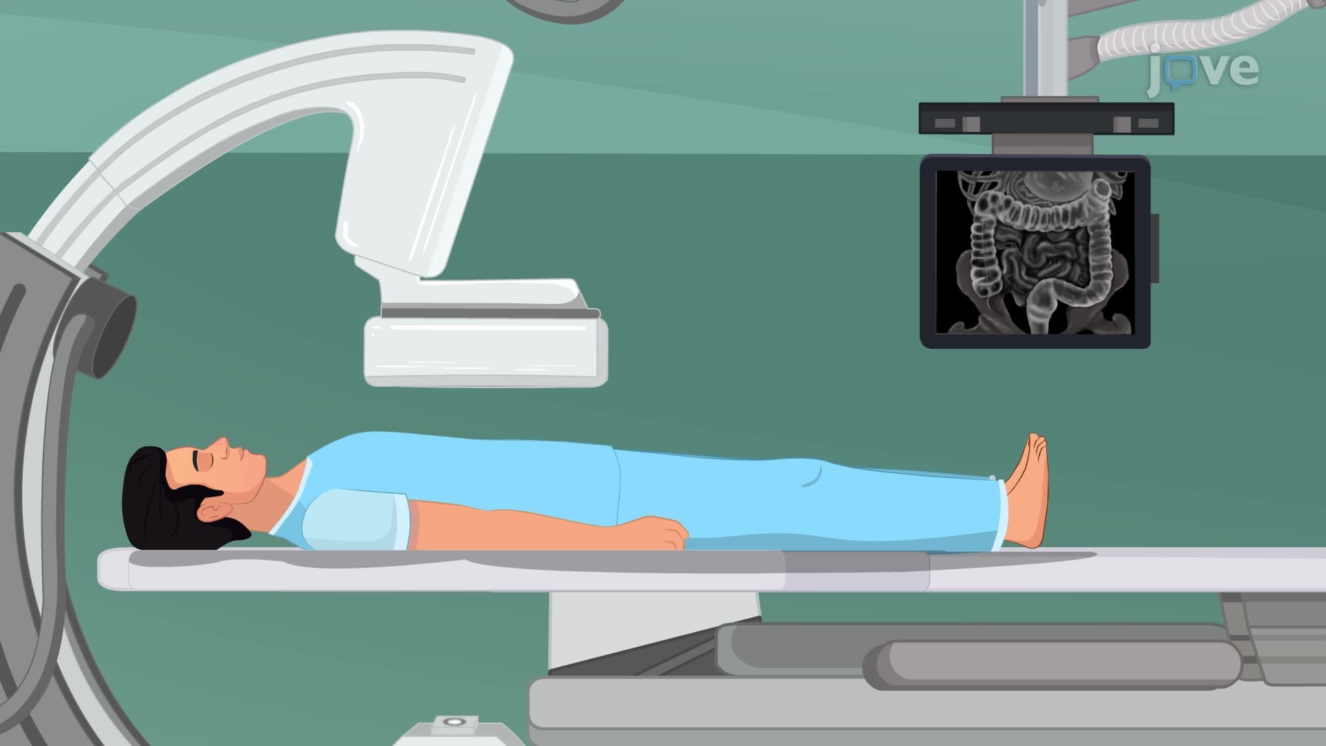
Colonic transit studies utilize radiopaque markers to evaluate large bowel function over several days. Patients ingest capsules containing 20 small markers visible on standard X-rays. Normal colonic transit sees all markers eliminated within 5 days, with specific regional timing patterns. The ascending colon typically clears within 24-48 hours, while the rectosigmoid region may retain markers for 72-96 hours in healthy individuals.
This technique proves invaluable for diagnosing functional constipation, differentiating slow-transit constipation from outlet obstruction disorders. Major academic medical centers like Stanford Medicine use these studies to guide treatment decisions, distinguishing patients who benefit from dietary modifications versus those requiring surgical intervention.
Virtual colonoscopy represents the convergence of advanced CT imaging with sophisticated computer processing. This technique generates three-dimensional reconstructions of the colon interior, allowing radiologists to navigate virtually through the bowel. The American College of Radiology recognizes virtual colonoscopy as an acceptable screening method for colorectal cancer, particularly beneficial for patients unable to undergo traditional colonoscopy due to anticoagulation therapy or anatomical constraints.
Related Micro-courses