Loading video…


 17 Concepts
 9.8K views


Video Summary: Ultrasound Ii Endoscopic Ultrasound and Fibroscan Explained
Did you know that doctors can examine your liver's health through your skin or look inside your digestive system using sound waves? Ultrasound II Endoscopic Ultrasound And Fibroscan represents two revolutionary diagnostic techniques that help physicians at leading medical centers like Mayo Clinic detect pancreatic cancer, liver cirrhosis, and gastrointestinal disorders without major surgery. These minimally invasive procedures combine traditional ultrasound technology with specialized equipment to provide detailed internal imaging. Watch the full video on JoVE Coach to master this concept with expert-led visuals and step-by-step explanations.
Modern diagnostic medicine relies heavily on non-invasive imaging techniques that provide detailed internal views without surgical intervention. Ultrasound II Endoscopic Ultrasound And Fibroscan encompasses two sophisticated ultrasound applications that have transformed how physicians diagnose digestive and liver disorders. These technologies demonstrate practical applications of wave physics principles students encounter in AP Physics courses, where sound wave frequency, reflection, and tissue penetration become critical factors in medical diagnostics.
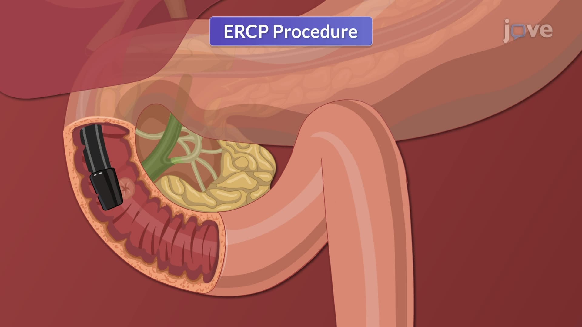
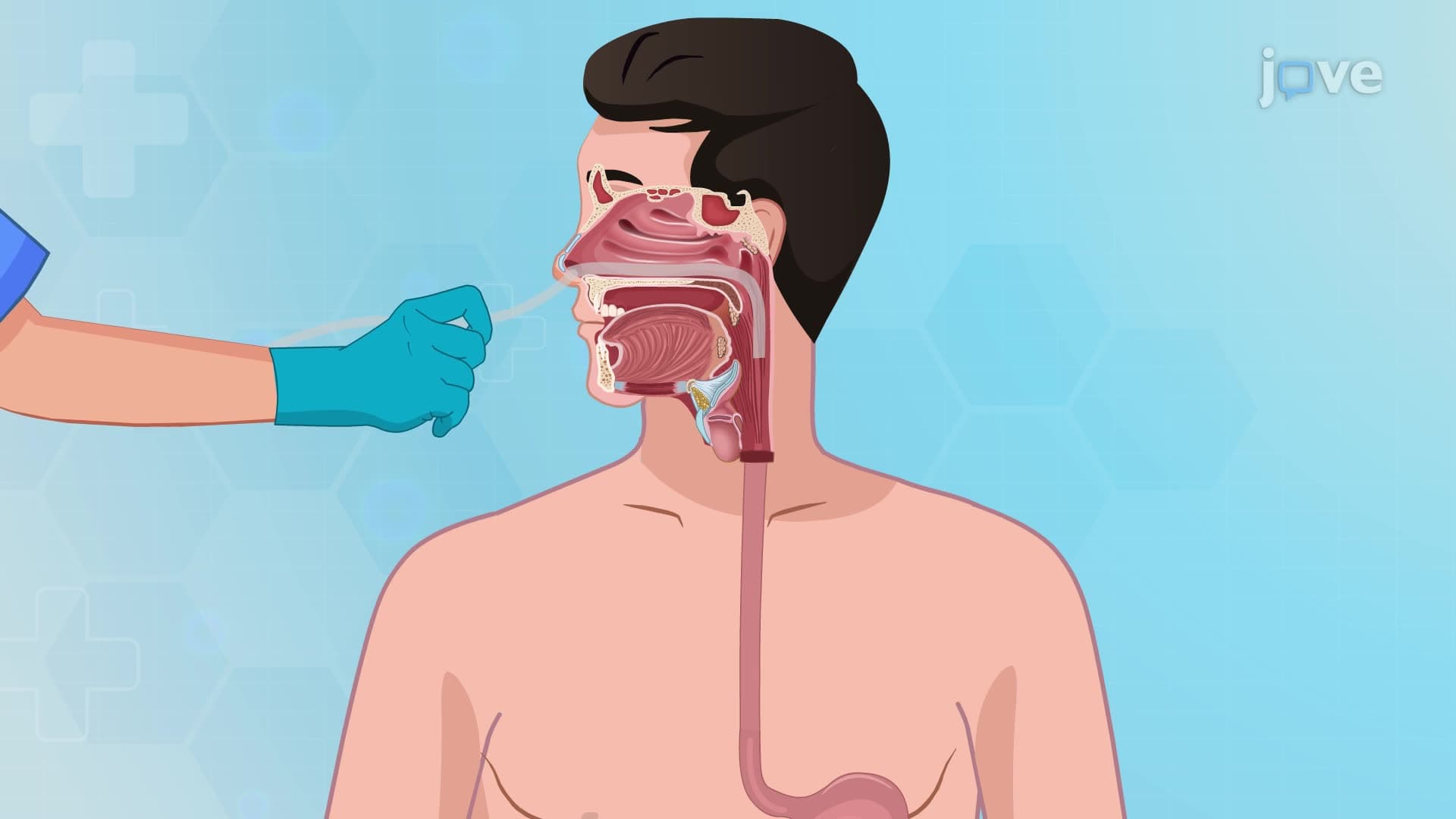
Endoscopic ultrasound (EUS) represents a breakthrough in gastrointestinal diagnostics by combining flexible endoscopy with high-frequency ultrasound imaging. The echoendoscope, a specialized instrument used at major US medical centers like Johns Hopkins and Cleveland Clinic, contains both a camera and ultrasound transducer. This dual-functionality allows physicians to visualize the digestive tract's interior walls while simultaneously examining surrounding tissues and organs. The procedure involves two primary approaches: transoral insertion through the mouth to examine upper gastrointestinal structures, and transrectal insertion to evaluate lower digestive tract abnormalities. Students preparing for the MCAT will recognize this as an excellent example of how physics principles apply directly to medical practice.
FibroScan technology utilizes controlled attenuation parameter (CAP) measurements and liver stiffness evaluation to assess hepatic health without tissue sampling. This technique has become standard practice at US hepatology centers for monitoring patients with chronic liver diseases. The procedure requires specific patient positioning-supine with right arm abducted-allowing optimal access to the liver through the intercostal spaces. Low-frequency ultrasound waves penetrate liver tissue, with wave velocity and attenuation patterns indicating fibrosis severity and fatty infiltration levels. This application demonstrates wave physics concepts that appear frequently on standardized exams, including wave speed calculations and tissue interaction principles.
Both technologies offer significant advantages over traditional diagnostic methods. Endoscopic ultrasound enables early detection of pancreatic adenocarcinoma, the fourth-leading cause of cancer deaths in the United States, with detection rates significantly higher than conventional imaging. FibroScan provides quantitative liver assessment crucial for managing the estimated 4.5 million Americans with chronic liver disease. These procedures exemplify evidence-based medicine principles emphasized in pre-medical curricula, demonstrating how technological advancement improves patient outcomes while reducing procedural risks and healthcare costs.
Related Micro-courses