Loading subject page…

- Medical Surgical Nursing
- Diagnostic and Radiological Studies of the Gi System
- Upper GI Series: Barium Swallow
Micro-course: Diagnostic and Radiological Studies of the GI System
9,805 views
Learn key concepts and earn your certificate.









Upper GI Series: Barium Swallow


00:00
01:24
Video Summary: Upper Gi Series Barium Swallow Explained
Ever wondered how doctors can see inside your digestive system without surgery? An upper GI series barium swallow uses a special contrast drink to create detailed X-ray images of your esophagus, stomach, and small intestine. At major medical centers like Mayo Clinic, this procedure helps diagnose conditions ranging from acid reflux to stomach ulcers in thousands of patients annually. Understanding the Upper GI Series Barium Swallow Explained process is essential for healthcare students and anyone preparing for medical imaging procedures. Watch the full video on JoVE Coach to master this concept with expert-led visuals and step-by-step explanations.
- Understand the complete Upper GI Series Barium Swallow Explained procedure from patient preparation through post-procedure care
- Identify the anatomical structures visualized during the examination, including esophagus, stomach, and duodenum
- Analyze how barium sulfate contrast enhances X-ray visibility of soft tissue structures in the digestive tract
- Evaluate appropriate patient preparation protocols, including NPO status and pre-procedure instructions
- Apply knowledge of fluoroscopy technology to understand real-time imaging during the procedure
- Recognize common diagnostic findings such as esophageal strictures, hiatal hernias, and gastric abnormalities
- Compare barium swallow procedures with other upper GI diagnostic methods like endoscopy
- Explore post-procedure patient education requirements and potential side effects
Understanding the Upper GI Series Barium Swallow Procedure
The upper GI series barium swallow represents a cornerstone diagnostic tool in gastroenterology and radiology. This imaging procedure provides detailed visualization of the upper gastrointestinal tract's structure and function, making it invaluable for diagnosing various digestive disorders. Unlike CT scans or MRIs, this procedure specifically focuses on the dynamic movement of contrast material through the digestive system.
Clinical Applications and Diagnostic Capabilities
Healthcare providers utilize the how upper gi series barium swallow works principle to diagnose numerous conditions affecting the upper digestive tract. Common applications include detecting esophageal motility disorders like achalasia, identifying structural abnormalities such as strictures or tumors, and evaluating gastroesophageal reflux disease (GERD). At institutions like Cleveland Clinic and Johns Hopkins, radiologists interpret thousands of these studies annually to guide treatment decisions.
The procedure's diagnostic power lies in its ability to reveal both anatomical and functional abnormalities. While static X-rays show structure, fluoroscopy during barium transit reveals how effectively the esophagus propels food toward the stomach and identifies areas of abnormal narrowing or dilation.
Technical Methodology and Patient Experience
The upper gi series barium swallow tutorial process begins with careful patient preparation. The eight-hour fasting period ensures optimal imaging quality by eliminating food particles that could obscure findings. Patients consume approximately 16-20 ounces of barium sulfate suspension, which tastes chalky but remains generally well-tolerated.
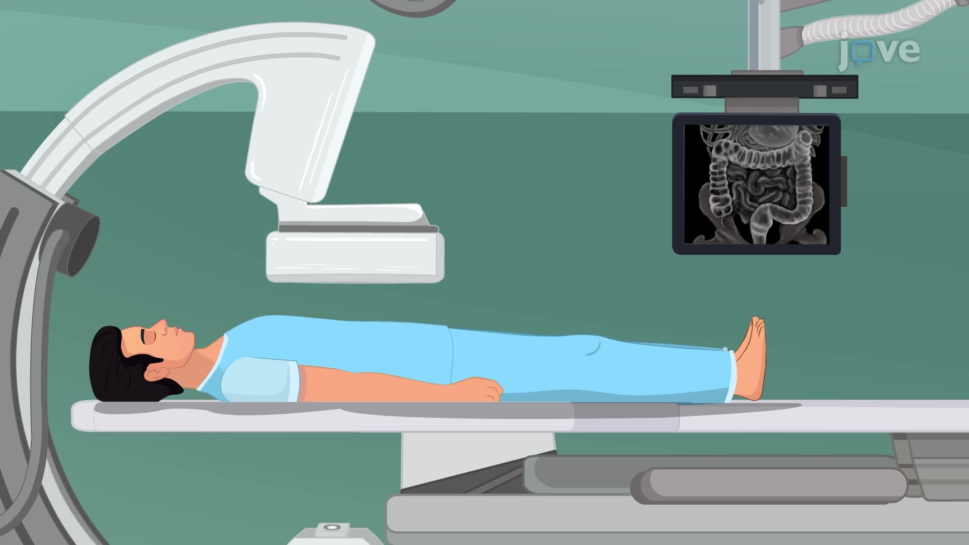
During fluoroscopy, patients assume various positions-upright, lateral, and prone-allowing comprehensive visualization of all upper GI structures. The radiologist observes barium flow in real-time, capturing images at specific intervals to document normal and abnormal findings.
Educational Relevance and Exam Preparation
For students preparing for healthcare careers, understanding the upper gi series barium swallow overview proves essential for various standardized examinations. The MCAT includes questions about diagnostic imaging principles, while nursing students encounter this topic on NCLEX and HESI A2 examinations. Pre-med students should understand contrast mechanisms, radiation safety principles, and clinical correlations.
This procedure exemplifies fundamental concepts in medical physics, particularly X-ray attenuation and contrast enhancement. Barium's high atomic number creates excellent radiopacity, making it ideal for outlining hollow organs that would otherwise remain invisible on plain radiographs.
Frequently Asked Questions
An upper GI series barium swallow is a diagnostic X-ray examination using barium contrast to visualize the esophagus, stomach, and small intestine. It's performed to diagnose swallowing disorders, ulcers, tumors, hernias, and other structural or functional problems in the upper digestive tract. The procedure provides detailed images that help doctors determine the best treatment approach for digestive symptoms.
MCAT questions often focus on the physics principles behind contrast imaging and diagnostic reasoning. You might encounter scenarios asking why barium enhances X-ray visibility or questions about radiation safety during fluoroscopy. Understanding the procedure's clinical applications and contraindications is essential for success on medical school examinations.
Yes, nursing entrance exams frequently include questions about diagnostic procedures and patient care protocols. Expect questions about pre-procedure preparation, patient education, and post-procedure monitoring. Understanding NPO requirements and recognizing potential complications demonstrates essential nursing knowledge for these standardized tests.
Major medical centers like Massachusetts General Hospital and UCLA Medical Center perform hundreds of these procedures monthly. Emergency departments use them to evaluate suspected esophageal perforations, while gastroenterology clinics employ them for routine GERD evaluation. The procedure remains cost-effective compared to more advanced imaging, making it accessible in community hospitals nationwide.
No advanced chemistry background is required to grasp the basic principles. Understanding that barium has a high atomic number, making it radiopaque (blocks X-rays), provides sufficient foundation. High school chemistry knowledge about atomic structure and periodic table trends adequately prepares students for learning about contrast mechanisms in medical imaging.
Create visual diagrams showing barium's path through the digestive system and practice identifying normal versus abnormal findings on sample images. Focus on correlating anatomical structures with their radiographic appearance. Review patient preparation protocols and post-procedure care, as these topics frequently appear on practical examinations and clinical scenarios.
Absolutely! Understanding barium swallows provides an excellent foundation for learning about lower GI series (barium enema), CT enterography, and endoscopic procedures. These related topics build upon the same contrast principles while expanding your knowledge of comprehensive gastrointestinal diagnostics. This progression prepares you well for advanced healthcare courses and clinical rotations.
Related Micro-courses

15 Concepts