Loading video…


 17 Concepts
 9.8K views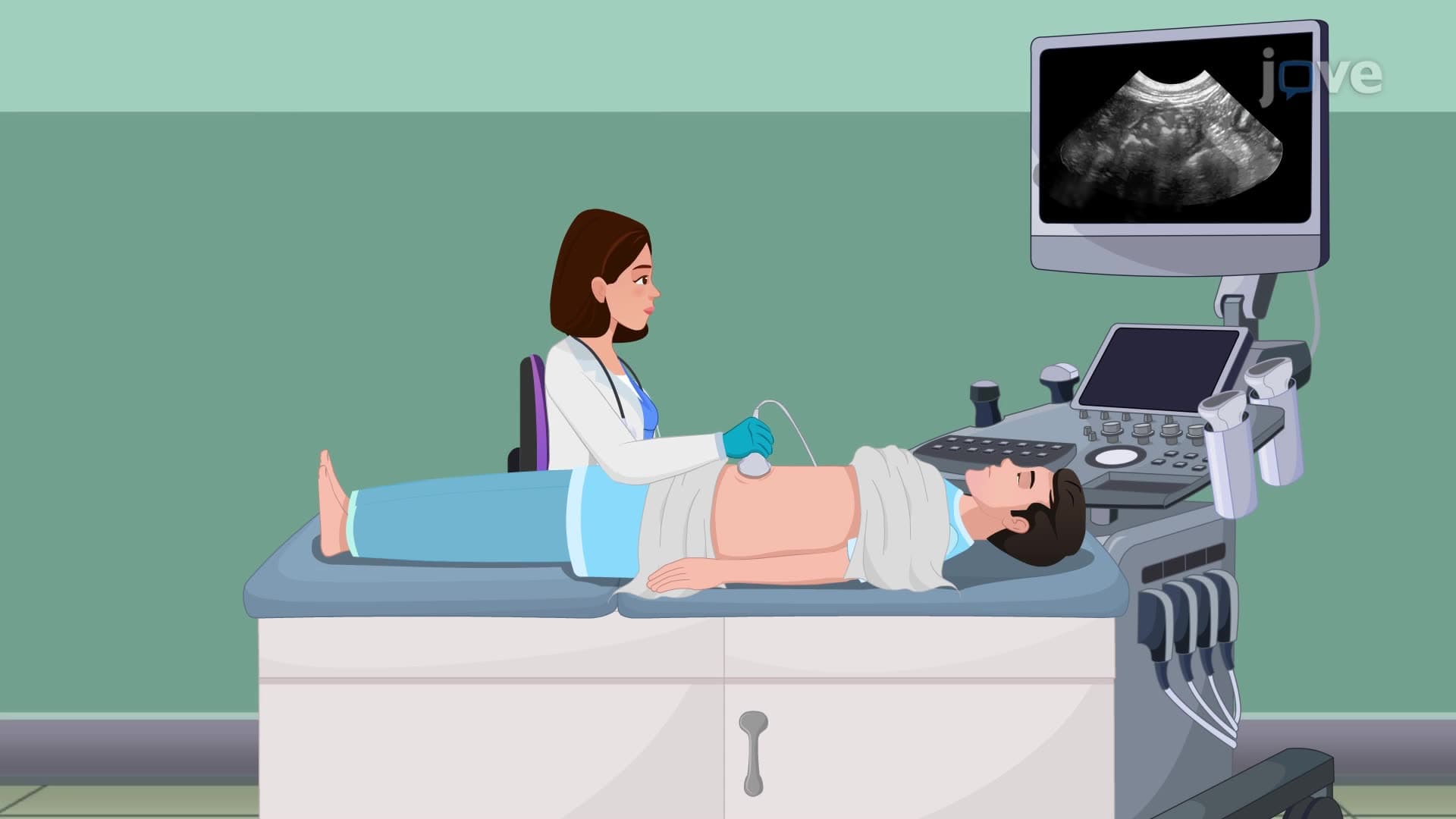

Video Summary: What Is Ultrasound I Abdominal Ultrasonography
Did you know that ultrasound technology can peer inside your abdomen without making a single cut? Ultrasound I abdominal ultrasonography uses high-frequency sound waves-similar to how dolphins navigate underwater-to create detailed images of internal organs. At hospitals like Mayo Clinic, this non-invasive procedure helps diagnose everything from gallstones to liver disease by bouncing sound waves off tissues and converting the echoes into visual images. Watch the full video on JoVE Coach to master this concept with expert-led visuals and step-by-step explanations.
Ultrasound I abdominal ultrasonography represents one of medicine's most versatile diagnostic tools, utilizing the physics of sound wave propagation to visualize internal structures without radiation exposure. Unlike X-rays or CT scans, this imaging modality harnesses acoustic energy-sound waves with frequencies between 1-20 MHz, far beyond human hearing range-to penetrate tissue and generate real-time images.
The technology operates on the principle of echolocation, similar to how bats navigate in darkness. When high-frequency sound waves encounter tissue boundaries with different acoustic properties, they reflect back to the transducer at varying intensities and time intervals. Dense structures like bone appear bright (hyperechoic) on the screen, while fluid-filled areas like the bladder appear dark (anechoic).
In US healthcare settings, abdominal ultrasound serves as a first-line diagnostic tool for numerous conditions. Emergency departments at hospitals like Johns Hopkins use bedside ultrasound to rapidly assess trauma patients for internal bleeding. The procedure excels at evaluating the liver, gallbladder, kidneys, pancreas, and major blood vessels, making it invaluable for detecting gallstones, kidney stones, liver masses, and abdominal aortic aneurysms.
For pre-med students preparing for the MCAT, understanding ultrasound physics connects directly to wave behavior and acoustic properties tested in the Physical Sciences section. The relationship between frequency, wavelength, and penetration depth demonstrates fundamental wave principles crucial for exam success.
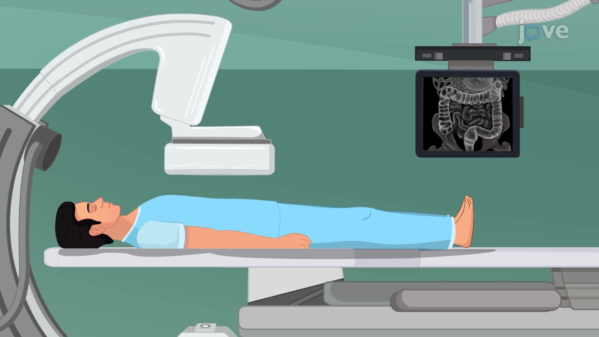
Healthcare professionals performing abdominal ultrasounds must master both technical skills and patient communication. The 8-12 hour fasting requirement ensures optimal image quality by reducing intestinal gas interference-a critical detail for nursing students preparing for NCLEX examinations. Patient positioning in the supine position with potential left lateral decubitus positioning optimizes organ visualization.
Modern ultrasound systems at facilities like Cleveland Clinic integrate advanced imaging modes including Doppler ultrasound for blood flow assessment and elastography for tissue stiffness evaluation, expanding diagnostic capabilities beyond traditional grayscale imaging.
Related Micro-courses