Loading subject page…


6,519 views

Video Summary: What Is Appendicitis I Introduction
Did you know that appendicitis affects approximately 1 in 20 Americans during their lifetime, making it the most common cause of emergency abdominal surgery? Understanding appendicitis I introduction begins with recognizing how a simple blockage in this small organ can rapidly progress to a life-threatening condition. At Johns Hopkins Hospital, emergency physicians diagnose over 300 cases annually, demonstrating the critical importance of early recognition. Watch the full video on JoVE Coach to master this concept with expert-led visuals and step-by-step explanations.
The appendicitis I introduction concept forms a cornerstone of emergency medicine education, particularly relevant for students preparing for healthcare careers. This condition represents one of the most frequently encountered surgical emergencies in US hospitals, with over 250,000 appendectomies performed annually according to the American College of Surgeons.
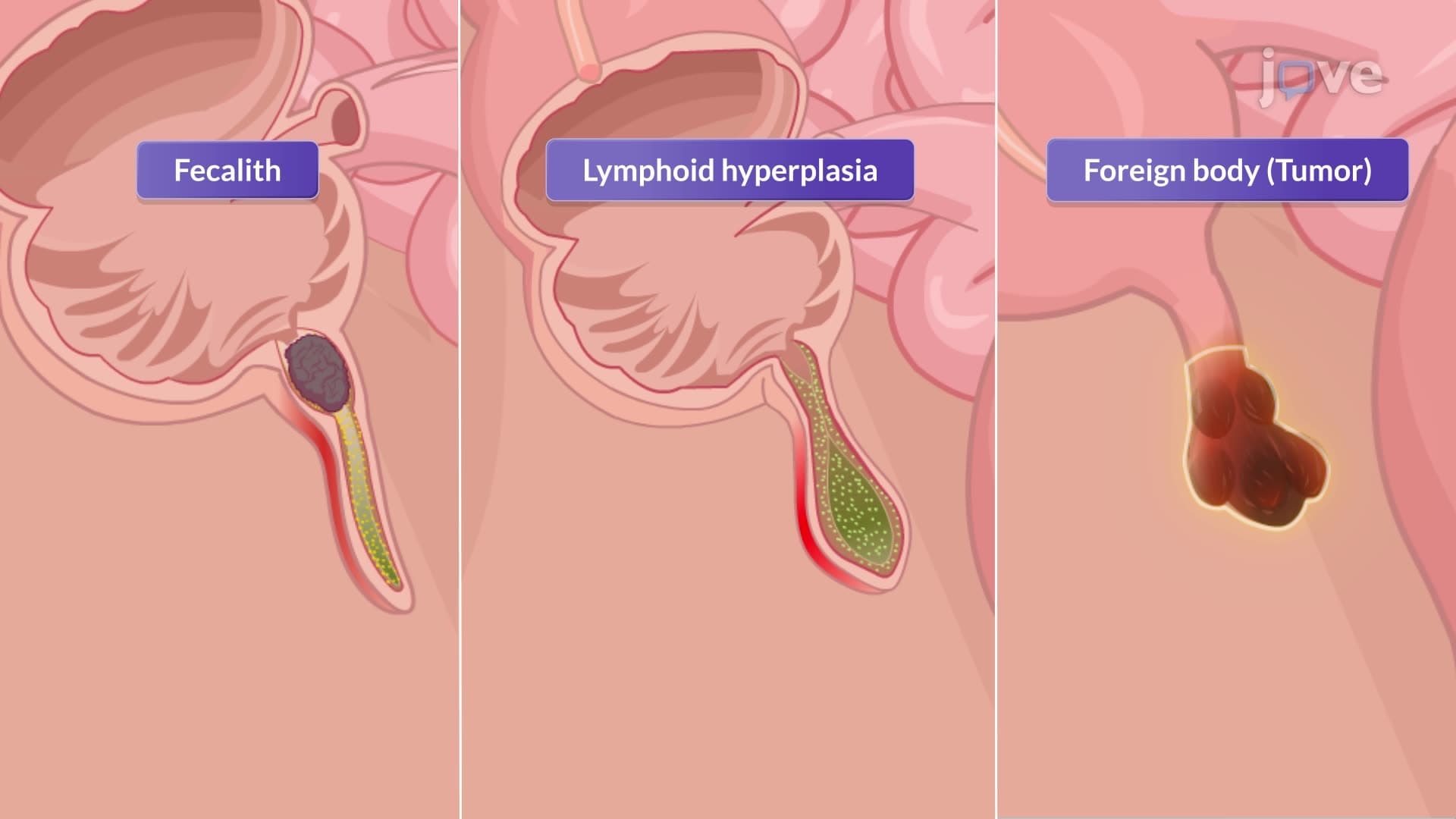
The appendicitis I introduction definition centers on understanding the sequential events that transform a healthy appendix into an inflamed, potentially life-threatening organ. The process begins when the narrow appendiceal lumen becomes obstructed. In pediatric patients, lymphoid hyperplasia accounts for approximately 60% of cases, while fecaliths (hardened stool deposits) predominate in adults. At major teaching hospitals like Mayo Clinic, medical students learn that tumors, though rare, can also cause obstruction, particularly in patients over 40 years old.
Once obstruction occurs, the appendiceal contents become trapped, creating a closed-loop system. This leads to progressive distention as secretions accumulate, dramatically increasing intraluminal pressure. The elevated pressure compromises blood flow, creating an ideal environment for bacterial proliferation. Common organisms include *Escherichia coli*, *Bacteroides fragilis*, and *Peptostreptococcus*, which rapidly multiply in the oxygen-depleted environment.
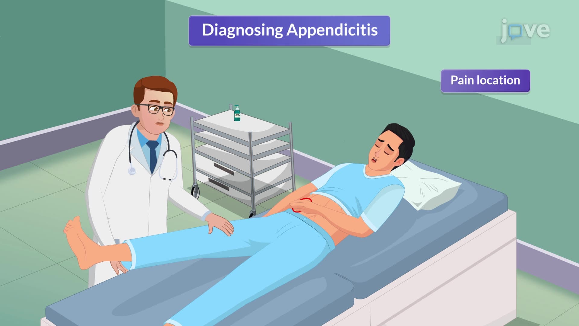
What is appendicitis I introduction in detail becomes clinically relevant through its characteristic presentation pattern. The classic symptom progression, taught extensively in MCAT preparation courses, begins with vague periumbilical pain that gradually migrates to McBurney's point-located one-third the distance from the anterior superior iliac spine to the umbilicus.
This pain migration reflects the embryological origin of the appendix from midgut structures, which initially causes referred visceral pain around the umbilicus. As inflammation progresses to involve the parietal peritoneum, pain becomes somatic and well-localized to the right iliac fossa. Students preparing for the USMLE Step 1 exam should note that approximately 80% of patients demonstrate this classic pattern, though variations exist based on appendiceal position.
The appendicitis I introduction study guide emphasizes several key physical findings that distinguish this condition from other abdominal pathologies. Rovsing's sign, positive in approximately 25% of cases, demonstrates the interconnected nature of peritoneal inflammation. When pressure applied to the left lower quadrant produces pain in the right lower quadrant, it suggests peritoneal irritation around the appendix.
Healthcare students at institutions like Harvard Medical School learn to assess for rebound tenderness, though this finding requires careful technique to avoid unnecessary patient discomfort. The presence of low-grade fever, typically 99-101°F, combined with gastrointestinal symptoms like anorexia and nausea, completes the classical triad that emergency physicians use for rapid triage decisions.
Understanding these foundational concepts prepares students for advanced topics in surgical decision-making and complications management, essential knowledge for success in medical school curricula and standardized examinations.
Related Micro-courses