Loading video…


 18 Concepts
 41.2K views

Video Summary: Assessment of Airway Skin Color Explained
Emergency responders at Houston Methodist Hospital frequently encounter patients where assessment of airway skin color reveals critical respiratory distress before other symptoms appear. This vital sign evaluation can mean the difference between life and death in emergency departments across America. The Assessment of Airway Skin Color Explained demonstrates how healthcare professionals identify oxygen deprivation through visual cues, particularly cyanosis-a bluish discoloration indicating dangerously low blood oxygen levels below 85%. Watch the full video on JoVE Coach to master this concept with expert-led visuals and step-by-step explanations.
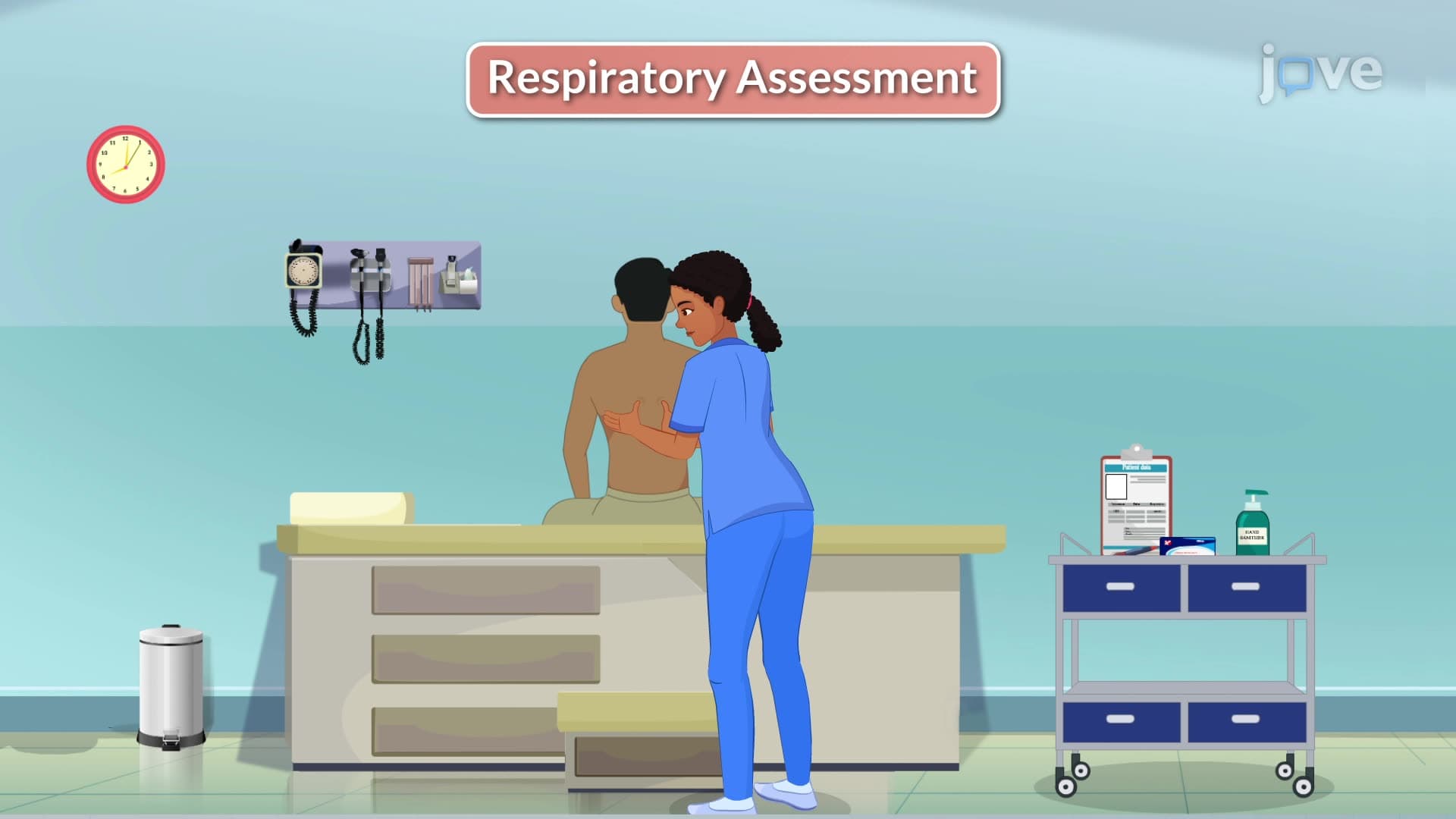
The assessment of airway skin color represents a fundamental yet sophisticated clinical skill that healthcare professionals use to evaluate respiratory status and tissue oxygenation. This non-invasive evaluation technique provides immediate visual feedback about a patient's oxygen-carrying capacity and circulatory efficiency. In emergency departments across major US medical centers like Johns Hopkins and Mayo Clinic, this assessment forms the cornerstone of rapid triage decisions.
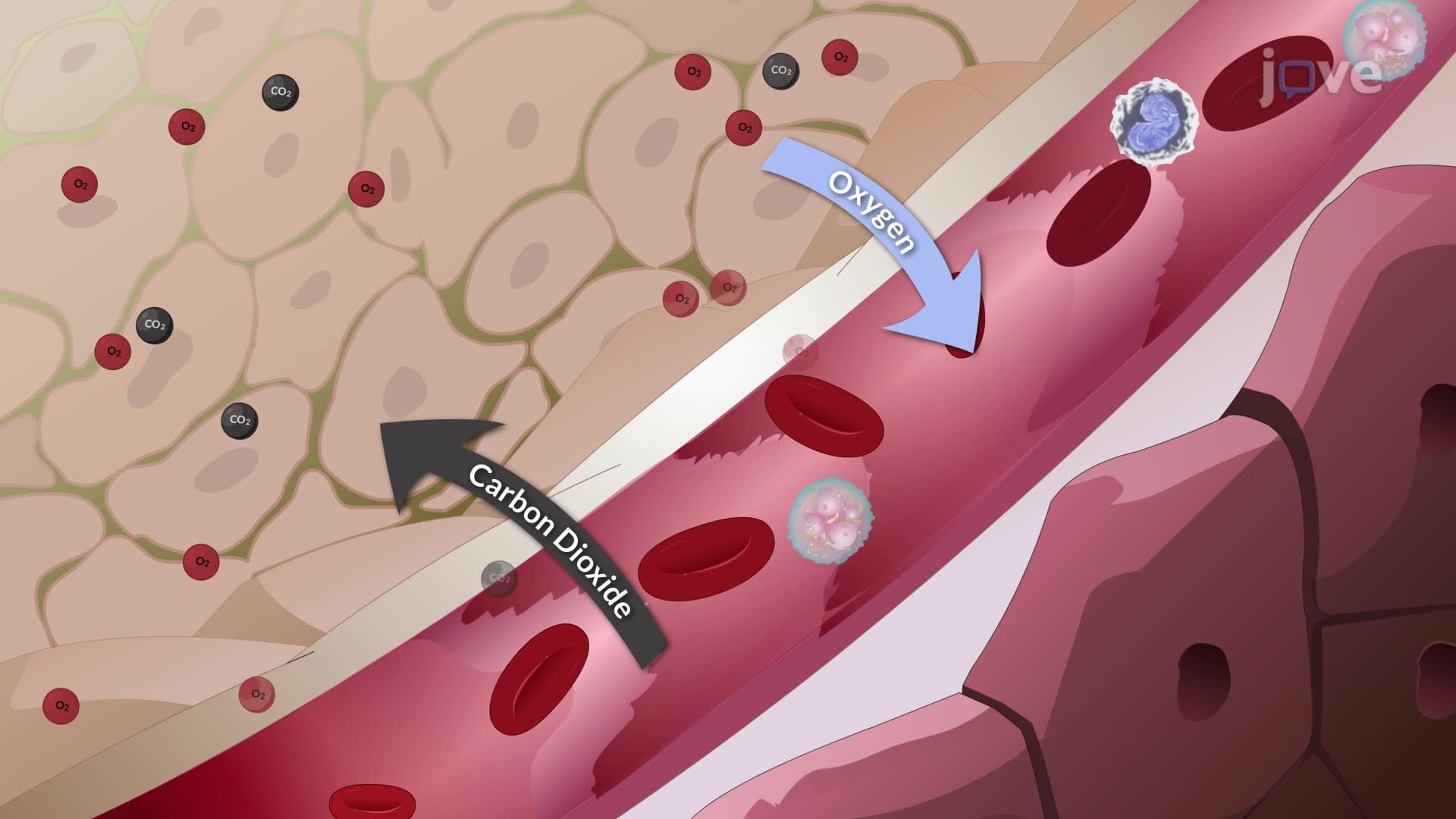
Cyanosis manifests as a bluish discoloration of skin and mucous membranes, occurring when arterial blood oxygen saturation falls below 85%. This threshold represents a critical point where tissue hypoxia becomes visually apparent. Central cyanosis appears in areas with rich blood supply-lips, tongue, and nail beds-indicating systemic hypoxemia. Peripheral cyanosis affects extremities like fingers and toes, often suggesting poor circulation or localized blood flow issues. Students preparing for the MCAT or NCLEX examinations must understand that cyanosis represents a late-stage finding, meaning significant respiratory compromise has already occurred.
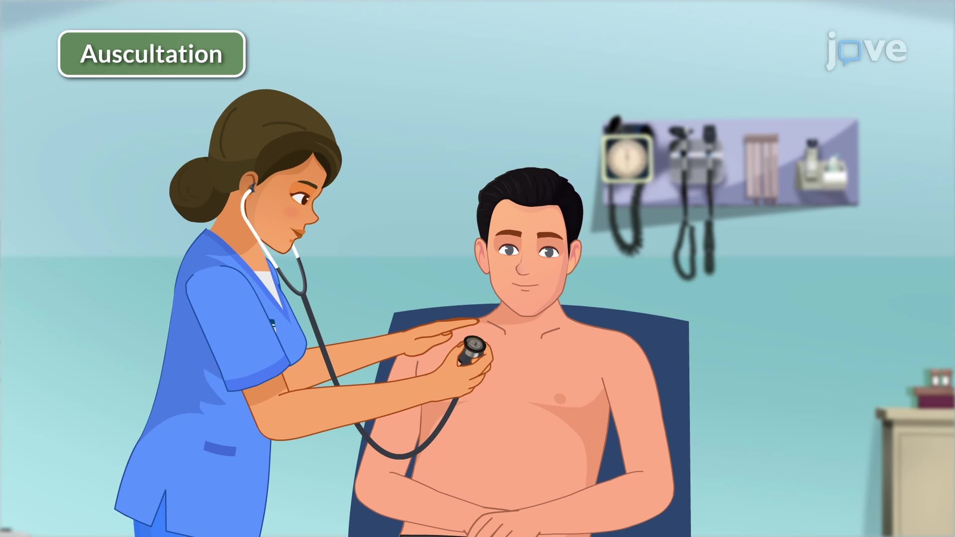
Healthcare professionals combine skin color assessment with multiple evaluation techniques for comprehensive airway analysis. Verbal responsiveness testing-asking patients simple questions-simultaneously assesses airway patency, breathing adequacy, and neurological function. A clear, appropriate verbal response indicates sufficient brain oxygenation and unobstructed airway. This technique proves invaluable in emergency medicine residencies at institutions like Harvard Medical School and UCSF.
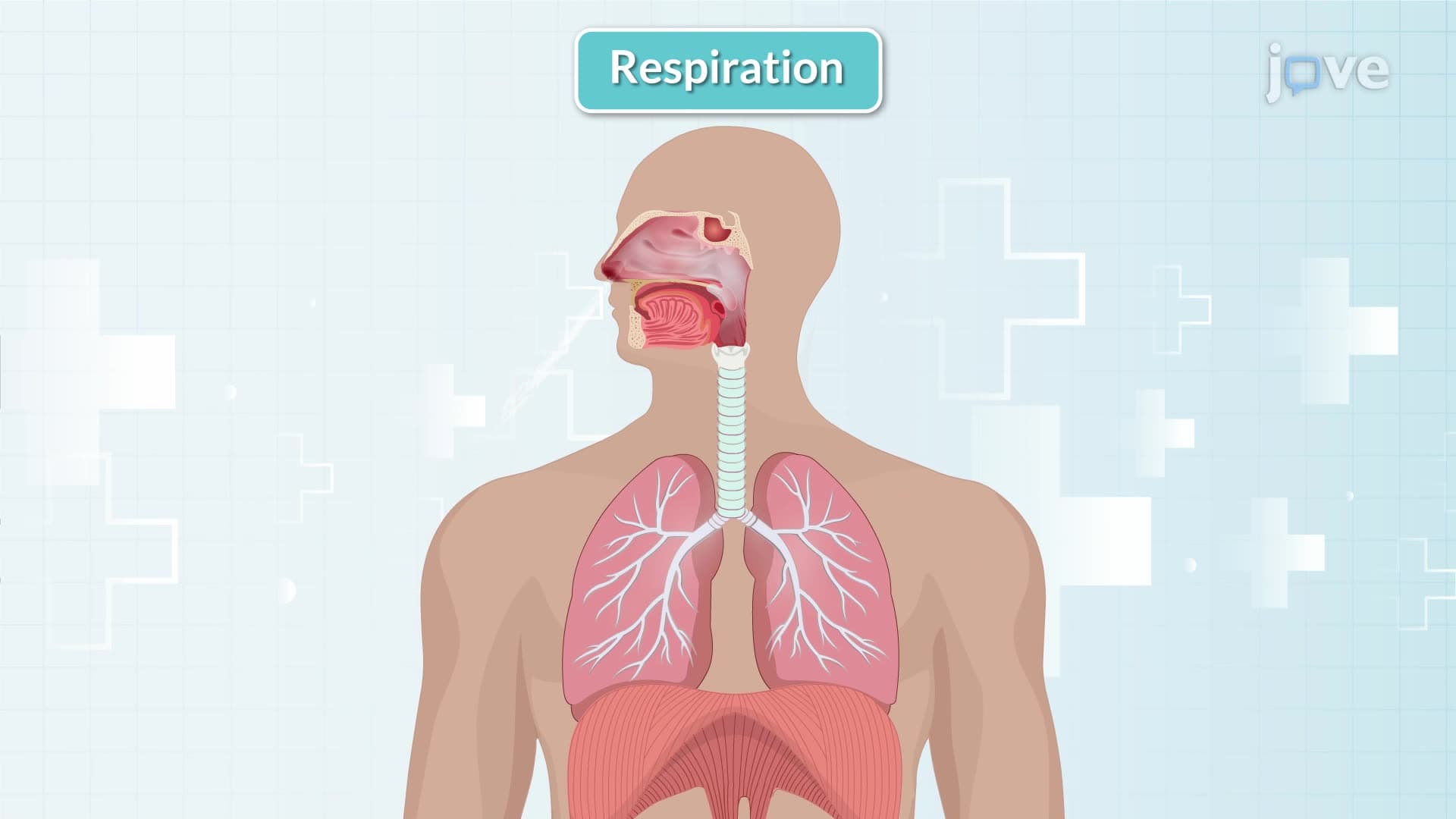
Accessory muscle recruitment during breathing signals respiratory compromise requiring immediate attention. Normal respiration primarily involves diaphragmatic and intercostal muscle action. When patients begin using sternocleidomastoid, scalene, or abdominal muscles for breathing, this indicates increased respiratory workload. College anatomy and physiology courses emphasize recognizing these compensatory mechanisms, particularly relevant for pre-med students tackling AP Biology or preparing for the HESI A2 entrance exam.
Systematic chest assessment includes observing anteroposterior diameter and expansion symmetry. Barrel chest configuration-increased front-to-back chest dimension-often indicates chronic obstructive pulmonary disease or severe asthma. Asymmetrical chest expansion may suggest pneumothorax, pleural effusion, or unilateral lung pathology. These findings frequently appear in USMLE Step 1 clinical vignettes and nursing school examinations.
Related Micro-courses