Loading subject page…


41,216 views

Video Summary: Assessment of Ventilation Ii Respiratory Explained
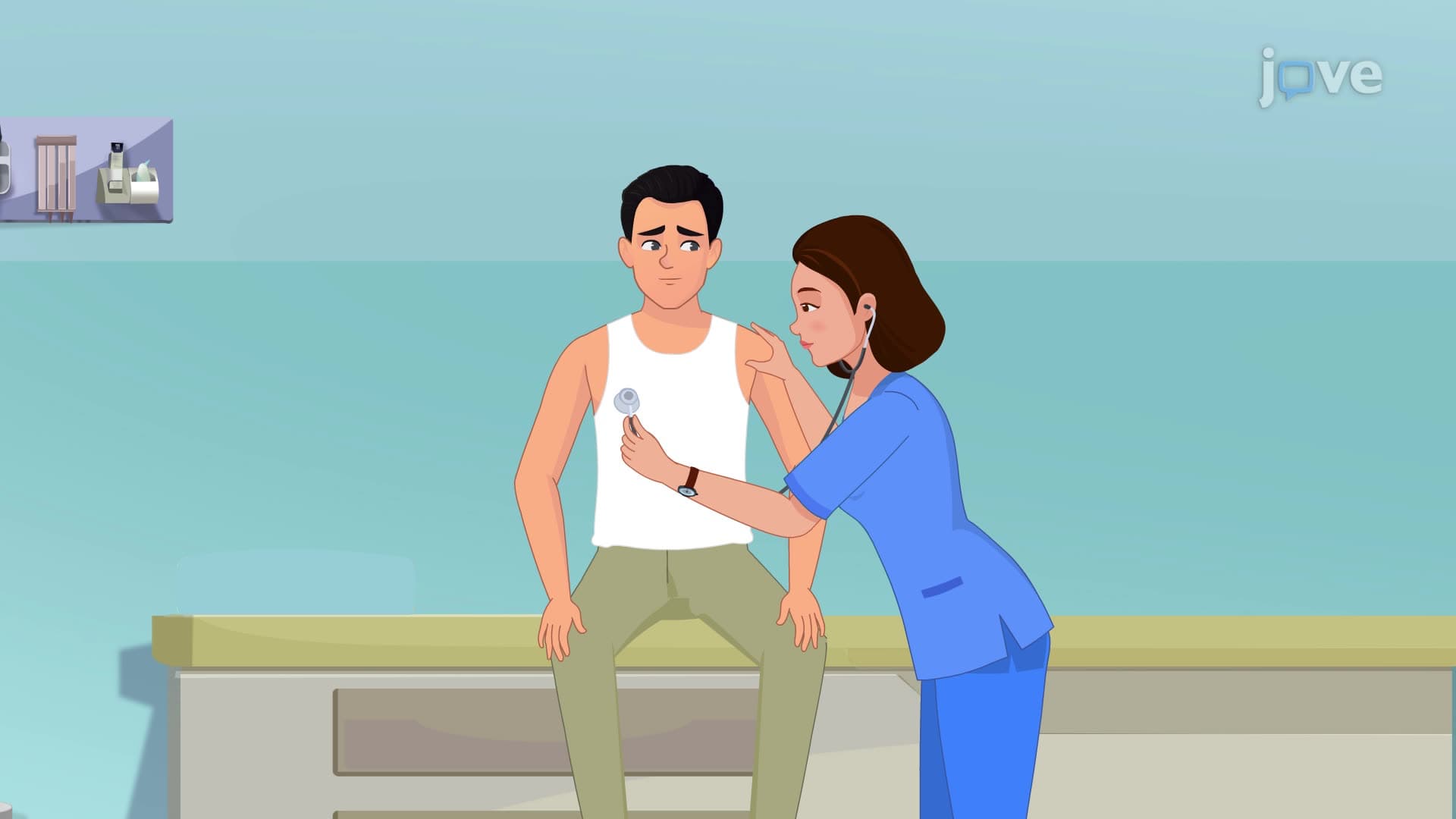
Did you know that emergency room physicians can diagnose life-threatening conditions just by watching how someone breathes? Assessment of ventilation ii respiratory techniques reveal critical information about lung function through careful observation of breathing patterns. For instance, when paramedics in Chicago assess car accident victims, they immediately evaluate respiratory depth and rhythm to determine injury severity. Assessment of Ventilation II Respiratory Explained provides the foundational knowledge healthcare professionals need to make these rapid, potentially life-saving assessments. Watch the full video on JoVE Coach to master this concept with expert-led visuals and step-by-step explanations.
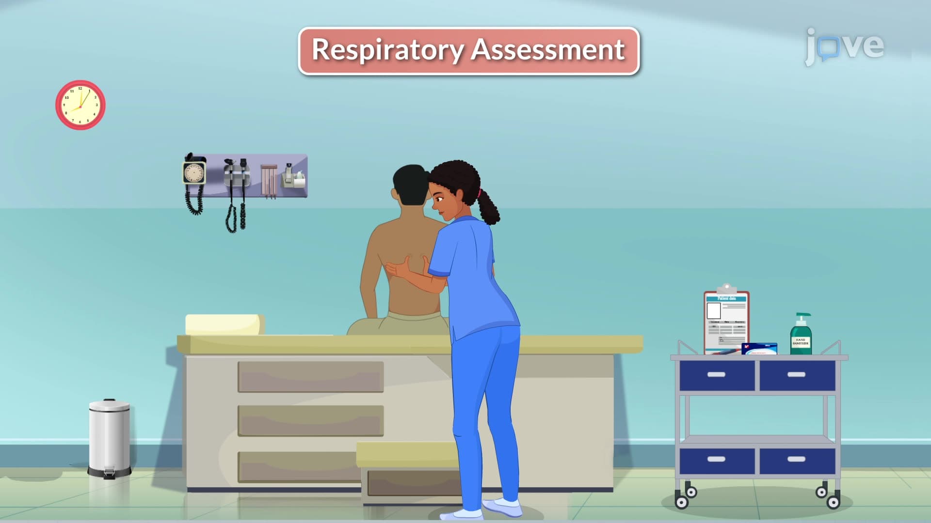
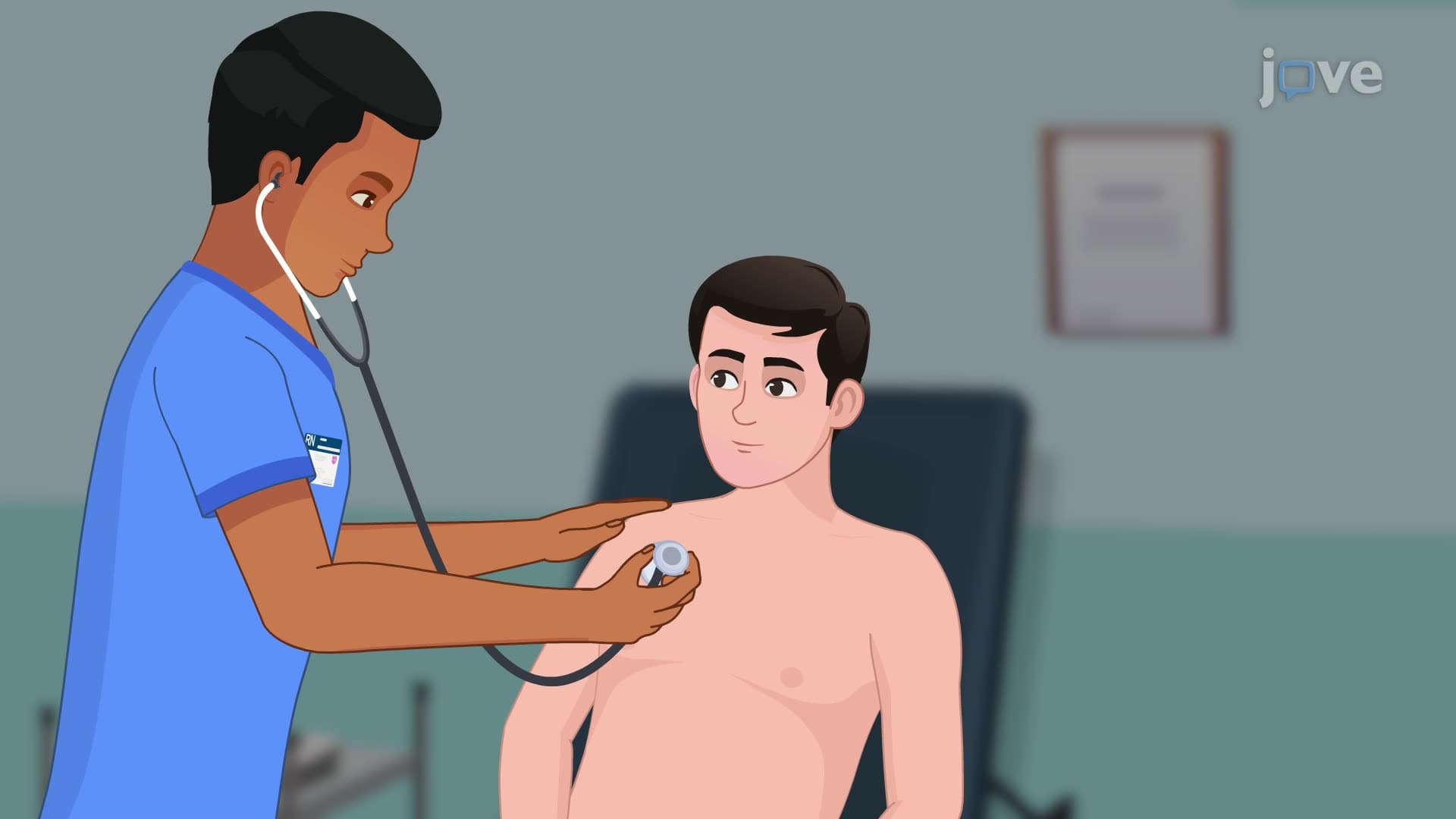
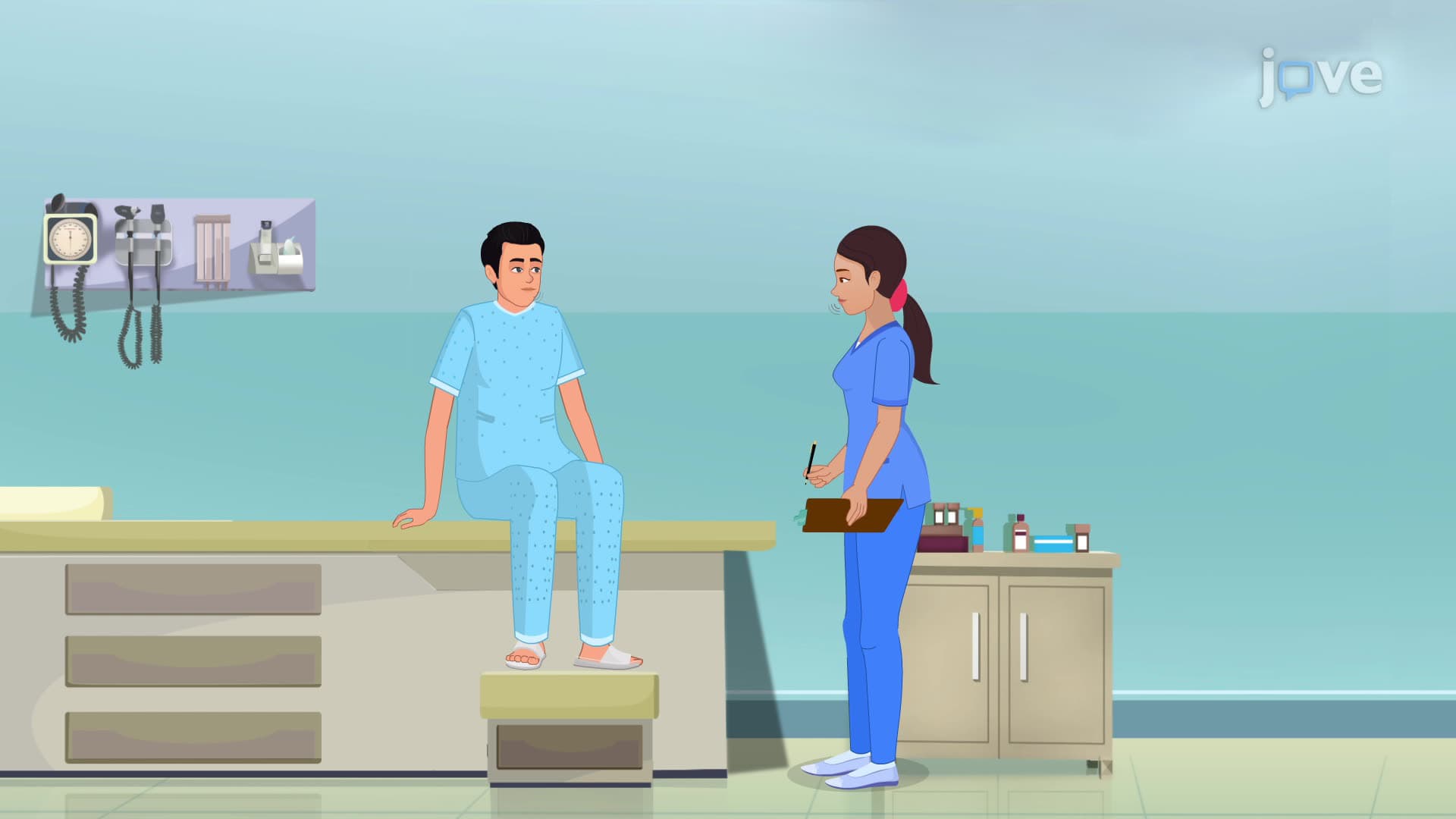
Assessment of Ventilation II Respiratory Explained encompasses the systematic evaluation of breathing patterns that healthcare professionals use to gauge patient respiratory status. This assessment involves two primary components: respiratory depth and respiratory rhythm, each providing crucial diagnostic information.
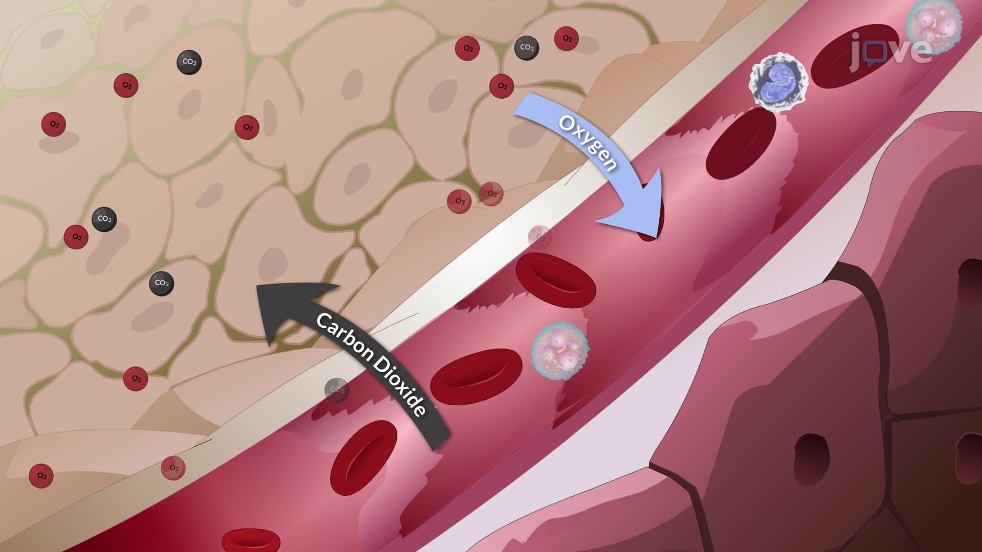
Respiratory depth measures the volume of air inhaled or exhaled during each breath cycle. Healthcare providers classify depth along a spectrum from shallow to deep breathing. Shallow respirations involve minimal air exchange with barely perceptible chest movement, often indicating respiratory compromise or patient fatigue. Conversely, deep respirations demonstrate full lung expansion and complete exhalation, maximizing oxygen intake and carbon dioxide elimination.
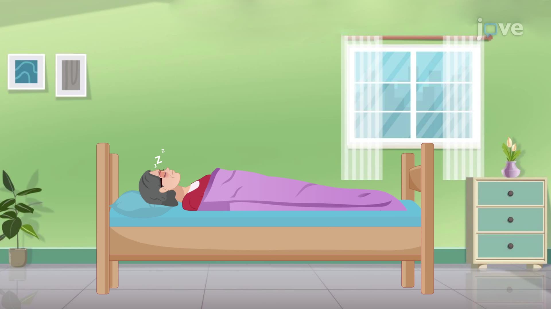
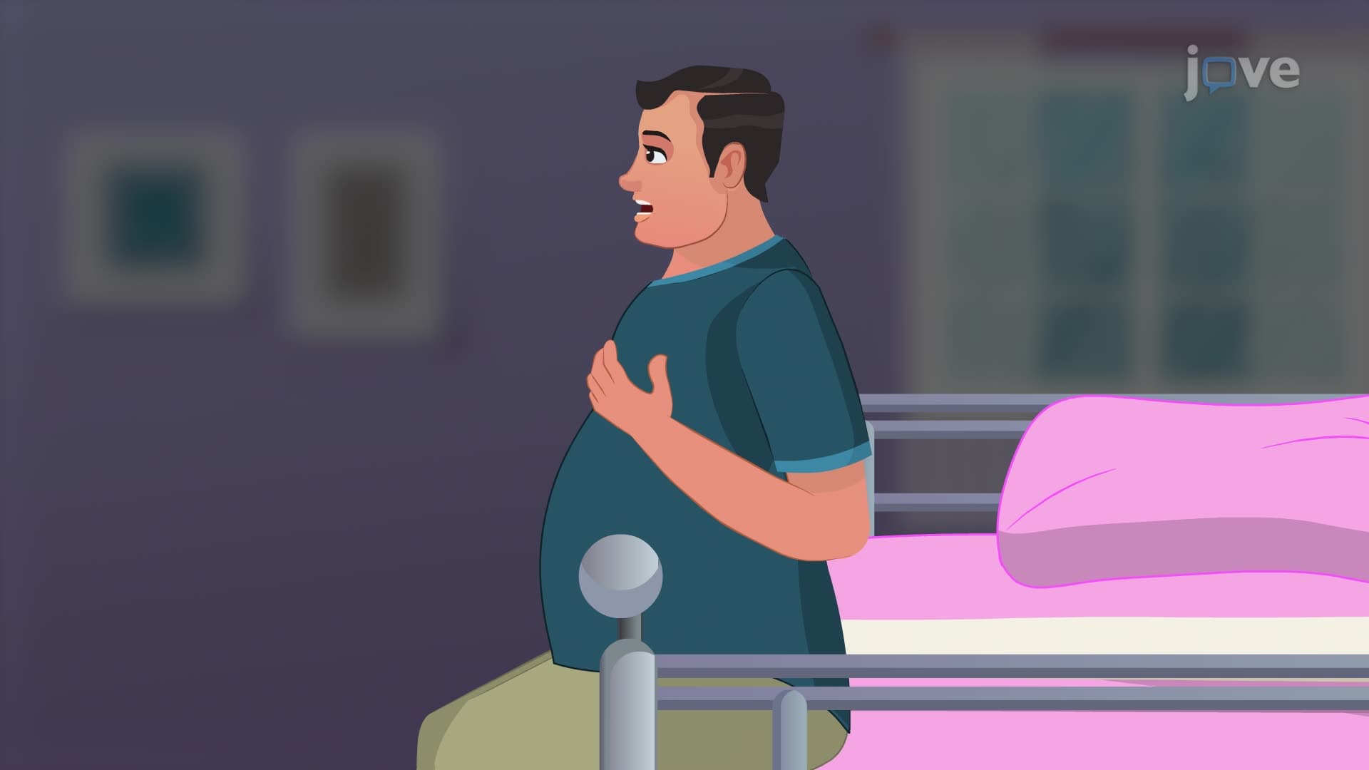
Clinical terminology for respiratory depth includes several key descriptors. Apnea refers to the temporary cessation of breathing, commonly observed in sleep disorders or neurological conditions. Dyspnea describes difficult or labored breathing, frequently seen in patients with heart failure or chronic obstructive pulmonary disease (COPD). Orthopnea specifically indicates breathing difficulty when lying flat, prompting patients to sleep propped up on pillows-a classic sign of congestive heart failure.
Respiratory rhythm encompasses the timing, regularity, and effort associated with breathing cycles. Normal respiratory rhythm demonstrates consistent intervals between breaths with minimal visible effort. Healthcare providers assess rhythm by observing chest and abdominal movements, noting any irregularities that might indicate underlying pathology.
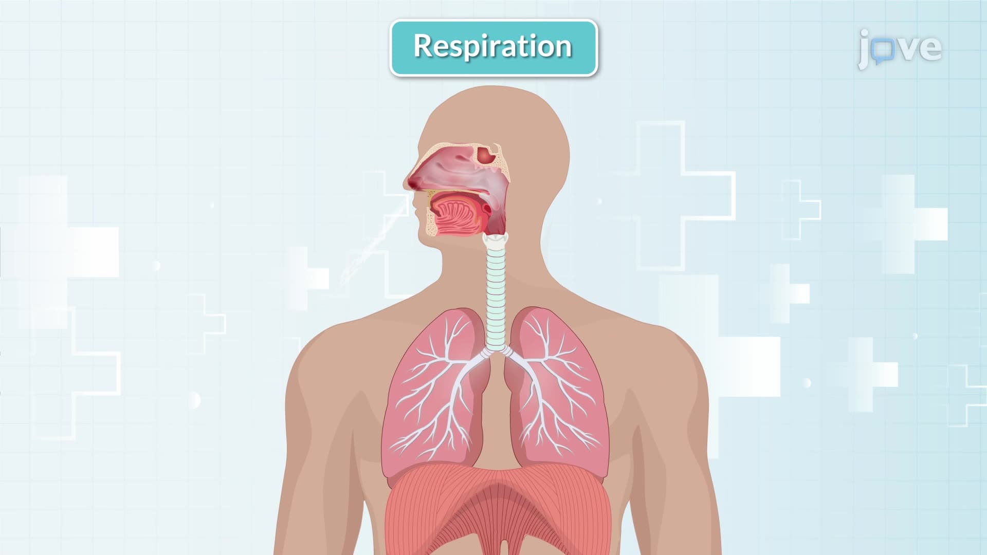
Diaphragmatic breathing occurs when the primary respiratory muscle-the diaphragm-contracts and relaxes rhythmically. This pattern appears as abdominal rising and falling rather than chest movement. Medical professionals often observe this by watching the patient's stomach area during assessment.
Gender differences in breathing patterns significantly impact assessment techniques. Women typically utilize thoracic muscles for breathing, creating visible upper chest movement during respiration. This anatomical difference stems from structural variations in rib cage configuration and respiratory muscle development.
Labored respirations indicate respiratory distress and involve accessory muscles beyond the diaphragm. These muscles, located in the neck and shoulder regions, become visibly active during difficult breathing episodes. Patients with severe asthma attacks or pneumonia often demonstrate this pattern.
Intercostal retractions represent another critical assessment finding. When foreign objects or severe inflammation block airflow, the spaces between ribs pull inward during inspiration attempts. This phenomenon indicates significant airway obstruction requiring immediate intervention.
Prolonged expiration phases suggest obstructed outward airflow, commonly observed in asthma, COPD, or bronchospasm conditions. Students preparing for nursing licensure exams like NCLEX-RN frequently encounter scenarios requiring recognition of these patterns. Similarly, pre-med students studying for the MCAT must understand how these assessment techniques correlate with respiratory physiology and pathophysiology concepts.
Related Micro-courses