Loading video…


 18 Concepts
 41.2K views

Video Summary: Mechanism of Breathing I Inspiration Explained
Ever wonder how your lungs automatically fill with air over 20,000 times daily without conscious effort? The mechanism of breathing I inspiration involves a precise coordination of muscles and pressure changes that create the driving force for airflow into your lungs. When a marathon runner in Boston takes deep breaths at the starting line, their diaphragm and intercostal muscles work together to expand the chest cavity and reduce air pressure below atmospheric levels. Watch the full video on JoVE Coach to master this concept with expert-led visuals and step-by-step explanations.
The mechanism of breathing I inspiration represents one of the most elegant examples of physics applied to human biology. This process relies on fundamental principles of pressure gradients and muscular mechanics that healthcare students encounter throughout their AP Biology, college physiology, and pre-medical coursework.
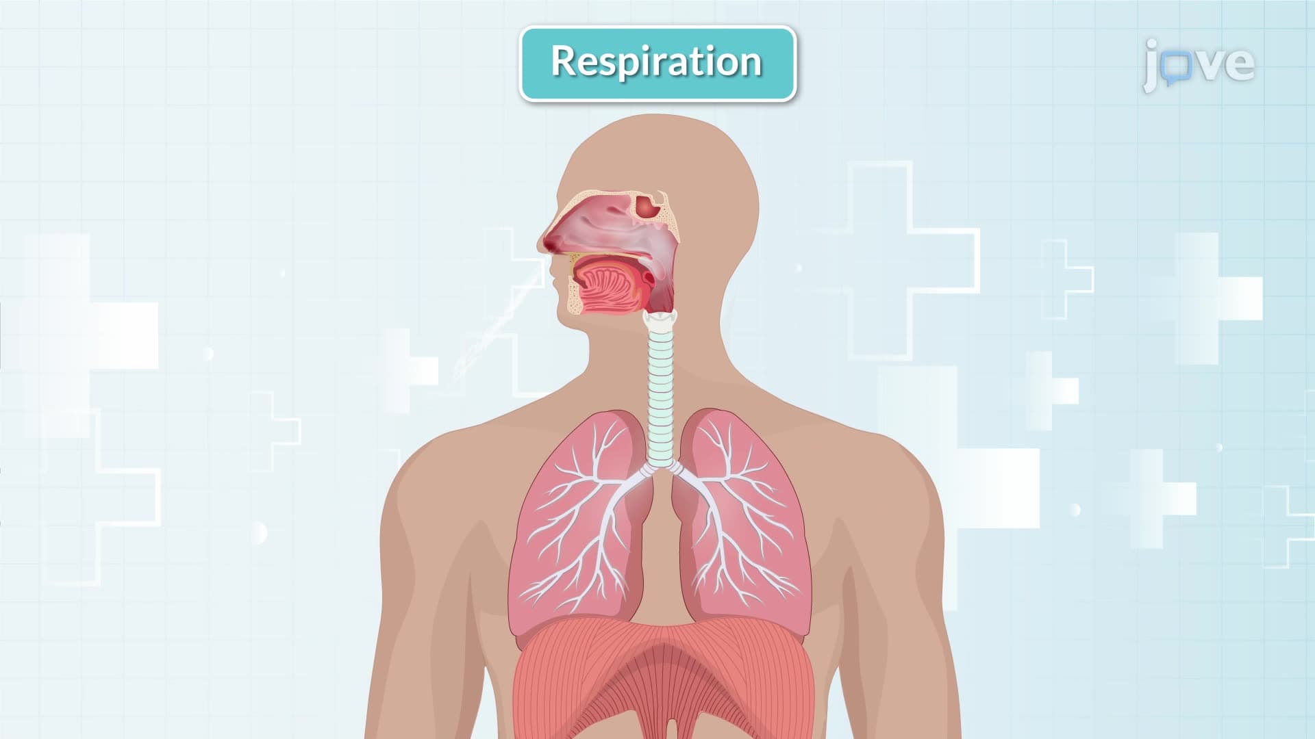
The diaphragm serves as the primary driver of inspiration, accounting for approximately 75% of the work during quiet breathing. When this dome-shaped muscle contracts, it flattens and moves downward, increasing the vertical dimension of the thoracic cavity. Simultaneously, the external intercostal muscles contract between the ribs, pulling them upward and outward in a motion similar to lifting a bucket handle. This coordinated action increases both the anterior-posterior and lateral dimensions of the chest cavity.
Students preparing for the MCAT or nursing entrance exams like the HESI A2 should understand that these muscle contractions don't directly move air-they create the conditions necessary for air movement by manipulating pressure relationships.
The genius of respiratory mechanics lies in creating a pressure gradient. As inspiratory muscles expand the thoracic cavity, the pleural pressure becomes more negative, causing the lungs to follow the chest wall expansion due to surface tension forces. This lung expansion increases alveolar volume, which decreases alveolar pressure below atmospheric pressure (approximately 760 mmHg at sea level). Air naturally flows from high to low pressure, driving atmospheric air into the lungs until pressures equalize.
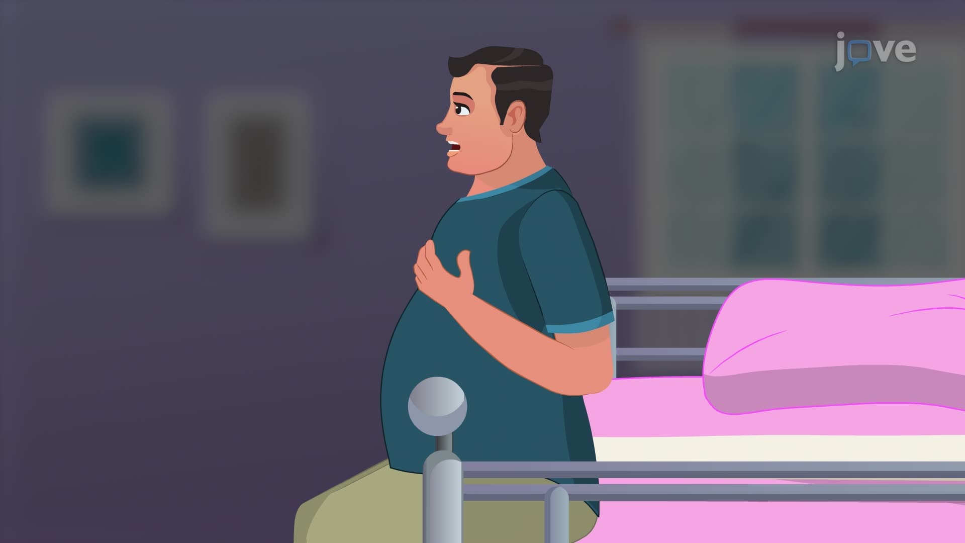
During exercise or respiratory distress-conditions relevant to clinical scenarios nurses encounter-accessory muscles activate to enhance inspiration. The sternocleidomastoid elevates the sternum, while the scalenes lift the upper ribs. Muscles like the pectoralis major and minor, originally designed for arm movement, can assist in chest expansion when breathing demands increase. Understanding these compensatory mechanisms helps explain why patients with respiratory compromise often assume specific postures that optimize accessory muscle function.
This knowledge directly applies to clinical assessment skills tested on nursing exams and provides the foundation for understanding pathological conditions like asthma or COPD that affect inspiratory mechanics.
Related Micro-courses