Loading video…


 18 Concepts
 41.2K views

Video Summary: What Is Mechanism of Breathing Iii
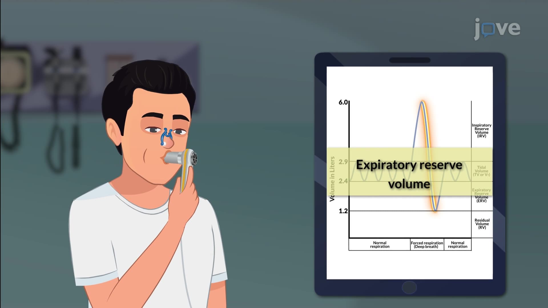
Did you know that when Olympic swimmer Katie Ledecky takes a breath during her record-breaking freestyle, her diaphragm creates negative pressure strong enough to draw air into lungs that can hold up to 6 liters? The mechanism breathing process involves complex muscle coordination where the diaphragm contracts and descends while intercostal muscles expand the rib cage during inhalation. Understanding what is mechanism of breathing iii reveals how accessory muscles like the sternocleidomastoid activate during deep breathing or respiratory distress. Watch the full video on JoVE Coach to master this concept with expert-led visuals and step-by-step explanations.
The mechanism breathing process represents one of the most sophisticated automated functions in human physiology. While basic respiration involves simple diaphragmatic movement, advanced breathing mechanics-what we call the mechanism of breathing iii-encompasses the intricate coordination of multiple muscle groups working in precise synchronization.
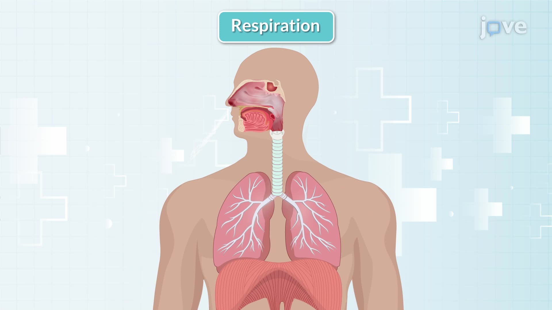
During normal inspiration, the diaphragm serves as the primary driver, contracting and flattening to increase thoracic cavity volume by approximately 500 mL. This creates a pressure gradient of about -3 to -5 mmHg relative to atmospheric pressure. Simultaneously, the external intercostal muscles contract, lifting and expanding the rib cage outward and upward in a "bucket handle" motion. This dual action creates the negative intrathoracic pressure essential for air intake.
Students preparing for the MCAT or AP Biology exams should understand that this process follows Boyle's Law: as volume increases, pressure decreases proportionally. This fundamental relationship appears frequently in standardized test questions about respiratory physiology.
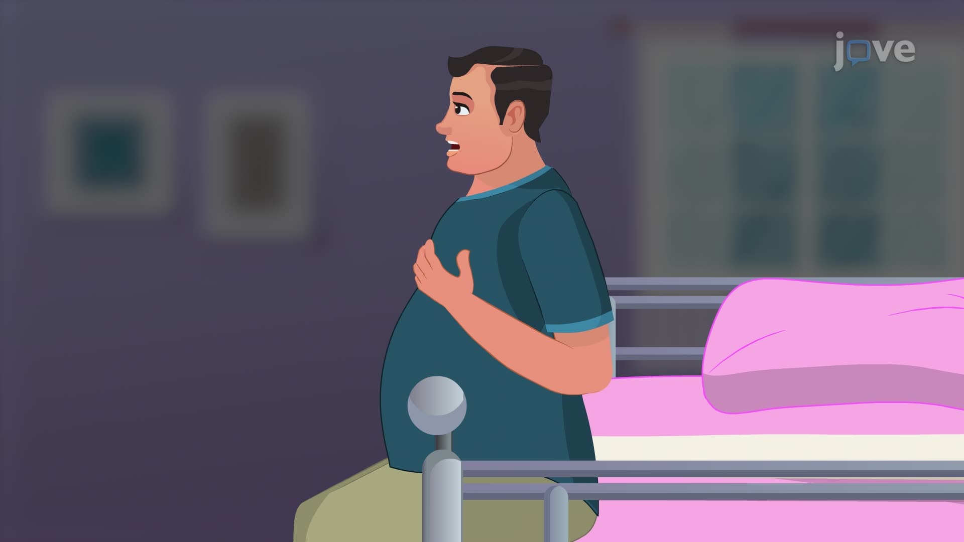
The mechanism of breathing definition explained becomes more complex during periods of increased respiratory demand. Accessory muscles activate during exercise, emotional stress, or pathological conditions. The sternocleidomastoid muscles elevate the sternum and first rib, while scalene muscles lift the upper ribs, creating additional thoracic expansion. The pectoralis minor contributes by elevating ribs 3-5, particularly important during deep inspiration.
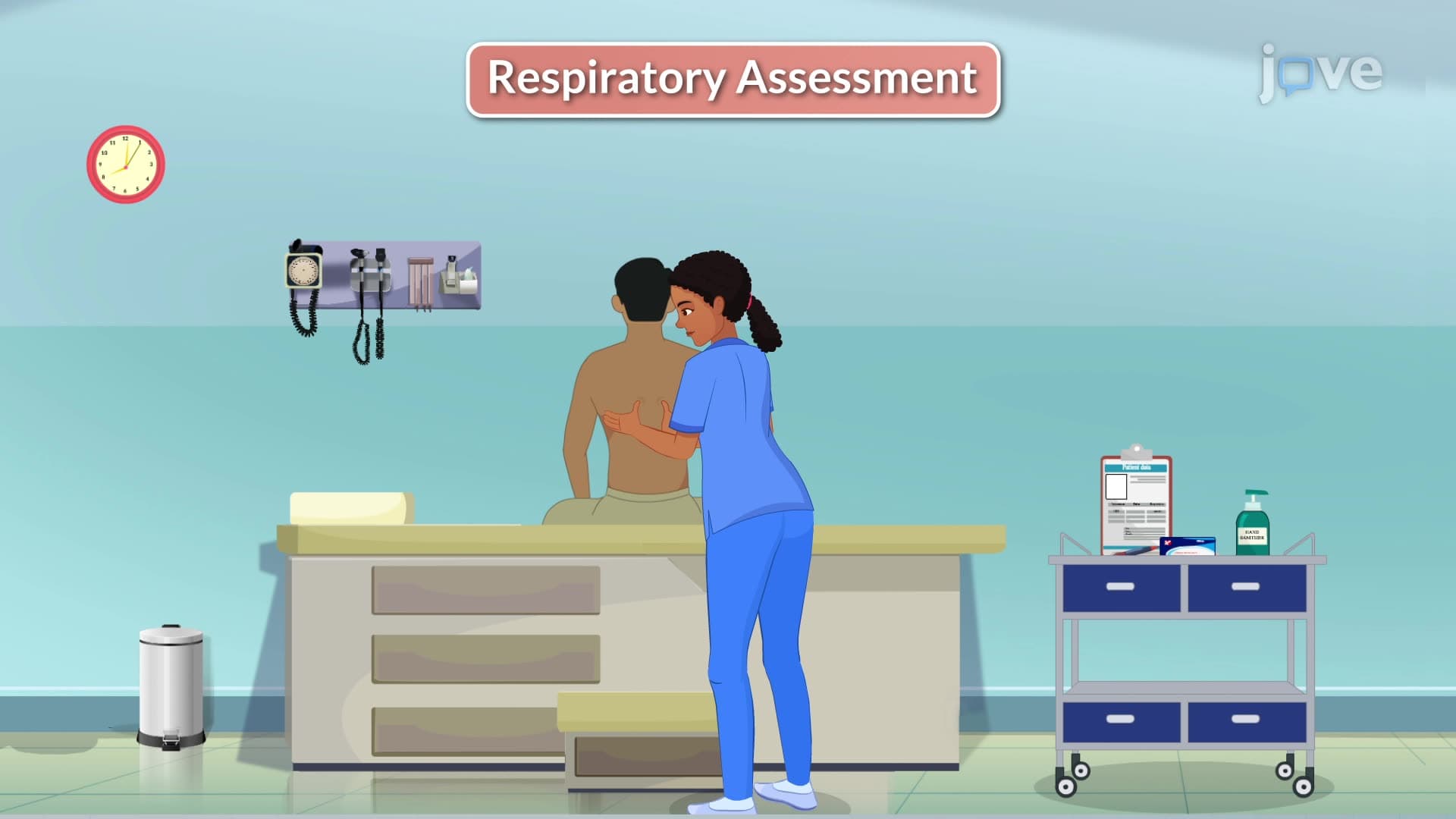
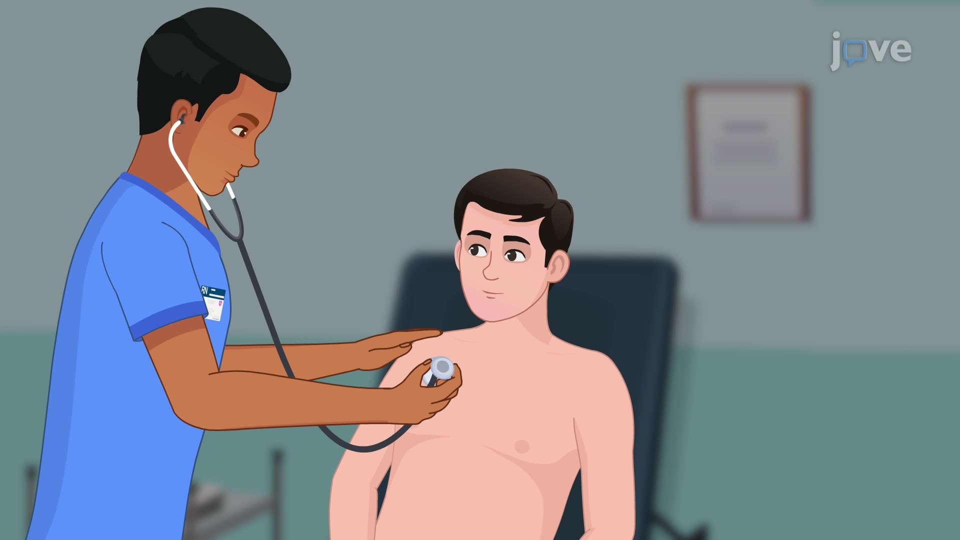
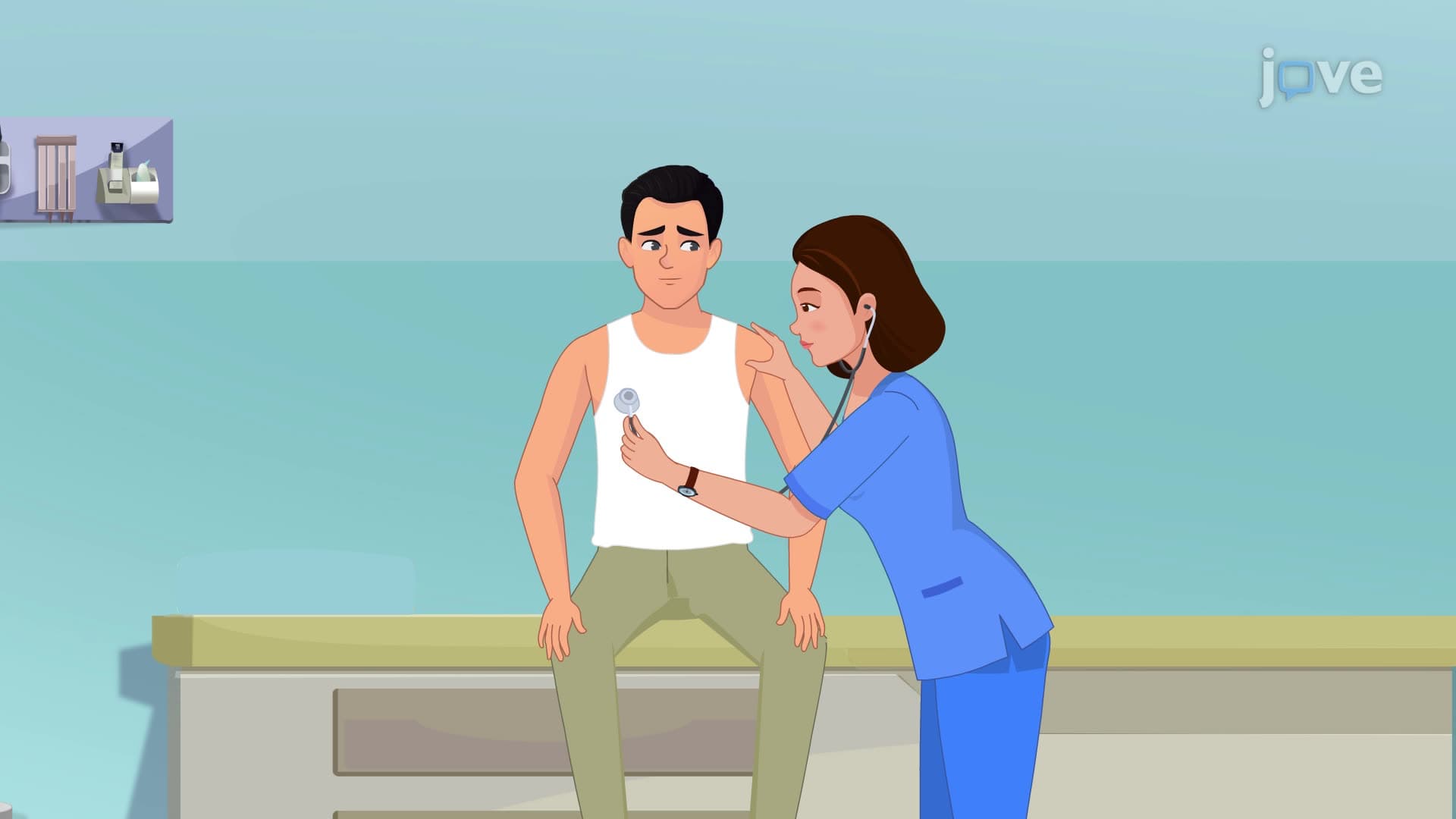
Healthcare students studying for NCLEX or HESI A2 exams encounter these concepts when learning about respiratory assessment and identifying signs of respiratory distress, where accessory muscle use becomes visibly apparent.
Normal expiration relies on elastic recoil of lung tissue and relaxation of inspiratory muscles. However, during forced expiration-such as during athletic performance or coughing-the internal intercostal muscles and abdominal muscles (rectus abdominis, external obliques) actively contract. This active expiration can generate positive pressures exceeding 100 mmHg, crucial for effective coughing and airway clearance.
Understanding these mechanisms proves essential for college-level anatomy and physiology courses and prepares students for advanced study in respiratory therapy, nursing, or medical school.
Related Micro-courses