Loading subject page…


41,216 views

Video Summary: What Is Physiological Control of Respiration
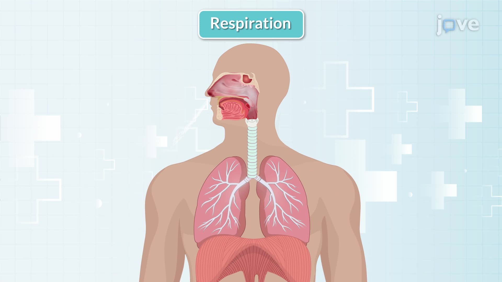
Ever wonder why you breathe faster during a pop quiz? The physiological control of respiration automatically adjusts your breathing rate based on stress and metabolic demands. This intricate system involves brain centers in the medulla and pons working with chemoreceptors throughout your body. Consider how EMTs monitor respiratory patterns in emergency patients to assess neurological function. Watch the full video on JoVE Coach to master this concept with expert-led visuals and step-by-step explanations.
The physiological control of respiration represents one of the body's most sophisticated regulatory systems, automatically adjusting breathing to maintain optimal blood chemistry. Unlike simple reflexes, this control system integrates multiple inputs to ensure cellular oxygen delivery while eliminating carbon dioxide waste. Medical students at Johns Hopkins University and other leading institutions study this concept extensively, as respiratory control dysfunction underlies conditions from sleep apnea to altitude sickness.
The medulla oblongata houses the primary respiratory control center, containing neurons that generate the basic breathing rhythm. These medullary neurons fire approximately 12-15 times per minute in healthy adults, establishing the baseline respiratory rate. The pons modifies this rhythm through the pneumotaxic and apneustic centers, fine-tuning breathing patterns based on physiological demands. When students at UCLA study for finals, stress hormones trigger the pons to increase breathing rate, preparing the body for heightened mental activity.
The neural pathways involve specific cranial and spinal nerves. The phrenic nerve (C3-C5) controls diaphragmatic movement, while intercostal nerves regulate the muscles between ribs. Paramedics often assess phrenic nerve function when evaluating patients with cervical spine injuries, understanding that damage above C3 can compromise breathing independence.
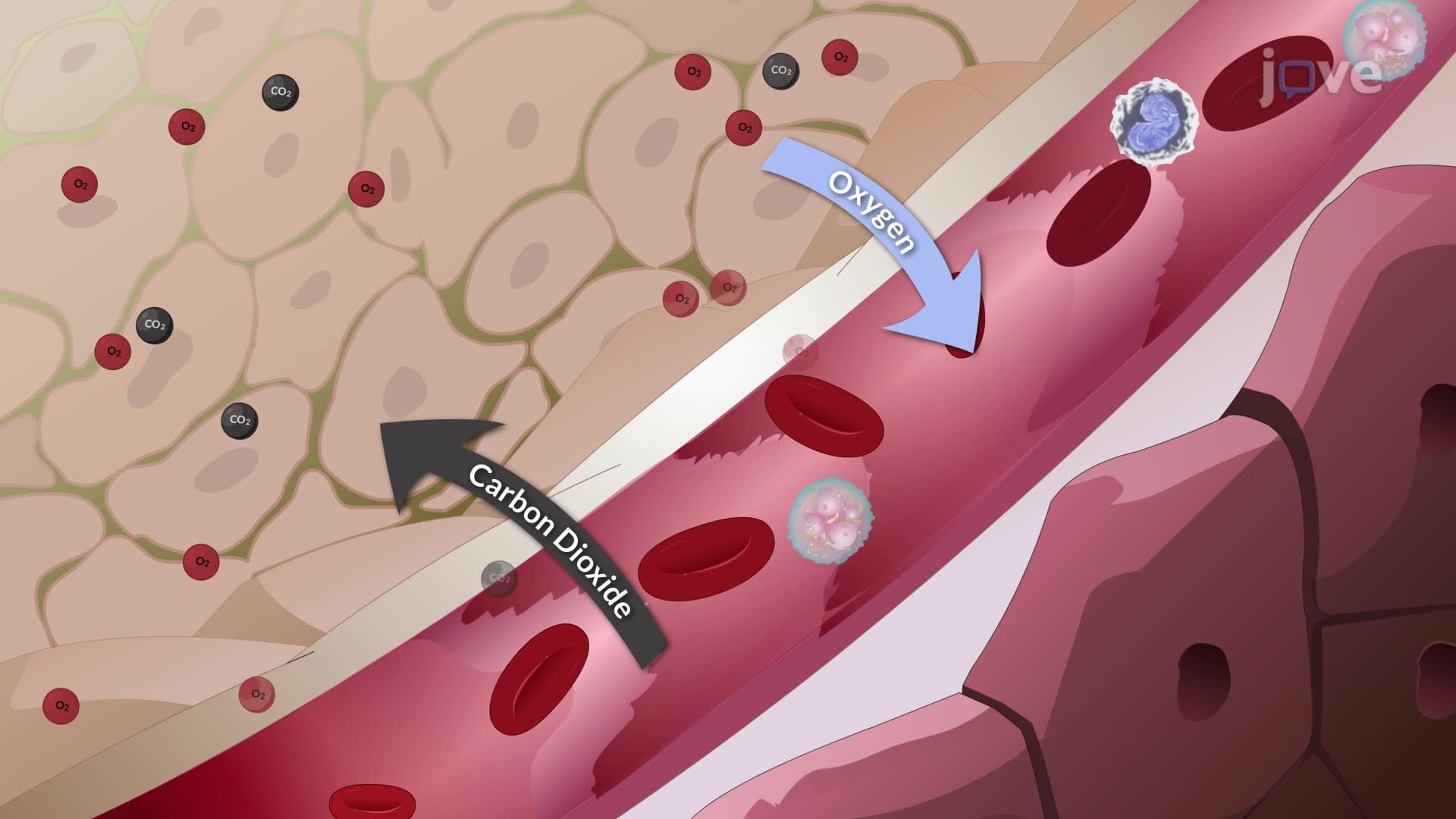
Carbon dioxide serves as the primary respiratory drive, monitored by central chemoreceptors in the medulla and peripheral chemoreceptors in carotid and aortic bodies. When CO2 levels rise, these receptors detect the resulting pH decrease and stimulate increased ventilation. This explains why holding your breath eventually becomes impossible-rising CO2 levels override voluntary control.
Oxygen levels provide secondary control through peripheral chemoreceptors, becoming significant only when arterial oxygen drops below 60 mmHg. Respiratory therapists at major hospitals like Mayo Clinic monitor these parameters in patients with chronic obstructive pulmonary disease (COPD), where altered chemoreceptor sensitivity affects breathing patterns.
This concept appears frequently on AP Biology exams and MCAT questions, often testing students' understanding of negative feedback loops and homeostatic control. Pre-med students should recognize that respiratory control abnormalities can indicate neurological damage, metabolic disorders, or drug effects. Emergency physicians assess respiratory patterns to evaluate conditions ranging from diabetic ketoacidosis to opioid overdose, making this knowledge clinically essential.
Related Micro-courses