Loading video…


 26 Concepts
 99.2K views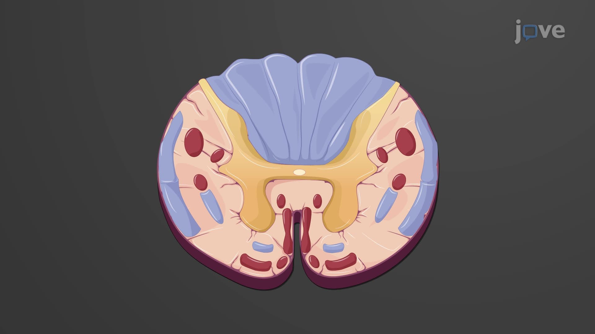

Video Summary: Spinal Cord Cross Sectional Anatomy Explained
Ever wondered why a spinal cord injury at the neck level, like those seen in American football accidents, affects both arms and legs while lower back injuries might only impact the legs? The spinal cord cross section anatomy reveals a butterfly-shaped gray matter core surrounded by white matter tracts that carry signals between your brain and body. This intricate organization explains how neurologists at major US medical centers like Johns Hopkins can predict which body functions will be affected based on injury location. Watch the full video on JoVE Coach to master this concept with expert-led visuals and step-by-step explanations.
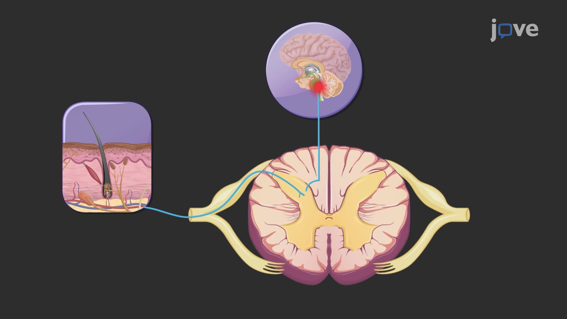
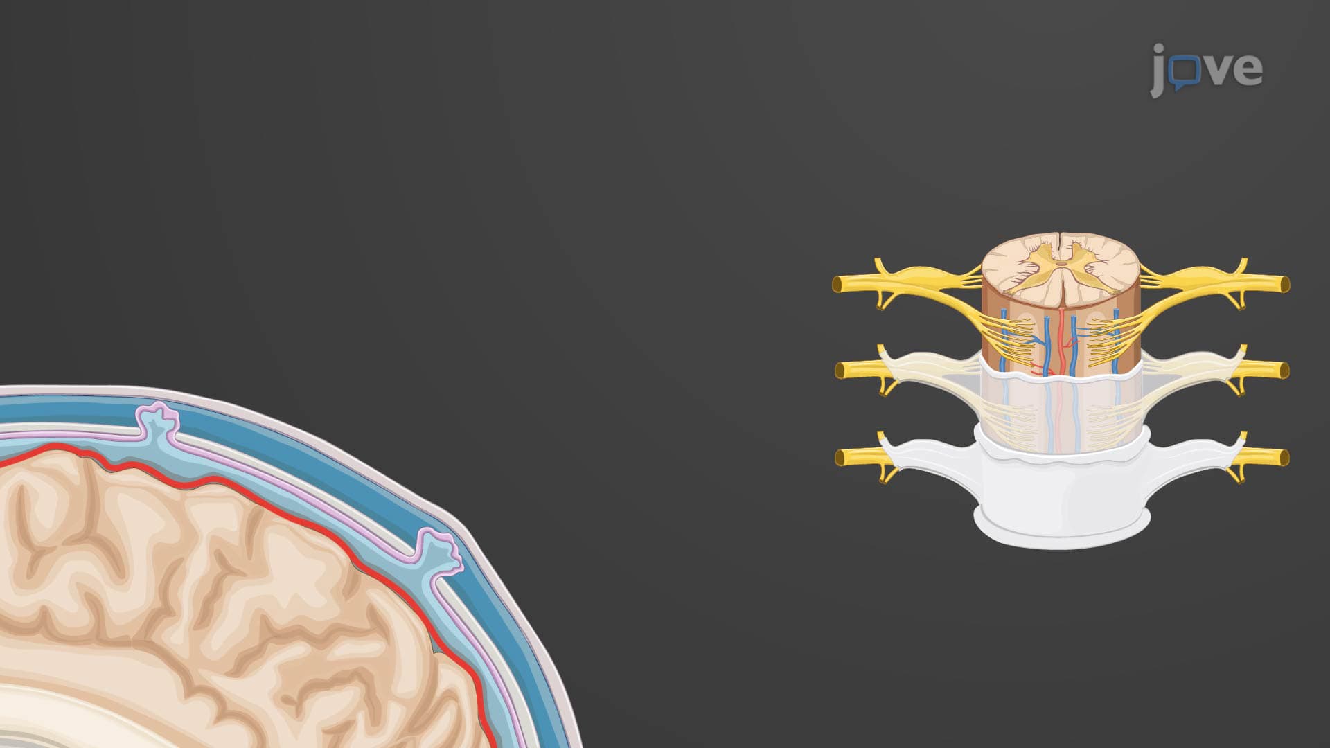
The spinal cord cross section anatomy reveals a highly organized structure that resembles a butterfly when viewed in transverse section. This distinctive pattern results from the central gray matter being completely surrounded by white matter, creating a system optimized for both local processing and long-distance communication between the brain and peripheral body regions.
The gray matter forms the characteristic butterfly or H-shaped central region, with the gray commissure connecting the two halves around the cerebrospinal fluid-filled central canal. The dorsal horns, pointing toward the back of the body, serve as the primary sensory processing centers. Here, interneurons receive and integrate signals from sensory neurons entering through the dorsal roots. Students preparing for the MCAT should note that pain and temperature sensations synapse in the superficial layers of the dorsal horn, while touch and proprioceptive information synapses in deeper layers.
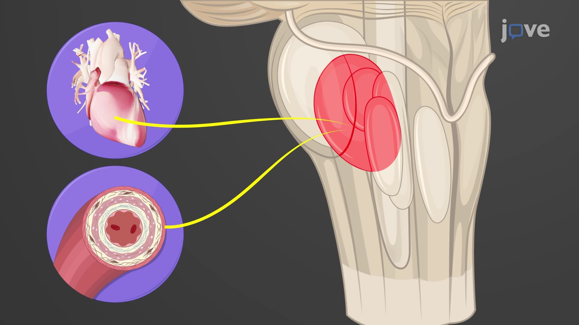
The ventral horns contain the cell bodies of somatic motor neurons that control skeletal muscle movement. These large multipolar neurons send their axons out through the ventral roots to innervate muscles throughout the body. In thoracic and upper lumbar regions (T1-L2), additional lateral horns appear, housing autonomic motor neurons that control involuntary functions like heart rate, breathing, and digestion - crucial concepts for AP Biology students studying the autonomic nervous system.
The white matter is systematically organized into three funiculi: dorsal, ventral, and lateral. Each funiculus contains specific ascending and descending tracts. The dorsal funiculus carries fine touch and proprioceptive information upward to the brain, while the lateral funiculus contains both ascending pain/temperature pathways and descending motor control tracts. This organization explains why spinal cord injuries produce predictable patterns of sensory and motor loss that neurologists can map precisely.
Understanding spinal cord cross-sectional anatomy is essential for pre-med students taking the MCAT, as questions frequently test knowledge of tract locations and functions. In clinical practice, this knowledge helps healthcare professionals at institutions like the Mayo Clinic determine the extent and location of spinal injuries. For example, damage to the lateral funiculus might preserve fine touch (dorsal funiculus intact) while eliminating pain sensation and voluntary movement on the same side below the injury level.
Related Micro-courses