Loading video…


 26 Concepts
 99.2K views


Video Summary: What Is Spinal Nerves Anatomy
Ever wonder how your brain instantly knows when you touch a hot stove and commands your hand to pull away? Spinal nerve anatomy biology reveals this intricate communication system through 31 pairs of mixed nerves that connect your spinal cord to every part of your body. When a medical student at Johns Hopkins dissects cadaveric specimens, they discover how spinal nerves structure enables both sensation and movement through specialized root systems. Watch the full video on JoVE Coach to master this concept with expert-led visuals and step-by-step explanations.
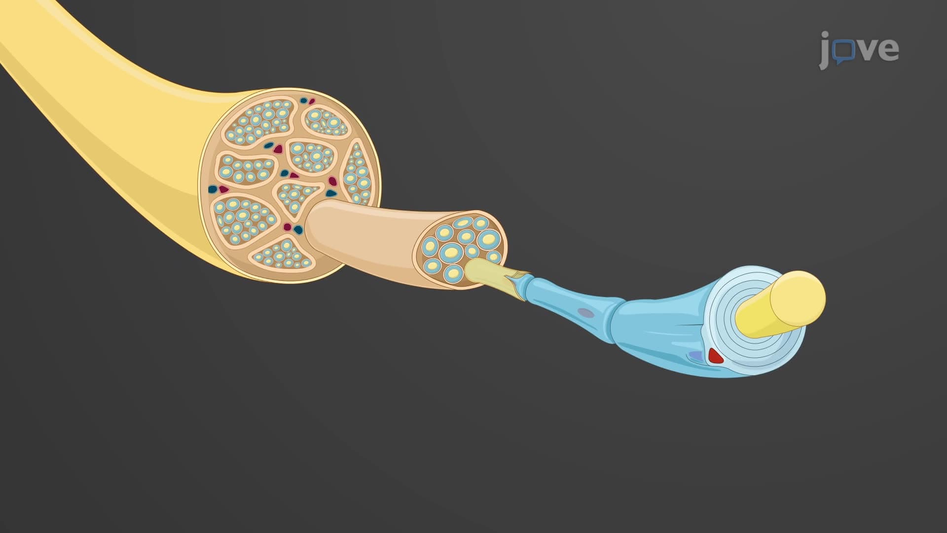
Spinal nerves anatomy biology represents one of the most elegant examples of biological organization in the human nervous system. Unlike pure sensory or motor nerves found elsewhere in the body, spinal nerves are mixed nerves that simultaneously carry sensory information toward the central nervous system and motor commands away from it. This dual functionality makes them essential for coordinated movement and sensory perception.
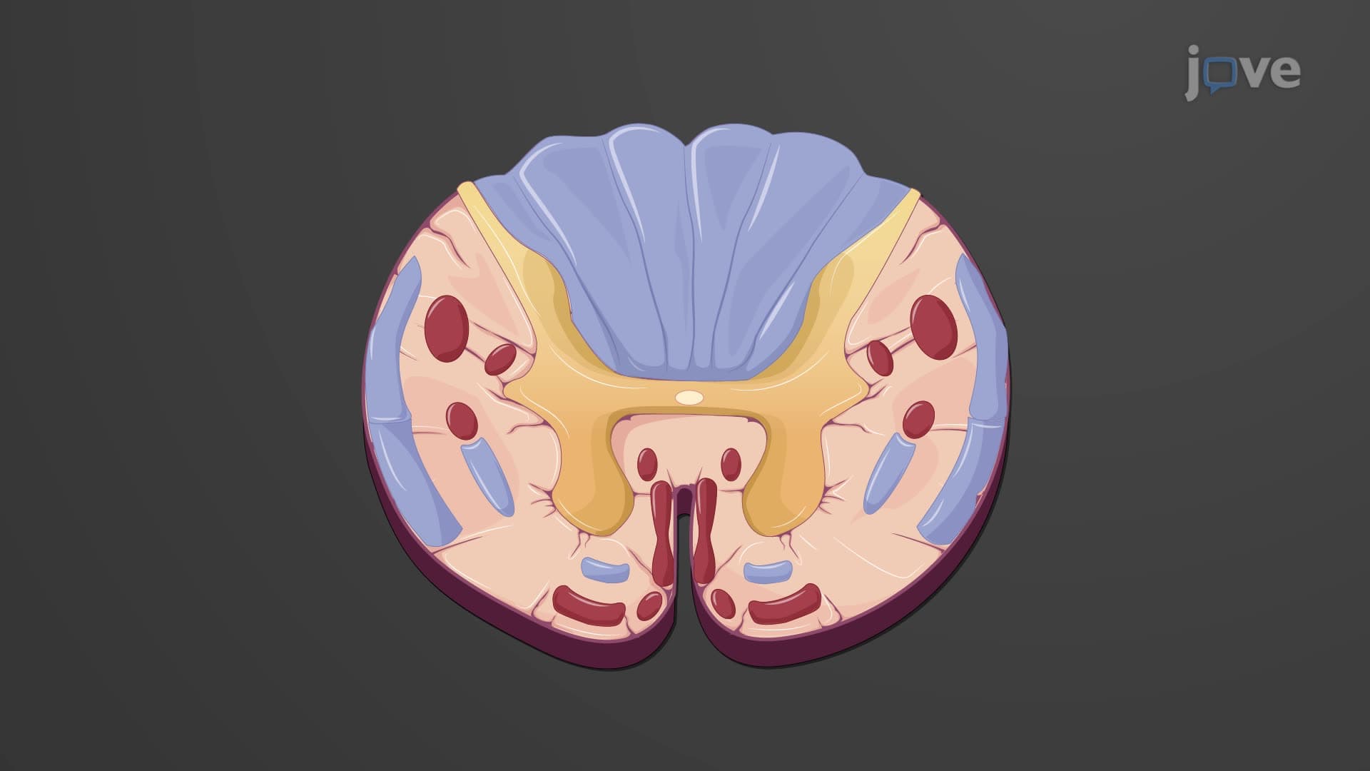
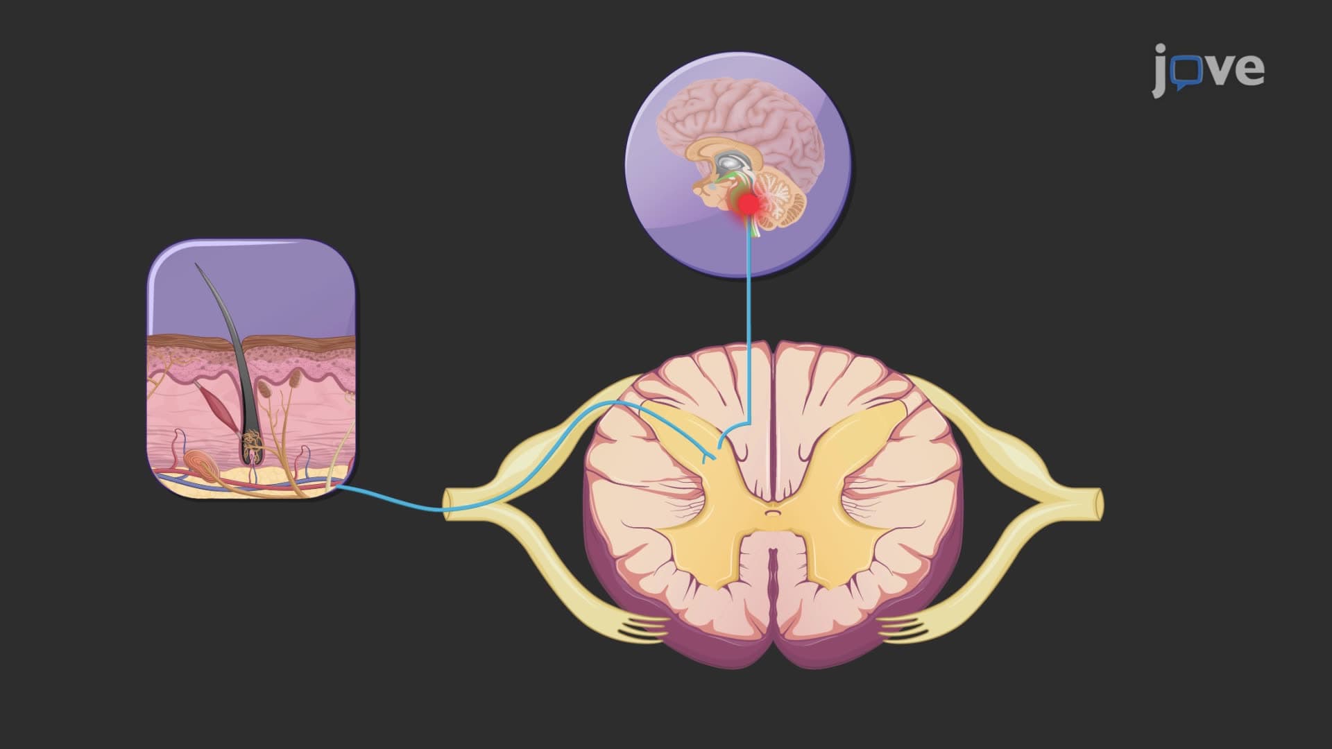
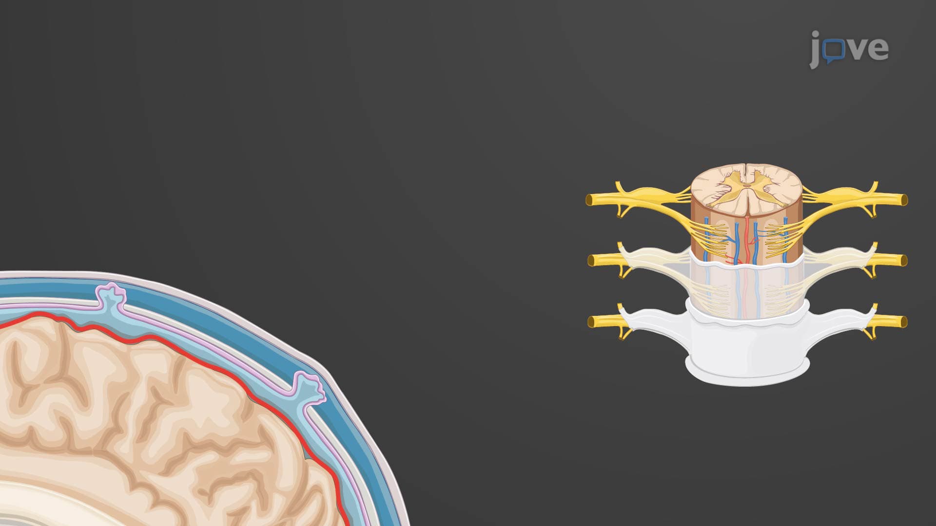
The formation of each spinal nerve begins within the vertebral column, where dorsal root ventral root systems converge. The dorsal root contains exclusively sensory (afferent) fibers that transmit information about touch, temperature, pain, and proprioception from peripheral receptors to the spinal cord. Conversely, the ventral root houses motor (efferent) fibers that carry commands from the spinal cord to muscles and glands. When these roots merge just outside the intervertebral foramen, they create a complete spinal nerve capable of bidirectional communication.
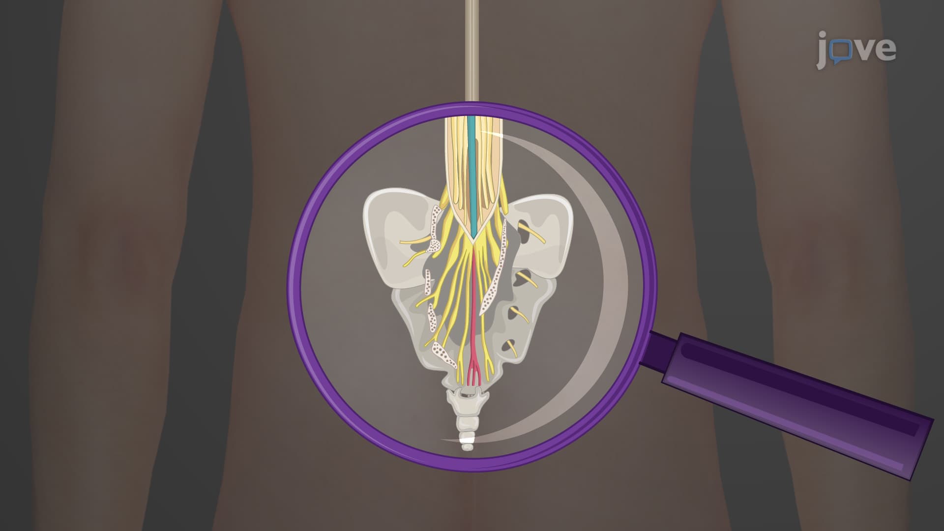
The spinal nerve 31 pairs follow a precise anatomical organization that corresponds directly to vertebral segments. This includes eight cervical nerves (C1-C8), twelve thoracic nerves (T1-T12), five lumbar nerves (L1-L5), five sacral nerves (S1-S5), and one coccygeal nerve (Co1). Interestingly, there are eight cervical spinal nerves but only seven cervical vertebrae because C1 exits above the first cervical vertebra, while C8 exits below the seventh cervical vertebra.
Each ramus branch spinal nerve serves specific anatomical territories. The smaller posterior ramus innervates the deep muscles of the back and overlying skin, explaining why back muscle strains can cause both muscular pain and skin sensitivity. The larger anterior ramus serves the limbs and the anterior/lateral trunk, making it clinically significant for conditions affecting arm and leg function.
Understanding spinal nerve anatomy structure becomes crucial for medical professionals diagnosing neurological conditions. For instance, when a patient presents with numbness following a specific dermatome spinal nerve pattern, physicians can localize the affected spinal nerve level. This principle guides diagnosis in conditions ranging from shingles (which follows dermatome distributions) to spinal cord injuries.
The formation of nerve plexuses represents an evolutionary adaptation that increases functional redundancy and precision. The brachial plexus (C5-T1) gives rise to major arm nerves like the median and ulnar nerves, while the lumbosacral plexus (L1-S4) forms the sciatic nerve. Students preparing for the MCAT or AP Biology exams should understand that plexus formation allows for more complex movement patterns and provides backup innervation pathways.
In clinical settings like those at Mayo Clinic or Cleveland Clinic, neurologists routinely use spinal nerve anatomy knowledge to perform nerve conduction studies and interpret MRI findings. Physical therapists apply this knowledge when designing rehabilitation programs for patients with radiculopathies or peripheral nerve injuries.
Related Micro-courses