Loading subject page…


442,001 views

Video Summary: What Is Neurulation
Ever wonder how your brain and spinal cord first formed during your earliest weeks of development? Neurulation is the fascinating embryonic process that transforms a flat sheet of cells into the hollow neural tube that becomes your entire nervous system. This critical developmental stage occurs around week 3-4 of human pregnancy, similar to what researchers study in mouse models at leading US institutions like Harvard Medical School. Watch the full video on JoVE Coach to master this concept with expert-led visuals and step-by-step explanations.
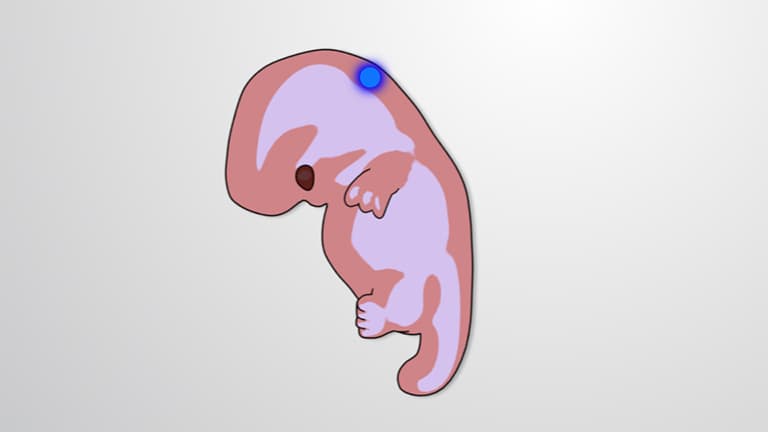
Neurulation represents one of embryology's most elegant transformations-converting a simple flat sheet of cells into the complex three-dimensional structure that will become the brain and spinal cord. This process occurs during the third and fourth weeks of human development, making it a critical period that determines proper nervous system formation.
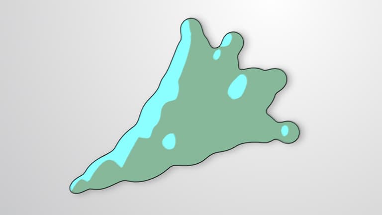
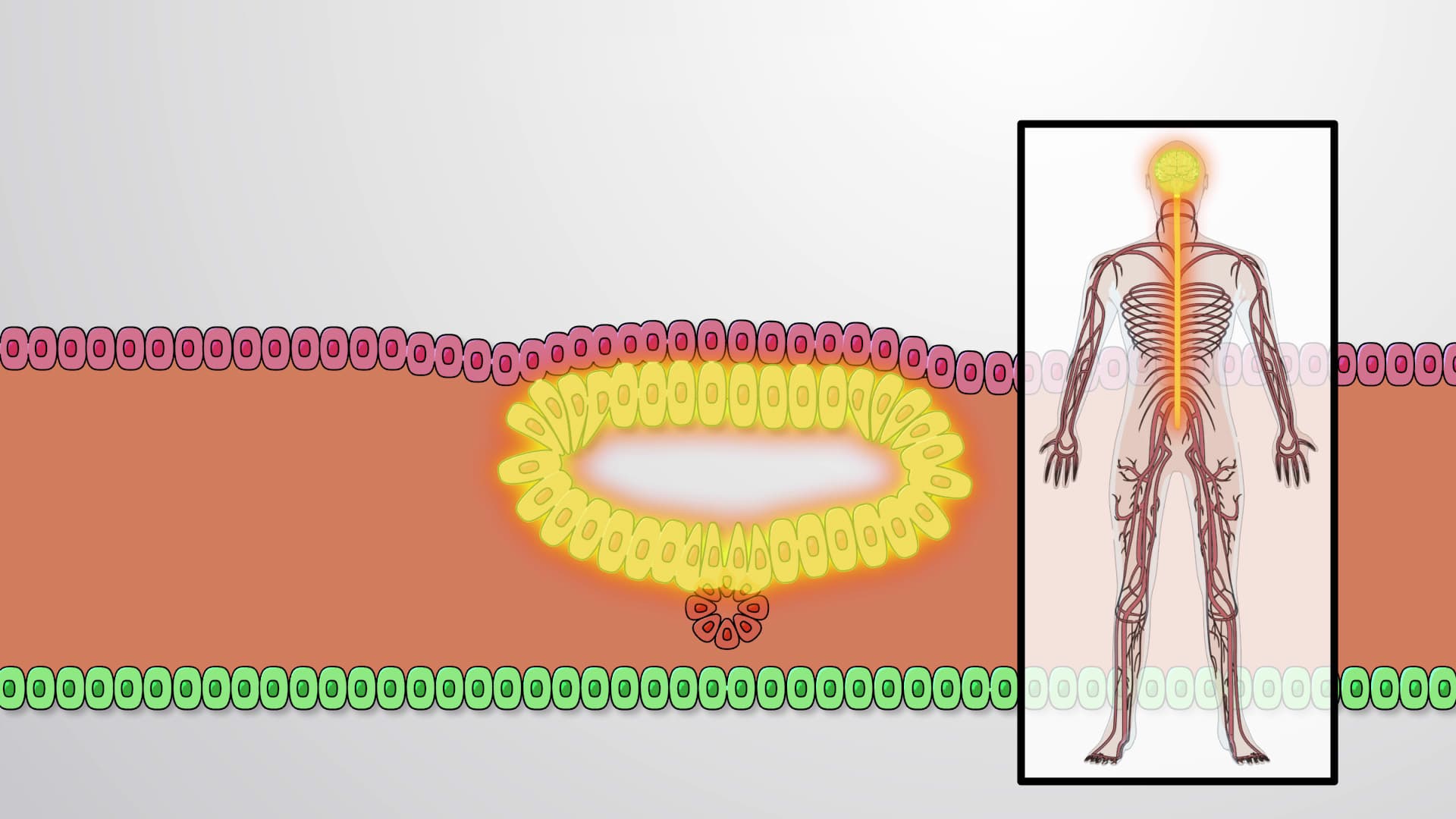
The process begins with the neural plate, a thickened region of ectoderm that forms along the embryo's midline. Beneath this plate lies the notochord, a rod-like structure of mesodermal cells that serves as both a scaffold and signaling center. The notochord releases crucial molecular signals, including proteins from the Sonic hedgehog pathway, that guide the overlying neural tissue through its dramatic transformation.
At the cellular level, neurulation involves remarkable shape changes. Initially, the neural plate cells appear columnar-tall and narrow like miniature columns. As neurulation progresses, these cells undergo apical constriction, transforming into wedge shapes that create the mechanical forces needed for tissue folding. This cellular choreography resembles origami performed at the microscopic level.
Primary neurulation unfolds through a series of coordinated folding events. The neural plate first develops a central groove, then its edges-called neural folds-begin rising upward. Three distinct hinge points drive this process: one central hinge anchored to the notochord, and two lateral hinges attached to the surrounding surface ectoderm.
As the neural folds approach each other, specialized cells at their tips facilitate the final fusion step. Once complete, the newly formed neural tube separates from the surface ectoderm, which will go on to form skin. The hollow neural tube then develops into either spinal cord (in the trunk region) or brain (in the head region).
Understanding neurulation proves essential for medical students preparing for the MCAT or USMLE Step 1, where neural tube defects frequently appear as exam topics. In clinical practice, disrupted neurulation leads to conditions like spina bifida, affecting roughly 1 in 1,000 births in the United States according to CDC data.
Folic acid supplementation during pregnancy helps prevent neural tube defects, demonstrating neurulation's practical medical importance. Research institutions like Johns Hopkins and the University of California system continue investigating neurulation mechanisms, contributing to our understanding of both normal development and birth defect prevention strategies.
Related Micro-courses